. 2022 Jun 22;96(12):e0050822.
doi: 10.1128/jvi.00508-22. Epub 2022 May 24.

DLK-Dependent Biphasic Reactivation of Herpes Simplex Virus Latency Established in the Absence of Antivirals

Affiliations

DLK-Dependent Biphasic Reactivation of Herpes Simplex Virus Latency Established in the Absence of Antivirals

Sara Dochnal et al. J Virol. .

Abstract

Understanding the molecular mechanisms of herpes simplex virus 1 (HSV-1) latent infection and reactivation in neurons requires the use of in vitro model systems. Establishing a quiescent infection in cultured neurons is problematic, as any infectious virus released can superinfect the cultures. Previous studies have used the viral DNA replication inhibitor acyclovir to prevent superinfection and promote latency establishment. Data from these previous models have shown that reactivation is biphasic, with an initial phase I expression of all classes of lytic genes, which occurs independently of histone demethylase activity and viral DNA replication but is dependent on the cell stress protein DLK. Here, we describe a new model system using HSV-1 Stayput-GFP, a reporter virus that is defective for cell-to-cell spread and establishes latent infections without the need for acyclovir. The establishment of a latent state requires a longer time frame than previous models using DNA replication inhibitors. This results in a decreased ability of the virus to reactivate using established inducers, and as such, a combination of reactivation triggers is required. Using this system, we demonstrate that biphasic reactivation occurs even when latency is established in the absence of acyclovir. Importantly, phase I lytic gene expression still occurs in a histone demethylase and viral DNA replication-independent manner and requires DLK activity. These data demonstrate that the two waves of viral gene expression following HSV-1 reactivation are independent of secondary infection and not unique to systems that require acyclovir to promote latency establishment. IMPORTANCE Herpes simplex virus-1 (HSV-1) enters a latent infection in neurons and periodically reactivates. Reactivation manifests as a variety of clinical symptoms. Studying latency and reactivation in vitro is invaluable, allowing the molecular mechanisms behind both processes to be targeted by therapeutics that reduce the clinical consequences. Here, we describe a novel in vitro model system using a cell-to-cell spread-defective HSV-1, known as Stayput-GFP, which allows for the study of latency and reactivation at the single neuron level. We anticipate this new model system will be an incredibly valuable tool for studying the establishment and reactivation of HSV-1 latent infection in vitro. Using this model, we find that initial reactivation events are dependent on cellular stress kinase DLK but independent of histone demethylase activity and viral DNA replication. Our data therefore further validate the essential role of DLK in mediating a wave of lytic gene expression unique to reactivation.

Keywords: dual leucine zipper kinase; herpes simplex virus; human herpesviruses; in vitro model systems; latent infection; reactivation.


Conflict of interest statement

The authors declare no conflict of interest.

Figures
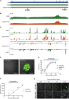
FIG 1
FIG 1
Stayput-GFP replicates as wild type but is unable to spread. (A) Schematic overview of HSV-1 strain SC16, the gH-deletion mutant SCgHZ, and Stayput-GFP. The gH deletion/LacZ insertion and Us11-GFP insertion sites are shown by blue and green triangles, respectively. (B and C) Coverage plots derived from (B) nanopore gDNA sequencing and (C) nanopore direct RNA sequencing of SCgHZ and Stayput-GFP. Sequence read data were aligned against the SC16 reference genome and demonstrate a drop in coverage at the gH locus. (D) Plaque-forming assay of Stayput-GFP on Vero (left) or gH-complementing Vero-F6 (right) cells 48 h postinfection at a 10−7 dilution. The GFP-positive area in the example image is reported on the top left of each image. Titer of viral stock reported bottom left of each image. (E) Vero-F6 cells were infected with Stayput-GFP, SCgHZ, or Us11-GFP at an MOI of 5. Infectious virus was collected over time and titrated on Vero-F6 cells (n = 3 biological replicates). (F and G) Neonatal sympathetic neurons were infected at an MOI of 0.5 PFU/cell with Stayput-GFP or Us11-GFP in the absence of DNA replication inhibitors. Us11-GFP-positive neurons were counted over time (n = 3 biological replicates). Shapiro-Wilk normality test. Unpaired Student’s t test between Us11-GFP and Stayput-GFP. ****, P < 0.0001. The means and standard errors of the means (SEMs) are shown.
FIG 2
FIG 2
Stayput-GFP in a latency and reactivation model using ACV to promote latency establishment. (A) The latency and reactivation model scheme. Neonatal sympathetic neurons were infected with Stayput-GFP, parent virus SCgHZ, or wild-type Us11-GFP at an MOI of 7.5 PFU/cell in the presence of ACV (50 μM). Then, 6 days later, ACV was removed, and 2 days later, cultures were reactivated with LY294002 (20 μM). (B) The numbers of GFP-positive neurons in a single well (containing approximately 5,000 neurons) for Stayput-GFP and wild-type Us11-GFP were counted over time. (C to E) Viral gene expression also was quantified by RT-qPCR for immediate early (ICP27), early (ICP8), and late (gC) genes at 20 h (C), 48 h (D), and 72 h (E) poststimulus. Relative expression to unreactivated samples and cellular control (mGAPDH). n = 6 biological replicates from 3 litters. Normality was determined by Kolmogorov-Smirnov test in panels B to E. Mann-Whitney (B) or Kruskal-Wallis with comparison of means (C to E); *, P < 0.05; **, P < 0.01; ***, P < 0.001. The means and SEMs are represented. Individual biological replicates are indicated in panels C to E.
FIG 3
FIG 3
Stayput-GFP can be used to create a quiescence model in the absence of viral DNA replication inhibitors in neonatal sympathetic neurons. Neonatal sympathetic neurons were infected with Stayput-GFP at an MOI of 7.5 PFU/cell, and the numbers of Us11-GFP-positive neurons were quantified. (A) n = 9 biological replicates from 3 litters. (B) SYTOX Orange-positive neurons were also quantified over time (n = 3). (C) Following infection, the same field of view was imaged to track GFP and SYTOX Orange (250 μm scale bar for field of view [FOV], 25 μm scale bar for zoom) over time. (D to G) Lytic (D to F) and latent (G) viral transcripts (n = 6) were quantified up to 40 days postinfection. (H) Viral DNA load (n = 6) was also quantified up to 40 days postinfection. Individual biological replicates along with the means and SEMs are shown.
FIG 4
FIG 4
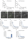
Reactivation decreases with length of time infected. Sympathetic neurons were infected with Stayput-GFP at an MOI of 7.5 PFU/cell and were treated with LY294002 when GFP-positive neurons were no longer detected (approximately 30 days postinfection). GFP-positive neurons were quantified over time; (A) peak GFP (48 h poststimulus) is represented. Neonatal SCGs were infected at age postnatal day 8 (P8) with Stayput-GFP in the presence of ACV. (B) ACV was removed 6 days postinfection, and reactivation was triggered at the indicated times postinfection. (C) Neonatal SCGs were infected as described above after different lengths of time in vitro, representing indicated postnatal ages, and reactivated 8 days postinfection with LY294002. n = 12 biological replicates from 3 litters. Normality was determined by the Kolmogorov-Smirnov test. Unpaired Student’s t test (B) or the Mann-Whitney test (A and C) was used based on normality of data. **, P < 0.01; ***, P < 0.001; ****, P < 0.0001. Individual biological replicates along with the means and SEMs are shown.
FIG 5
FIG 5
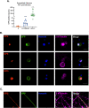
Viral protein synthesis can be induced from neurons quiescently infected with Stayput-GFP using a combination of stimuli. Neonatal sympathetic neurons or adult sensory neurons were infected at an MOI of 7.5 PFU/cell with Stayput-GFP in the absence of viral DNA replication inhibitors. Following the loss of GFP, signaling quiescence of the culture, wells were reactivated with a variety of triggers, including combinations of LY294002 (20 μM), forskolin (60 μM), and heat shock (43°C for 3 h), as well as a superinfection with untagged F strain at an MOI of 10 PFU/cell. (A) GFP was quantified over time, and the peak GFP, at 48 h poststimulus, is depicted. n = 12 biological replicates. (B to C) Neurons were fixed at 48 h poststimulus and stained with viral immediate early (ICP4), early (ICP8) (B), or late (ICP5) (C) protein (red), GFP (green), Hoescht (blue), and β II tubulin (magenta). 25-μm scale bar.
FIG 6
FIG 6
Viral gene expression can be restarted following latency establishment. Neonatal sympathetic neurons or adult sensory neurons were infected at an MOI of 7.5 PFU/cell with Stayput-GFP in the absence of viral DNA replication inhibitors. Following the loss of GFP, signaling quiescence of the culture, wells were reactivated with a variety of triggers, including combinations of LY294002 (20 μM), forskolin (60 μM), and heat shock (43°C for 3 h). (A to F) Immediate early (A), early (B), and late (C to F) viral transcripts were investigated over time following the stimulus. Reactivated samples were compared to latent samples described as 0 h poststimulus. n = 9 biological replicates from 3 litters. Mann-Whitney against 0 h (A to D). Normality was determined by the Kolmogorov-Smirnov test; *, P < 0.05; **,P < 0.01; ***, P < 0.001; ****, P < 0.0001. Individual biological replicates along with the means and SEMs are shown.
FIG 7
FIG 7
Reactivation from quiescently infected adult sensory trigeminal ganglia neurons. Neurons isolated from the trigeminal ganglia (TG) of female mice were infected with Stayput-GFP at an MOI of 7.5 PFU/cell. (A) The resolution of lytic infection was monitored over time by imaging and counting GFP-positive neurons. n = 12 biological replicates from 3 dissections. The mean and SEM are shown. Following the loss of GFP, signaling quiescence of the culture, wells were reactivated with a combination of LY294002 (20 μM), forskolin (60 μM), and heat shock (43°C for 3 h), as well as a superinfection with untagged F strain at an MOI of 10 PFU/cell. (B) GFP was quantified over time, and the peak GFP, at 48 h poststimulus, is depicted. n = 9 biological replicates. (C to F) Immediate early (C), early (D), and late (E and F) viral transcripts were investigated over time following the stimulus. n = 6 replicates. Mann-Whitney test against 0 h; *, P < 0.05; **, P < 0.01; ***, P < 0.001; ****, P < 0.0001. Individual biological replicates along with the means and SEMs are shown.
FIG 8
FIG 8
Reactivation is dependent on DLK. Cultures were infected with Stayput-GFP at an MOI of 7.5 PFU/cell in the absence of ACV. Following loss of GFP, cultures were reactivated with a combination of LY294002, forskolin, and heat shock in the presence of DLK inhibitor GNE-3511 (4 μM). (A to D) Immediate early (ICP27) and early (ICP8/UL30) viral genes (A to C) were investigated at 12.5 h poststimulus, and GFP was counted over time (D). Peak GFP, consistently around 48 h poststimulus, is presented. n = 9 biological replicates from 3 litters. Normality was determined by the Kolmogorov-Smirnov test. Mann-Whitney test; *, P < 0.05; **, P < 0.01; ***, P < 0.001; ****, P < 0.0001. The mean and SEM are shown.
FIG 9
FIG 9
The early phase of lytic gene expression following a reactivation stimulus is independent of demethylase activity. Cultures were infected with Stayput-GFP at an MOI of 7.5 PFU/cell. Following loss of GFP, cultures were reactivated with a combination of LY294002, forskolin, and heat shock in the presence of H3K27 demethylase inhibitor GSK-J4 (2 μM) or H3K9 demethylase inhibitor OG-L002 (20 μM). Immediate early (ICP27) and early (ICP8/UL30) viral genes (A to C) were investigated at 12.5 h poststimulus, and GFP was counted over time. Peak GFP is presented. n = 3 biological replicates from 3 litters. Normality was determined by the Kolmogorov-Smirnov test. Mann-Whitney; *, P < 0.05; **, P < 0.01; ***, P < 0.001; ****, P < 0.0001. The mean and SEM are shown.
FIG 10
FIG 10
Differential dependence on viral DNA replication between phase I and II reactivation. Cultures were infected with Stayput-GFP at an MOI of 7.5 PFU/cell in the absence of ACV. Following loss of GFP, cultures were reactivated with a combination of LY294002, forskolin, and heat shock in the presence of ACV (50 μM). (A to D) Late (VP16, gC, UL10) genes (A to C) were investigated at 22 h poststimuli. GFP was counted over time, and peak GFP is presented (D). n = 12 biological replicates. Normality was determined by the Kolmogorov-Smirnov test. Mann-Whitney; *, P < 0.05; **, P < 0.01; ***, P < 0.001; ****, P < 0.0001. The mean and SEM are shown.

Similar articles

Cited by

References

    1. Richter ER, Dias JK, Gilbert JE, II, Atherton SS. 2009. Distribution of herpes simplex virus type 1 and varicella zoster virus in ganglia of the human head and neck. J Infect Dis 200:1901–1906. 10.1086/648474. - DOI - PMC - PubMed
    1. Baringer JR, Pisani P. 1994. Herpes simplex virus genomes in human nervous system tissue analyzed by polymerase chain reaction. Ann Neurol 36:823–829. 10.1002/ana.410360605. - DOI - PubMed
    1. Baringer JR, Swoveland P. 1973. Recovery of herpes-simplex virus from human trigeminal ganglions. N Engl J Med 288:648–650. 10.1056/NEJM197303292881303. - DOI - PubMed
    1. Warren KG, Brown SM, Wroblewska Z, Gilden D, Koprowski H, Subak-Sharpe J. 1978. Isolation of latent herpes simplex virus from the superior cervical and vagus ganglions of human beings. N Engl J Med 298:1068–1069. 10.1056/NEJM197805112981907. - DOI - PubMed
    1. Singh N, Tscharke DC. 2020. Herpes simplex virus latency is noisier the closer we look. J Virol 94:e01701-19. 10.1128/JVI.01701-19. - DOI - PMC - PubMed

Publication types