. 2022 May 19;18(5):e1010437.
doi: 10.1371/journal.ppat.1010437. eCollection 2022 May.

Comparison of herpes simplex virus 1 genomic diversity between adult sexual transmission partners with genital infection

Affiliations

Comparison of herpes simplex virus 1 genomic diversity between adult sexual transmission partners with genital infection

Molly M Rathbun et al. PLoS Pathog. .

Abstract

Herpes simplex virus (HSV) causes chronic infection in the human host, characterized by self-limited episodes of mucosal shedding and lesional disease, with latent infection of neuronal ganglia. The epidemiology of genital herpes has undergone a significant transformation over the past two decades, with the emergence of HSV-1 as a leading cause of first-episode genital herpes in many countries. Though dsDNA viruses are not expected to mutate quickly, it is not yet known to what degree the HSV-1 viral population in a natural host adapts over time, or how often viral population variants are transmitted between hosts. This study provides a comparative genomics analysis for 33 temporally-sampled oral and genital HSV-1 genomes derived from five adult sexual transmission pairs. We found that transmission pairs harbored consensus-level viral genomes with near-complete conservation of nucleotide identity. Examination of within-host minor variants in the viral population revealed both shared and unique patterns of genetic diversity between partners, and between anatomical niches. Additionally, genetic drift was detected from spatiotemporally separated samples in as little as three days. These data expand our prior understanding of the complex interaction between HSV-1 genomics and population dynamics after transmission to new infected persons.


Conflict of interest statement

I have read the journal’s policy and the authors of this manuscript have the following competing interests: CJ reports funding from UpToDate (royalties), AbbVie (consulting), Gilead (consulting), MedPace (DSMB), NIH and CDC. AW reports funding from NIH, Sanofi (grants), Aicuris (consulting), X-vax (consulting), Auritec (consulting), GSK (grants), Merck (DSMB), Crozet (consulting), VIR (consulting), and UptoDate (royalties).

Figures
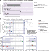
Fig 1
Fig 1. Overview of samples sequenced from each transmission pair during a one-year period.
(A) Participants were enrolled for clinical study after the detection of first-episode genital infections. Transmission partners were referred to the study through their partners. In panel (A) the position of each grey bar indicates the relative calendar time frame of sampling for each participant, and the bar length reflects how long they were enrolled in the study. E.g., Participant 40 was referred as a source partner and thus completed only one month of daily swabs, whereas participant 41 enrolled with first-episode genital infection and completed a full year of the study. LTFU indicates participants lost to follow-up. (B) Sequenced samples are plotted by participant, according to the number of days since each participant’s first reported symptoms. Dot color denotes sample type: oral area (blue), genital area (red), or site-specific genital lesion (green). Larger shaded bars early and late in each participant’s first year of infection denote the 30-day sessions of daily self-collected swabs. Lesion samples were also collected at clinic visits at intervening times. Magnified panels in (B) highlight selected time frames, including the two sessions of daily self-collected swabs. Dots stacked vertically in these panels indicate samples collected on the same day. Open circles designate samples from pre-existing multi-year infections (v40 and v46 oral). Participant 40 (Pair 1, source) presented with a pre-existing oral infection, and a genital infection with an unknown start date. Participant 46 (Pair 4, source) entered the study with a pre-existing oral infection.
Fig 2
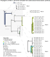
Fig 2. Network graph comparing 65 globally sampled HSV-1 genomes and 33 viral genomes from new adult sexual transmission pairs (n = 10 participants).
The starburst pattern in this network graph (which excluded gaps) exemplifies the unique genetic diversity in each randomly sampled HSV-1 genome from around the globe (black branches). Consensus genomes sampled over time from individual participants, or between transmission partners, indicated near complete nucleotide conservation (≥ 98% identity) and formed thick clusters of branches. Genome names in black denote well-known strains or isolates that are nearest-neighbors to the new transmission pair samples (highlighted in magenta). Genome names highlighted in blue indicate previously published data on viral genomes from parent-child familial transmission pairs. The gray scale bar indicates approximately 0.1% nucleotide divergence. Internal reticulations within the network reflect likely historical recombination events. All strain names and prior references for these are provided in S1 Table.
Fig 3
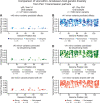
Fig 3. Neighbor-joining phylogram reveals consensus-level differences among HSV-1 genomes from five transmission pairs (n = 10 individuals).
Single nucleotide differences or variants (SNVs) in the consensus-level viral genomes occur in three out of five transmission pairs (Pairs 1, 2, and 4; excluding those that occurred at repetitive elements). These SNVs demonstrate that genetic diversity may arise transiently between transmission partners and/or within individual infections. Branches in the phylogram are shaded according to the range of SNVs detected between genomes. Comparisons with a small number of SNVs (1 ≤ n ≤ 10, light green) are consistent with the genome conservation observed in cases of familial transmission. Comparisons with a larger number of SNVs (100 ≤ n ≤ 300, dark blue) are more similar to the level of divergence between globally sampled genomes in the larger network graph analysis (Fig 2). The gray scale bar indicates approximately 0.1% nucleotide divergence.
Fig 4
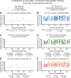
Fig 4. A homopolymer frameshift mutation in viral gene UL22, encoding glycoprotein H (gH), occurs in both partners of Pair 3.
The consensus viral genomes from participants 44 and 45 (Pair 3) are identical, as there were no consensus-level single-nucleotide differences between these partners, outside of repetitive elements in the genome. The consensus sequences for the UL22 gene encoding gH of viral genomes v44 and v45 were compared to the HSV-1 strain 17 reference genome. This revealed a T7 to T8 homopolymer frame-shift mutation affecting the C-terminus of UL22. The T-homopolymer is indicated with red lettering and a pink background shading. This frameshift mutation ablates the canonical stop codon for gH, alters the four amino acid C-terminal tail found in all other HSV-1 isolates, and extends the encoded protein by 14 amino acids. Thus, the reference strain encodes a viral gH protein of 838 amino acids in length, while the viral genomes of both Pair 3 partners encode a predicted length of 852 amino acids for gH. The nucleotide position is numbered based on the position of this frameshift within the UL22 gene.
Fig 5
Fig 5. With-host variation occurs in viral genome populations transmitted within and between partners over time.
(A) HSV-1 genomes were sequenced from Pair 2 specimens spanning multiple shedding episodes over the first year of infection. Analysis of within-host diversity in these samples revealed two MVs of interest within the gene encoding glycoprotein B (gB, UL27). (B) One variant (causing a synonymous mutation at amino acid (AA) position 877 in gB) was detected in four v42 samples spanning two shedding episodes of this source partner. Similar fluctuations at this locus were also detected in three v43 samples, spanning three shedding episodes of this recipient partner. In both partners, early samples showed a dominant G nucleotide, which shifted to a dominant A nucleotide in later samples. In later v43 samples (see Table 2), this position harbored 100% penetrance of the “A” variant. (C) A second site of within-host diversity in gB was specific to the viral population of v43. This minor variant encoded a nonsynonymous mutation at AA position 175 in gB. This variant was present at a high frequency (65%) in the site-specific lesion sample on day 349, but it was detectable at a far lower frequency (4%) in a genital area swab collected on the same day. The frequency of this variant dropped to 7% in the day 352 lesion sample, and it was undetectable in that day’s genital area swab.
Fig 6
Fig 6. Oral samples from Pair 1 indicate shared within-host genetic diversity.
Both the source and recipient partners in Pair 1 harbored high levels of within-host HSV-1 genetic diversity, or minor variants (MV), in at least one sample each. The source sample v40 had an average coverage depth of ~428X, allowing for MV analysis with a 2% threshold (v40_y14_oral1; 27 MVs detected). The recipient’s sample v41 had an average coverage depth of 28X, and thus a stringent threshold that required ≥ 20% MV frequency was applied (v41_d354_oral, 530 MVs detected). (A-B) In both cases, MVs were randomly distributed across the HSV-1 genome, with a range of impacts from synonymous to nonsynonymous to intergenic. (C-D) In the v41 sample, a majority of variants were observed to be connected to nearby variants on the same sequencing read. (E-F) Most MVs were shared between partners; colors indicate if each MV was above threshold, below threshold, or not detected in the transmission partner. See S2 Table for position and frequency of all MV loci.
Fig 7
Fig 7. Samples from Pair 4 indicate shared within-host genetic diversity across anatomical niches.
Both the source and recipient partners in Pair 4 harbored high within-host HSV-1 genetic diversity in at least one sample. In this comparison, both samples had an average coverage depth ~30X, so a stringent threshold requiring ≥ 20% MV frequency was applied when detecting MVs (v46_y16_oral had 34X coverage and 5 MV detected, while v47_d61-79_gen had 31X coverage and 256 MV detected). (A-B) MVs and their predicted effects were randomly distributed across the HSV-1 genome. (C-D) In the v47 sample, a majority of variants were connected to other nearby variants by the same sequencing read. (E-F) Most MVs were shared between transmission partners; colors indicate if each MV was above threshold, below threshold, or not detected in the transmission partner. See S2 Table for position and frequency of all MV loci.
Fig 8
Fig 8. Within-host diversity in source participant v48 varies over time within a single shedding episode.
Within-host diversity analysis of v48 oral samples on days 100–103 since first symptoms revealed minor variants (MV) only on days 100 and 101. (A) The overall number of minor variants increased from days 100 to 101 (v48_d100_oral had 350X coverage and 62 MV detected, while v48_d101_oral had 466X coverage and 234 MV detected). (B) The individual frequencies of these MVs remained between 2–20%. (C) These samples were collected at peak viral load (indicated by lavender shading), at the end of this shedding episode for participant 48. The following two oral samples (on days 102–103) had zero MVs detected. (D-E) The MVs in samples from day 100 and 101 were randomly distributed across the HSV-1 genome. Many of the same MV loci detected in the d100 sample were also detected on d101. See S2 Table for position and frequency of all MV loci.

Similar articles

Cited by

References

    1. James C, Harfouche M, Welton NJ, Turner KM, Abu-Raddad LJ, Gottlieb SL, et al.. Herpes simplex virus: global infection prevalence and incidence estimates, 2016. Bull World Health Organ. 2020;98: 315–329. doi: 10.2471/BLT.19.237149 - DOI - PMC - PubMed
    1. Kriesel JD, Bhatia A, Thomas A. Cold sore susceptibility gene-1 genotypes affect the expression of herpes labialis in unrelated human subjects. Hum Genome Var. 2014;1: 14024. doi: 10.1038/hgv.2014.24 - DOI - PMC - PubMed
    1. Kleinstein SE, Shea PR, Allen AS, Koelle DM, Wald A, Goldstein DB. Genome-wide association study (GWAS) of human host factors influencing viral severity of herpes simplex virus type 2 (HSV-2). Genes Immun. 2018; 1. doi: 10.1038/s41435-018-0013-4 - DOI - PMC - PubMed
    1. Ramchandani MS, Jing L, Russell RM, Tran T, Laing KJ, Magaret AS, et al.. Viral Genetics Modulate Orolabial Herpes Simplex Virus Type 1 Shedding in Humans. J Infect Dis. 2019;219: 1058–1066. doi: 10.1093/infdis/jiy631 - DOI - PMC - PubMed
    1. Shipley MM, Renner DW, Ott M, Bloom DC, Koelle DM, Johnston C, et al.. Genome-wide surveillance of genital herpes simplex virus type 1 from multiple anatomic sites over time. J Infect Dis. 2018;218: 595–605. doi: 10.1093/infdis/jiy216 - DOI - PMC - PubMed

Publication types