. 2022 May 7;11(9):1581.
doi: 10.3390/cells11091581.

Multifaceted and Intricate Oncogenic Mechanisms of NDRG1 in Head and Neck Cancer Depend on Its C-Terminal 3R-Motif

Affiliations

Multifaceted and Intricate Oncogenic Mechanisms of NDRG1 in Head and Neck Cancer Depend on Its C-Terminal 3R-Motif

Guo-Rung You et al. Cells. .

Abstract

N-Myc downstream-regulated 1 (NDRG1) has inconsistent oncogenic functions in various cancers. We surveyed and characterized the role of NDRG1 in head and neck cancer (HNC). Cellular methods included spheroid cell formation, clonogenic survival, cell viability, and Matrigel invasion assays. Molecular techniques included transcriptomic profiling, RT-qPCR, immunoblotting, in vitro phosphorylation, immunofluorescent staining, and confocal microscopy. Prognostic significance was assessed by Kaplan-Meier analysis. NDRG1 participated in diverse oncogenic functions in HNC cells, mainly stress response and cell motility. Notably, NDRG1 contributed to spheroid cell growth, radio-chemoresistance, and upregulation of stemness-related markers (CD44 and Twist1). NDRG1 facilitated cell migration and invasion, and was associated with modulation of the extracellular matrix molecules (fibronectin, vimentin). Characterizing the 3R-motif in NDRG1 revealed its mechanism in the differential regulation of the phenotypes. The 3R-motif displayed minimal effect on cancer stemness but was crucial for cell motility. Phosphorylating the motif by GSK3b at serine residues led to its nuclear translocation to promote motility. Clinical analyses supported the oncogenic function of NDRG1, which was overexpressed in HNC and associated with poor prognosis. The data elucidate the multifaceted and intricate mechanisms of NDRG1 in HNC. NDRG1 may be a prognostic indicator or therapeutic target for refractory HNC.

Keywords: 3R-motif; NDRG1; cancer stemness; cell motility; head and neck cancer; prognosis.


Conflict of interest statement

The authors declare that they have no conflict of interest.

Figures

Figure 1
Figure 1
Global survey of molecular mechanisms reveals NDRG1 functions mainly in the regulations of stemness property and invasion phenotype. (a) Successful transfection of NDRG1-FL in three HNC cell lines. After transfection of NDRG1 overexpression (FL) or the vector control (Vt) plasmid into HNC cell lines (OECM1, SAS, and FaDu), the NDRG1 protein expression was determined by Western blot analysis. Because the EGFP protein sequence was fused in-frame in the NDRG1 expressing plasmids, the NDRG1 protein in Western blot gel appeared by two bands: the lower one of the endogenous cellular NDRG1 and the higher one of the transfected NDRG1 protein. The GAPDH level was used as an internal control. (b) The morphology of OECM1 cells transfected with vector or the NDRG1-FL plasmids, scale bar 20 µm. (c) The transcriptomic and bioinformatic analyses for the overexpression of NDRG1 in OECM1 cells. Affymetrix Exon array was performed, and the differential expressed genes (DEGs) were identified by ANOVA analysis with the two-fold change in the gene expression level. Distribution of NDRG1 regulated downstream molecules with functional category, as determined by GO analysis from DAVID website. (d) The most significant KEGG functional pathways are regulated by NDRG1, as determined by KEGG enrichment analysis from the DAVID website. (e) GSEA analysis of the stemness property and invasion phenotype in vector control or NDRG1 overexpressed in the OECM1 cells.
Figure 2
Figure 2
NDRG1 promoted cancer stemness by cancer stemness markers. (a) Spheroid cell formation was increased in NDRG1 overexpressed in HNC cells. After transfection of NDRG1-FL (FL) or the vector (Vt) plasmid into HNC cells (OECM1, FaDu), the effect of tumorsphere number and size was determined by microscope. (b) Overexpression of NDRG1-induced radioresistant ability in HNC cells, as determined by clonogenic survival assay. The error bars shown in the relevant figures indicate the standard deviation of the quantification results in the experiments. (*** p < 0.001; ANOVA test). (c) NDRG1 protected cisplatin-induced cell death, as determined by the MTS assay. (d,e) OECM1 and FaDu cells were transfected with NDRG1-FL or vector control and cells were collected for RT-qPCR analysis (d) or the cellular proteins were extracted and subjected to Western blot analysis and determination of protein expressions by antibodies (e). The GAPDH was used as an internal control. All the experiments were performed three times independently and typical results were shown. The error bars shown in the relevant figures indicate the standard deviation of the quantification results in all experiments. (* p < 0.05; ** p < 0.01; *** p < 0.001; n.s., no significance; t-test).
Figure 2
Figure 2
NDRG1 promoted cancer stemness by cancer stemness markers. (a) Spheroid cell formation was increased in NDRG1 overexpressed in HNC cells. After transfection of NDRG1-FL (FL) or the vector (Vt) plasmid into HNC cells (OECM1, FaDu), the effect of tumorsphere number and size was determined by microscope. (b) Overexpression of NDRG1-induced radioresistant ability in HNC cells, as determined by clonogenic survival assay. The error bars shown in the relevant figures indicate the standard deviation of the quantification results in the experiments. (*** p < 0.001; ANOVA test). (c) NDRG1 protected cisplatin-induced cell death, as determined by the MTS assay. (d,e) OECM1 and FaDu cells were transfected with NDRG1-FL or vector control and cells were collected for RT-qPCR analysis (d) or the cellular proteins were extracted and subjected to Western blot analysis and determination of protein expressions by antibodies (e). The GAPDH was used as an internal control. All the experiments were performed three times independently and typical results were shown. The error bars shown in the relevant figures indicate the standard deviation of the quantification results in all experiments. (* p < 0.05; ** p < 0.01; *** p < 0.001; n.s., no significance; t-test).
Figure 3
Figure 3
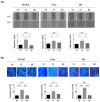
NDRG1 promoted cell motility via ECM mechanism. (a) NDRG1 facilitated cell migration in HNC cells. After seeding the NDRG1 overexpression (FL) or vector control (Vt) transfected cells into ibidi culture inserts for 8 h, cells were subjected to wound healing migration analysis. (b) NDRG1 increased cell invasion in HNC cells. After seeding the NDRG1 overexpression (FL) or vector control (Vt) transfected cells into Matrigel-coated membranes for 30 h, the cells were subjected to a Matrigel invasion assay. (c,d) OECM1 and FaDu cells were transfected with NDRG1-FL or vector control and cells were collected for RT-qPCR analysis (c) or the cellular proteins were extracted and subjected to Western blot analysis and determination of protein expressions by antibodies (d). The GAPDH was used as an internal control. All the experiments were performed three times independently and typical results were shown. The error bars shown in the relevant figures indicate the standard deviation of the quantification results in all experiments. (* p < 0.05; ** p < 0.01; *** p < 0.001; t-test).
Figure 3
Figure 3
NDRG1 promoted cell motility via ECM mechanism. (a) NDRG1 facilitated cell migration in HNC cells. After seeding the NDRG1 overexpression (FL) or vector control (Vt) transfected cells into ibidi culture inserts for 8 h, cells were subjected to wound healing migration analysis. (b) NDRG1 increased cell invasion in HNC cells. After seeding the NDRG1 overexpression (FL) or vector control (Vt) transfected cells into Matrigel-coated membranes for 30 h, the cells were subjected to a Matrigel invasion assay. (c,d) OECM1 and FaDu cells were transfected with NDRG1-FL or vector control and cells were collected for RT-qPCR analysis (c) or the cellular proteins were extracted and subjected to Western blot analysis and determination of protein expressions by antibodies (d). The GAPDH was used as an internal control. All the experiments were performed three times independently and typical results were shown. The error bars shown in the relevant figures indicate the standard deviation of the quantification results in all experiments. (* p < 0.05; ** p < 0.01; *** p < 0.001; t-test).
Figure 4
Figure 4
NDRG1-3R motif has minimal effect on cancer stemness. (a) Illustration of various NDRG1 truncated forms of EGFP tagged plasmid constructs (NDRG1-FL and NDRG1-dC). NDRG1-FL is composed of 394 amino acids; NDRG1-dC is composed of 337 amino acids, which lost the 3R motif. The mutations of GSK3b phosphorylation site of NDRG1 (NDRG1-3mt) are located at the 3R motif, which was made by triple serine-to-alanine substitutions (S342A, and S352A and S362A). (b) Confirmation of the various NDRG1 truncated forms’ expressions by Western blot analysis. After transfection of the NDRG1-FL, NDRG1-dC, or the vector plasmid into HNC cells, cellular proteins were extracted and subjected to Western blot analysis for NDRG1 expression. (c) Deletion of the C-terminal domain has no influence on NDRG1-induced cancer stemness. After transfection of the NDRG1-FL, NDRG1-dC, or the vector plasmid into HNC cells, the effect of NDRG1 on stem-like property was determined by spheroid cell formation. (d) Deletion of the C-terminal domain has no significant influence on NDRG1-induced radioresistance compared to NDRG1-FL, as determined by clonogenic survival assay in OECM1 cells. The error bars shown in the relevant figures indicate the standard deviation of the quantification results in the experiments. (* p < 0.05; n.s., no significance; ANOVA test). (e) After transfection of the NDRG1-FL, NDRG1-dC, or the vector plasmid into OECM1 cells, RNA was extracted and subjected to RT-qPCR analysis for cancer stemness markers. All the experiments were performed three times independently and typical results were shown. The error bars shown in the relevant figures indicate the standard deviation of the quantification results in all experiments. (* p < 0.05; ** p < 0.01; *** p < 0.001; n.s., no significance; t-test).
Figure 4
Figure 4
NDRG1-3R motif has minimal effect on cancer stemness. (a) Illustration of various NDRG1 truncated forms of EGFP tagged plasmid constructs (NDRG1-FL and NDRG1-dC). NDRG1-FL is composed of 394 amino acids; NDRG1-dC is composed of 337 amino acids, which lost the 3R motif. The mutations of GSK3b phosphorylation site of NDRG1 (NDRG1-3mt) are located at the 3R motif, which was made by triple serine-to-alanine substitutions (S342A, and S352A and S362A). (b) Confirmation of the various NDRG1 truncated forms’ expressions by Western blot analysis. After transfection of the NDRG1-FL, NDRG1-dC, or the vector plasmid into HNC cells, cellular proteins were extracted and subjected to Western blot analysis for NDRG1 expression. (c) Deletion of the C-terminal domain has no influence on NDRG1-induced cancer stemness. After transfection of the NDRG1-FL, NDRG1-dC, or the vector plasmid into HNC cells, the effect of NDRG1 on stem-like property was determined by spheroid cell formation. (d) Deletion of the C-terminal domain has no significant influence on NDRG1-induced radioresistance compared to NDRG1-FL, as determined by clonogenic survival assay in OECM1 cells. The error bars shown in the relevant figures indicate the standard deviation of the quantification results in the experiments. (* p < 0.05; n.s., no significance; ANOVA test). (e) After transfection of the NDRG1-FL, NDRG1-dC, or the vector plasmid into OECM1 cells, RNA was extracted and subjected to RT-qPCR analysis for cancer stemness markers. All the experiments were performed three times independently and typical results were shown. The error bars shown in the relevant figures indicate the standard deviation of the quantification results in all experiments. (* p < 0.05; ** p < 0.01; *** p < 0.001; n.s., no significance; t-test).
Figure 5
Figure 5
NDRG1-3R motif is critical for cellular motility. (a) Deletion of the C-terminal domain significantly attenuated NDRG1-induced cell migration. After transfection of the NDRG1-FL, NDRG1-dC, or the vector plasmid into HNC cells, the cells were subjected to wound healing migration analysis. (b) Deletion of the C-terminal domain significantly inhibited NDRG1-induced cell invasion. After transfection of the NDRG1-FL, NDRG1-dC, or the vector plasmid into HNC cells, the cells were subjected to Matrigel invasion assay. (c) Deletion of the C-terminal domain suppressed NDRG1-induced ECM-associated molecules. After transfection of the NDRG1-FL, NDRG1-dC, or the vector plasmids into HNC cells, cellular proteins were extracted and subjected to Western blot analysis. The GAPDH was used as an internal control. All the experiments were performed three times independently and typical results were shown. The error bars shown in the relevant figures indicate the standard deviation of the quantification results. (* p < 0.05; ** p < 0.01; *** p < 0.001; n.s., no significance; t-test).
Figure 5
Figure 5
NDRG1-3R motif is critical for cellular motility. (a) Deletion of the C-terminal domain significantly attenuated NDRG1-induced cell migration. After transfection of the NDRG1-FL, NDRG1-dC, or the vector plasmid into HNC cells, the cells were subjected to wound healing migration analysis. (b) Deletion of the C-terminal domain significantly inhibited NDRG1-induced cell invasion. After transfection of the NDRG1-FL, NDRG1-dC, or the vector plasmid into HNC cells, the cells were subjected to Matrigel invasion assay. (c) Deletion of the C-terminal domain suppressed NDRG1-induced ECM-associated molecules. After transfection of the NDRG1-FL, NDRG1-dC, or the vector plasmids into HNC cells, cellular proteins were extracted and subjected to Western blot analysis. The GAPDH was used as an internal control. All the experiments were performed three times independently and typical results were shown. The error bars shown in the relevant figures indicate the standard deviation of the quantification results. (* p < 0.05; ** p < 0.01; *** p < 0.001; n.s., no significance; t-test).
Figure 6
Figure 6
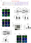
The 3R-motif of NDRG1 is actioned for nuclear translocation and phosphorylation. (a) Sequence homology of NDRG1-3R motif with SR protein family. The sequences of 3R motif and SR family proteins were aligned using Pileup program in Chang Gung University. The conserved amino acids are blocked in the indicated boxes. (b) Depletion of NDRG-3R motif significantly inhibited nuclear translocation in OECM1and FaDu cells. After transfection of NDRG1-FL, NDRG1-dC, or the vector plasmid into HNC cells, the cells were subjected to immunofluorescent and confocal microscopic analysis. Because the EGFP protein sequence was fused in-frame in the NDRG1 expression plasmids, the localization of EGFP indicated the specific localization of NDRG1. Blue staining with DAPI dye was used to indicate nuclei, scale bar 10 µm. (c) Depletion of 3R motif significantly inhibited nuclear translocation. After transfection of NDRG1-FL, NDRG1-dC, or the vector plasmid into HNC cells, the subcellular compartment proteins were isolated and separated to nuclear and cytosolic fractions. The NDRG1 expression level in various subcellular fractions was determined by Western blot analysis. The HDAC1 and GAPDH were used as internal controls for nuclear and cytosolic compartment proteins. (d) GSK3b phosphorylated NDRG1 at the serine residues of its C-terminal domain. After transfection of the NDRG1-FL or NDRG1-dC expression plasmids into OECM1 cells, the cellular proteins were extracted and subjected to an in vitro phosphorylation assay with recombinant GSK3b kinase. These NDRG1 proteins were then pulled down with anti-GFP antibody, and their phosphorylation status was revealed by immunoblotting with phosphoserine-specific antibody. Expression of the EGFP protein in each transfected sample was used as an internal control. (e) Treatment of GSK3b inhibitor suppressed nuclear translocation of NDRG1 in HNC cells. The OECM1 cells were treated with GSK3b-specific inhibitor (SB21673) at various doses as indicated for 24 h. The subcellular compartment proteins were isolated. The endogenous NDRG1 expression level in various subcellular fractions was determined by Western blot analysis. The HDAC1 and GAPDH were used as internal controls for nuclear and cytosolic compartment proteins, respectively. (f) Treatment of GSK3b inhibitor attenuated nuclear translocation of NDRG1 in HNC cells. After transfection of the NDRG1-FL or NDRG1-dC expression plasmids into HNC cells, transfected cells were treated with GSK3b-specific inhibitor (SB21673) at 3 μM for 24 h. These cells were then subjected to immunofluorescence analysis and confocal microscopy, as described in the method section, scale bar 10 µm. The error bars shown in the relevant figures indicate the standard deviation of the quantification results. (* p < 0.05; *** p < 0.001; n.s., no significance; t-test).
Figure 7
Figure 7
The phosphorylation of 3R-motif by GSK3b is critical for cell motility in HNC cells. (a) Dephosphorylation of NDRG1 by GSK3b inhibitor reversed the effect of NDRG1-induced cell invasion. The HNC cells (OECM1, SAS) were transfected with NDRG1-FL or the vector plasmids, with or without the addition of GSK3b inhibitors (SB21673, 3 μM) for 24 h. These cells were then subjected to Matrigel invasion assay. (b) Mutation of GSK3b phosphorylation sites in NDRG1-3R motif reversed the effect of NDRG1-induced cell invasion. After transfection of various NDRG1 truncated plasmids (-FL, -dC, -3mt) into OECM1 cells for 30 h, these cells were subject to Matrigel invasion assay. All the experiments were performed three times independently and typical results were shown. The error bars shown in the relevant figures indicate the standard deviation of the quantification results in all experiments. (** p < 0.01; *** p < 0.001; n.s., no significance; t-test).
Figure 8
Figure 8
High level of NDRG1 is significantly associated with poor survival in HNC patients. (a) In HNC patients, NDRG1 is overexpressed in the tumors compared to the normal tissues from TCGA-HNSC datasets generated by GEPIA2 website. The relative expression levels were shown. (b) Higher level of NDRG1 expression is associated with poor prognosis in HNC patients, as determined by Kaplan–Meier survival analysis via KM-Plotter online tool from the HNC dataset (N = 500). * p < 0.05; t-test.

Similar articles

Cited by

References

    1. Sung H., Ferlay J., Siegel R.L., Laversanne M., Soerjomataram I., Jemal A., Bray F. Global Cancer Statistics 2020: GLOBOCAN Estimates of Incidence and Mortality Worldwide for 36 Cancers in 185 Countries. CA Cancer J. Clin. 2021;71:209–249. doi: 10.3322/caac.21660. - DOI - PubMed
    1. Meltzer C., Nguyen N.T., Zhang J., Aguilar J., Blatchins M.A., Quesenberry C.P., Jr., Wang Y., Sakoda L.C. Survival associated with consolidated multidisciplinary care in head and neck cancer: A retrospective cohort study. Otolaryngol. Head Neck Surg. 2021;9:1945998211057852. doi: 10.1177/01945998211057852. - DOI - PubMed
    1. Chang J.T., Wang H.M., Chang K.W., Chen W.H., Wen M.C., Hsu Y.M., Yung B.Y., Chen I.H., Liao C.T., Hsieh L.L., et al. Identification of differentially expressed genes in oral squamous cell carcinoma (OSCC): Overexpression of NPM, CDK1 and NDRG1 and underexpression of CHES1. Int. J. Cancer. 2005;114:942–949. doi: 10.1002/ijc.20663. - DOI - PubMed
    1. Lorini L., Ardighieri L., Bozzola A., Romani C., Bignotti E., Buglione M., Guerini A., Lombardi D., Deganello A., Tomasoni M., et al. Prognosis and management of recurrent and/or metastatic head and neck adenoid cystic carcinoma. Oral Oncol. 2021;115:105213. doi: 10.1016/j.oraloncology.2021.105213. - DOI - PubMed
    1. Tang E., Nguyen T.V., Clatot F., Rambeau A., Johnson A., Sun X.S., Tao Y., Thariat J. Radiation therapy on primary tumour of synchronous metastatic head and neck squamous cell carcinomas. Cancer Radiother. 2020;24:559–566. doi: 10.1016/j.canrad.2020.05.004. - DOI - PubMed

Publication types

MeSH terms

Substances

Grants and funding

This research was supported by grants from Chang Gung Memorial Hospital-Linko Medical Center in Taiwan (CMRPD1G0451~3 and CMRPD1M0191~2).