

The Role of KH-Type Splicing Regulatory Protein (KSRP) for Immune Functions and Tumorigenesis
- PMID: 35563788
- PMCID: PMC9104899
- DOI: 10.3390/cells11091482
The Role of KH-Type Splicing Regulatory Protein (KSRP) for Immune Functions and Tumorigenesis
Abstract
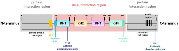
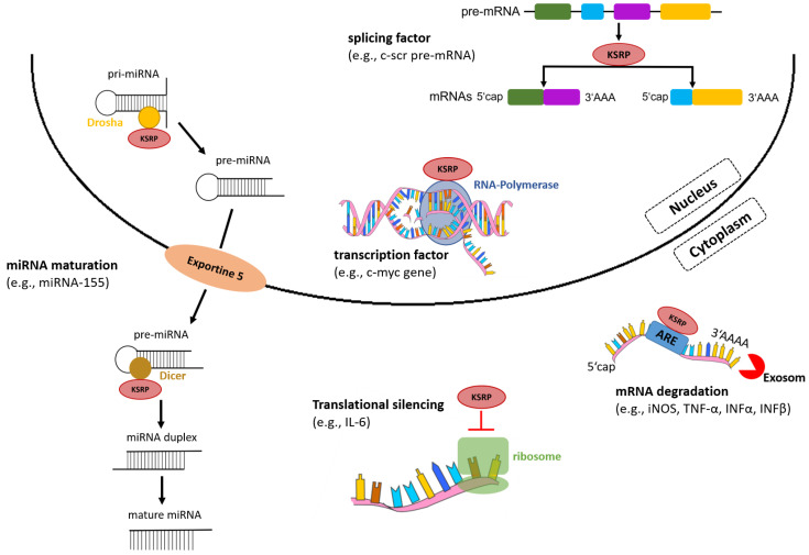
Post-transcriptional control of gene expression is one important mechanism that enables stringent and rapid modulation of cytokine, chemokines or growth factors expression, all relevant for immune or tumor cell function and communication. The RNA-binding protein KH-type splicing regulatory protein (KSRP) controls the mRNA stability of according genes by initiation of mRNA decay and inhibition of translation, and by enhancing the maturation of microRNAs. Therefore, KSRP plays a pivotal role in immune cell function and tumor progression. In this review, we summarize the current knowledge about KSRP with regard to the regulation of immunologically relevant targets, and the functional role of KSRP on immune responses and tumorigenesis. KSRP is involved in the control of myeloid hematopoiesis. Further, KSRP-mediated mRNA decay of pro-inflammatory factors is necessary to keep immune homeostasis. In case of infection, functional impairment of KSRP is important for the induction of robust immune responses. In this regard, KSRP seems to primarily dampen T helper cell 2 immune responses. In cancer, KSRP has often been associated with tumor growth and metastasis. In summary, aside of initiation of mRNA decay, the KSRP-mediated regulation of microRNA maturation seems to be especially important for its diverse biological functions, which warrants further in-depth examination.
Keywords: KH-type splicing regulatory protein; T helper cell; antiviral response; cytokine; mRNA decay; micro RNA; post-transcriptional gene regulation; tumorigenesis.
Conflict of interest statement
The authors declare no conflict of interest.
Figures

Similar articles
-
The role of KSRP in mRNA decay and microRNA precursor maturation.Wiley Interdiscip Rev RNA. 2010 Sep-Oct;1(2):230-9. doi: 10.1002/wrna.2. Epub 2010 May 6. Wiley Interdiscip Rev RNA. 2010. PMID: 21935887 Review.
-
The KH-type splicing regulatory protein (KSRP) regulates type III interferon expression post-transcriptionally.Biochem J. 2019 Jan 31;476(2):333-352. doi: 10.1042/BCJ20180522. Biochem J. 2019. PMID: 30578289
-
KSRP suppresses cell invasion and metastasis through miR-23a-mediated EGR3 mRNA degradation in non-small cell lung cancer.Biochim Biophys Acta Gene Regul Mech. 2017 Oct;1860(10):1013-1024. doi: 10.1016/j.bbagrm.2017.08.005. Epub 2017 Aug 25. Biochim Biophys Acta Gene Regul Mech. 2017. PMID: 28847731
-
Role of KSRP in control of type I interferon and cytokine expression.J Interferon Cytokine Res. 2014 Apr;34(4):267-74. doi: 10.1089/jir.2013.0143. J Interferon Cytokine Res. 2014. PMID: 24697204 Free PMC article. Review.
-
Upregulation of KSRP by miR-27b provides IFN-γ-induced post-transcriptional regulation of CX3CL1 in liver epithelial cells.Sci Rep. 2015 Dec 3;5:17590. doi: 10.1038/srep17590. Sci Rep. 2015. PMID: 26631623 Free PMC article.
Cited by
-
The RNA-binding protein KSRP aggravates malignant progression of clear cell renal cell carcinoma through transcriptional inhibition and post-transcriptional destabilization of the NEDD4L ubiquitin ligase.J Biomed Sci. 2023 Aug 14;30(1):68. doi: 10.1186/s12929-023-00949-9. J Biomed Sci. 2023. PMID: 37580757 Free PMC article.
-
Comparative Transcriptome Analysis of CCCH Family in Roles of Flower Opening and Abiotic Stress in Osmanthus fragrans.Int J Mol Sci. 2022 Dec 6;23(23):15363. doi: 10.3390/ijms232315363. Int J Mol Sci. 2022. PMID: 36499688 Free PMC article.
-
The mRNA-Binding Protein KSRP Limits the Inflammatory Response of Macrophages.Int J Mol Sci. 2024 Mar 30;25(7):3884. doi: 10.3390/ijms25073884. Int J Mol Sci. 2024. PMID: 38612694 Free PMC article.
-
KHSRP knockdown inhibits papillary renal cell carcinoma progression and sensitizes to gemcitabine.Front Pharmacol. 2024 Oct 8;15:1446920. doi: 10.3389/fphar.2024.1446920. eCollection 2024. Front Pharmacol. 2024. PMID: 39439895 Free PMC article.
-
RNA-Binding Proteins in the Regulation of Adipogenesis and Adipose Function.Cells. 2022 Jul 31;11(15):2357. doi: 10.3390/cells11152357. Cells. 2022. PMID: 35954201 Free PMC article. Review.
References
Publication types
MeSH terms
Substances
Grants and funding
LinkOut - more resources
Full Text Sources
