Expression Profiles of GILZ and Annexin A1 in Human Oral Candidiasis and Lichen Planus
- PMID: 35563776
- PMCID: PMC9100531
- DOI: 10.3390/cells11091470
Expression Profiles of GILZ and Annexin A1 in Human Oral Candidiasis and Lichen Planus
Abstract
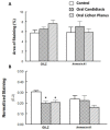
Adrenal glands are the major source of glucocorticoids, but recent studies indicate tissue-specific production of cortisol, including that in the oral mucosa. Both endogenous and exogenous glucocorticoids regulate the production of several proteins, including the glucocorticoid-induced leucine zipper (GILZ) and Annexin A1, which play important roles in the regulation of immune and inflammatory responses. Common inflammation-associated oral conditions include lichen planus and candidiasis, but the status of GILZ and Annexin A1 in these human conditions remains to be established. Accordingly, archived paraffin-embedded biopsy samples were subjected to immunohistochemistry to establish tissue localization and profile of GILZ and Annexin A1 coupled with the use of hematoxylin-eosin stain for histopathological assessment; for comparison, fibroma specimens served as controls. Histopathological examination confirmed the presence of spores and pseudohyphae for oral candidiasis (OC) specimens and marked inflammatory cell infiltrates for both OC and oral lichen planus (OLP) specimens compared to control specimens. All specimens displayed consistent and prominent nuclear staining for GILZ throughout the full thickness of the epithelium and, to varying extent, for inflammatory infiltrates and stromal cells. On the other hand, a heterogeneous pattern of nuclear, cytoplasmic, and cell membrane staining was observed for Annexin A1 for all specimens in the suprabasal layers of epithelium and, to varying extent, for inflammatory and stromal cells. Semi-quantitative analyses indicated generally similar fractional areas of staining for both GILZ and Annexin A1 among the groups, but normalized staining for GILZ, but not Annexin A1, was reduced for OC and OLP compared to the control specimens. Thus, while the cellular expression pattern of GILZ and Annexin A1 does not differentiate among these conditions, differential cellular profiles for GILZ vs. Annexin A1 are suggestive of their distinct physiological functions in the oral mucosa.
Keywords: Annexin A1; GILZ; human; immunohistochemistry; oral candidiasis; oral lichen planus.
Conflict of interest statement
The authors declare no conflict of interest.
Figures


Similar articles
-
Expression profiles of glucocorticoid-inducible proteins in human papilloma virus-related oropharyngeal squamous cell carcinoma.Front Oral Health. 2023 Oct 25;4:1285139. doi: 10.3389/froh.2023.1285139. eCollection 2023. Front Oral Health. 2023. PMID: 37954869 Free PMC article.
-
Expression Profiles of GILZ and SGK-1 in Potentially Malignant and Malignant Human Oral Lesions.Front Oral Health. 2021 Sep 16;2:675288. doi: 10.3389/froh.2021.675288. eCollection 2021. Front Oral Health. 2021. PMID: 35048019 Free PMC article.
-
Role of the glucocorticoid-induced leucine zipper gene in dexamethasone-induced inhibition of mouse neutrophil migration via control of annexin A1 expression.FASEB J. 2017 Jul;31(7):3054-3065. doi: 10.1096/fj.201601315R. Epub 2017 Apr 3. FASEB J. 2017. PMID: 28373208
-
Exploiting the pro-resolving actions of glucocorticoid-induced proteins Annexin A1 and GILZ in infectious diseases.Biomed Pharmacother. 2021 Jan;133:111033. doi: 10.1016/j.biopha.2020.111033. Epub 2020 Dec 8. Biomed Pharmacother. 2021. PMID: 33378946 Review.
-
Role of distinct CD4(+) T helper subset in pathogenesis of oral lichen planus.J Oral Pathol Med. 2016 Jul;45(6):385-93. doi: 10.1111/jop.12405. Epub 2015 Dec 23. J Oral Pathol Med. 2016. PMID: 26693958 Review.
References
MeSH terms
Substances
Grants and funding
LinkOut - more resources
Full Text Sources
Molecular Biology Databases
Research Materials
Miscellaneous

