. 2022 May 5;23(9):5137.
doi: 10.3390/ijms23095137.

DOT1L Methyltransferase Regulates Calcium Influx in Erythroid Progenitor Cells in Response to Erythropoietin

Affiliations

DOT1L Methyltransferase Regulates Calcium Influx in Erythroid Progenitor Cells in Response to Erythropoietin

Yi Feng et al. Int J Mol Sci. .

Abstract

Erythropoietin (EPO) signaling plays a vital role in erythropoiesis by regulating proliferation and lineage-specific differentiation of murine hematopoietic progenitor cells (HPCs). An important downstream response of EPO signaling is calcium (Ca2+) influx, which is regulated by transient receptor potential channel (TRPC) proteins, particularly TRPC2 and TRPC6. While EPO induces Ca2+ influx through TRPC2, TRPC6 inhibits the function of TRPC2. Thus, interactions between TRPC2 and TRPC6 regulate the rate of Ca2+ influx in EPO-induced erythropoiesis. In this study, we observed that the expression of TRPC6 in KIT-positive erythroid progenitor cells was regulated by DOT1L. DOT1L is a methyltransferase that plays an important role in many biological processes during embryonic development including early erythropoiesis. We previously reported that Dot1l knockout (Dot1lKO) HPCs in the yolk sac failed to develop properly, which resulted in lethal anemia. In this study, we detected a marked downregulation of Trpc6 gene expression in Dot1lKO progenitor cells in the yolk sac compared to the wild type (WT). The promoter and the proximal regions of the Trpc6 gene locus exhibited an enrichment of H3K79 methylation, which is mediated solely by DOT1L. However, the expression of Trpc2, the positive regulator of Ca2+ influx, remained unchanged, resulting in an increased TRPC2/TRPC6 ratio. As the loss of DOT1L decreased TRPC6, which inhibited Ca2+ influx by TRPC2, Dot1lKO HPCs in the yolk sac exhibited accelerated and sustained elevated levels of Ca2+ influx. Such heightened Ca2+ levels might have detrimental effects on the growth and proliferation of HPCs in response to EPO.

Keywords: DOT1L; TRPC6; calcium influx; erythroid progenitors; erythropoietin.


Conflict of interest statement

The authors declare that they have no conflict of interest.

Figures
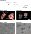
Figure 1
Figure 1
Dot1lKO progenitors failed to proliferate in response to EPO. (A) Schematic diagram showing the timeline of the embryo and yolk sac collection and in vitro culture of yolk-sac-derived erythroblasts. Representative images of E10.5 wild-type and Dot1lKO yolk sacs and embryos (B,C). Both the Dot1lKO yolk sacs as well as the embryos were smaller than those of the wild type (B,C). Cells were isolated from E10.5 wild-type and Dot1lKO yolk sacs and equal numbers were cultured in extensively self-renewing erythroblast (ESRE) culture medium as described. The cells were then differentiated from the definitive erythroid progenitors into erythroblasts (D,E). Compared to the wild-type erythroblasts (D), Dot1lKO erythroblasts had severely reduced numbers (E).
Figure 2
Figure 2
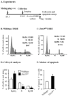
Dot1lKO progenitors underwent cell cycle arrest during in vitro erythroblast culture. (A) Schematic diagram showing the studies for cell cycle progression and apoptosis analysis. On day 4 of culture, cells were collected, fixed, and either stained with propidium iodide for cell cycle analyses or labeled with annexin V for signs of apoptosis. A large number of erythroid progenitors from Dot1lKO yolk sacs displayed G0/G1 arrest (BD). In addition, an increased percentage of Dot1lKO erythroid progenitors were found to be annexin V positive (E). Data are expressed as the mean ± SE; n > 6; * p < 0.05.
Figure 3
Figure 3
Equal numbers of wild-type and Dot1lKO E10.5 yolk sac cells were cultured in methylcellulose medium containing cytokines that promoted definitive erythroid and myeloid lineage differentiation. While Dot1lKO progenitors formed significantly smaller sized erythroid (BFU-E) colonies (A,B), the wild-type and Dot1lKO myeloid colonies (CFU-GM) were similar in size (C,D).
Figure 4
Figure 4
Expression of TRPC6 was markedly reduced in KIT-positive Dot1lKO hematopoietic progenitors. (A) Among the three genes in the Trpc family (i.e., Trpc1, Trpc2, and Trp6), only Trpc6 levels were dramatically reduced in Dot1lKO compared to WT, while the levels of the other members remained relatively unchanged. TRPC6 protein levels in KIT-positive progenitor cells from Dot1lKO yolk sacs were also significantly reduced (B,C). Data are expressed as the mean ± SE; n > 6; * p < 0.05.
Figure 5
Figure 5
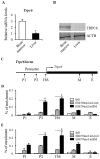
Enrichment of di- and tri-methyl H3K79 in Trpc6 gene loci. Trpc6 expression was tested by RT-qPCR in bone marrow (BM) and liver cells. Trpc6 mRNA levels were significantly higher in BM cells compared to that in liver cells (A). TRPC6 protein was also detected in BM and liver cells by Western blot analysis, which showed that TRPC6 protein was also markedly higher in BM cells (B). ChIP assays were performed on chromatin preparations from BM and liver cells to assess H3K79 di- and tri-methylation in the Trpc6 gene locus. (C) Several pairs of primers were designed covering the Trpc6 gene locus including the promoter region (P1, P2), transcription start site (TSS), and middle and end of the gene locus (M, E). ChIP assays were performed in the promoter region of Trpc6 gene locus for H3K79 di-methylation (D) and H3K79 tri-methylation (E). There was significantly greater enrichment of both H3K79 di- and tri-methylation at the P1, P2, and TSS sites of the Trpc6 gene in BM cells than that of liver cells. The ChIP-qPCR data represent 3 independent experiments. Data are expressed as the mean ± SE; n > 6; * p < 0.05.
Figure 6
Figure 6
Accelerated and enhanced calcium influx in Dot1lKO erythroid progenitors. E10.5 Dot1lKO and wild-type yolk sac cells were stained, treated with EPO, and the Ca2+ signals in individual cells were recorded using fluorescence microscopy. After EPO treatment, progenitor cells from Dot1lKO yolk sacs showed a sustained increase in Ca2+ levels. Data are expressed as the mean ± SE; n > 6; *p < 0.05.
Figure 7
Figure 7
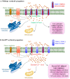
Dot1lKO erythroid progenitors exhibited increased calcium influx following decreased TRPC6 expression. TRPC2 allows Ca2+ influx into erythroid progenitors. TRPC6 forms a hetero-tetramer with TRPC2 and negatively regulates TRPC2-dependent Ca2+ influx to maintain the normal intracellular Ca2+ level (A). The loss of DOT1L decreases TRPC6 expression, resulting in an increased TRPC2/TRPC6 ratio. This favors the formation of TRPC2 homo-tetramers over TRPC2/TRPC6 hetero-tetramers leading to an excessive influx of Ca2+ into Dot1lKO erythroid progenitors (B). An elevated Ca2+ level results in toxic intracellular responses and aberrant cell signaling linked to cell cycle arrest and cell death observed in Dot1lKO erythroid progenitors.

Similar articles

Cited by

References

    1. Tsiftsoglou A.S., Vizirianakis I.S., Strouboulis J. Erythropoiesis: Model systems, molecular regulators, and developmental programs. IUBMB Life. 2009;61:800–830. doi: 10.1002/iub.226. - DOI - PubMed
    1. Ingley E., Tilbrook P.A., Klinken S.P. New insights into the regulation of erythroid cells. IUBMB Life. 2004;56:177–184. doi: 10.1080/15216540410001703956. - DOI - PubMed
    1. Lin C.S., Lim S.K., D’Agati V., Costantini F. Differential effects of an erythropoietin receptor gene disruption on primitive and definitive erythropoiesis. Genes Dev. 1996;10:154–164. doi: 10.1101/gad.10.2.154. - DOI - PubMed
    1. Wu H., Liu X., Jaenisch R., Lodish H.F. Generation of committed erythroid BFU-E and CFU-E progenitors does not require erythropoietin or the erythropoietin receptor. Cell. 1995;83:59–67. doi: 10.1016/0092-8674(95)90234-1. - DOI - PubMed
    1. Miller B.A., Bell L.L., Lynch C.J., Cheung J.Y. Erythropoietin modulation of intracellular calcium: A role for tyrosine phosphorylation. Cell Calcium. 1994;16:481–490. doi: 10.1016/0143-4160(94)90078-7. - DOI - PubMed

LinkOut - more resources