Review
. 2022 May 10;55(5):749-780.
doi: 10.1016/j.immuni.2022.04.013.

Mucosal immune responses to infection and vaccination in the respiratory tract

Affiliations
Review

Mucosal immune responses to infection and vaccination in the respiratory tract

Robert C Mettelman et al. Immunity. .

Abstract

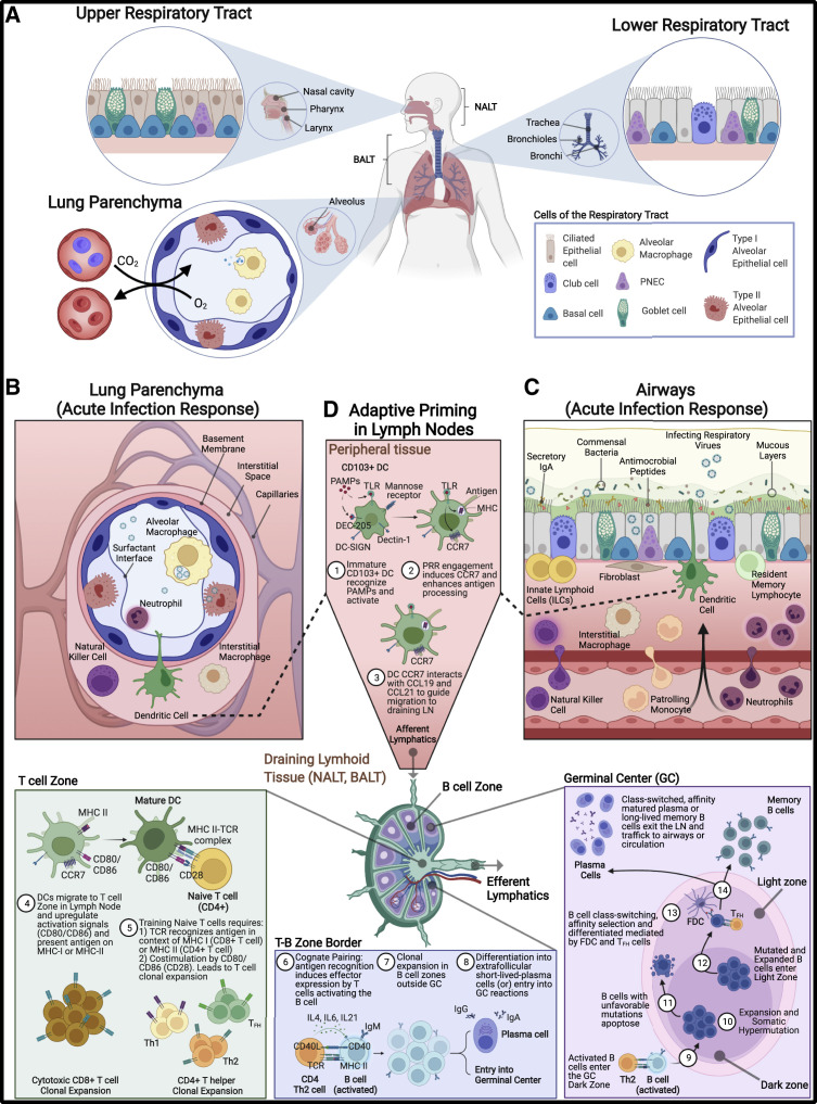
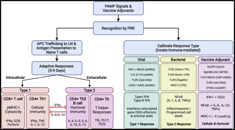
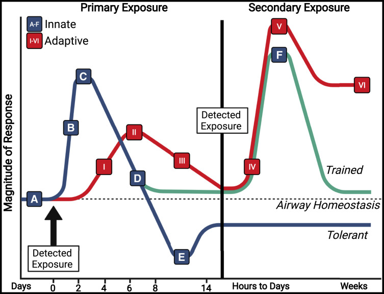
The lungs are constantly exposed to inhaled debris, allergens, pollutants, commensal or pathogenic microorganisms, and respiratory viruses. As a result, innate and adaptive immune responses in the respiratory tract are tightly regulated and are in continual flux between states of enhanced pathogen clearance, immune-modulation, and tissue repair. New single-cell-sequencing techniques are expanding our knowledge of airway cellular complexity and the nuanced connections between structural and immune cell compartments. Understanding these varied interactions is critical in treatment of human pulmonary disease and infections and in next-generation vaccine design. Here, we review the innate and adaptive immune responses in the lung and airways following infection and vaccination, with particular focus on influenza virus and severe acute respiratory syndrome coronavirus 2 (SARS-CoV-2). The ongoing SARS-CoV-2 pandemic has put pulmonary research firmly into the global spotlight, challenging previously held notions of respiratory immunity and helping identify new populations at high risk for respiratory distress.


Conflict of interest statement

Declaration of interests P.G.T. has consulted or received honorarium and/or travel support from Illumina, JNJ, and 10X. P.G.T. serves on the Scientific Advisory Board of ImmunoScape and CytoAgents.

Figures

Figure 1
Figure 1
Respiratory tract compartments and immune environment (A) The upper (nasal cavity, pharynx, larynx), lower (trachea, bronchi, bronchioles), and the lung parenchyma (alveoli and lung interstitial spaces) compartments of the respiratory tract. Specific structural cells in the URT include undifferentiated basal cells, ciliated epithelial, and secretory goblet cells. Ciliated epithelium in the URT along with mucus that is synthesized by goblet cells provide a mechanical means of removing debris, allergens, and potential pathogens from the URT. These cells are also present in LRT, with additional inclusion of club cells, non-ciliated epithelia that moderate tolerance responses. Like the URT, mechanical removal of materials from the LRT is accomplished by mucociliary activities of the ciliated epithelia and goblet cells along with coughing to dislodge materials impacted on bronchial branch points. (B and C) Cells and immune factors present during mucosal immune response and acute inflammation in the (B) lung parenchyma and in the (C) airway tissues. Embedded within the mucous layer are secreted innate immune factors including antimicrobial peptides, proteases, lactoferrin, and complement that act to further reduce passage of foreign material into the lungs. Secretory immunoglobulins (sIgA and sIgM), produced by sub-epithelial plasma cells, can also be found coating the surface of the URT and LRT in the mucous layer and provide antigen-specific targeting of foreign antigens in parallel to their innate immune counterparts. Among innate cellular responders during acute infection, neutrophils predominate and produce additional factors (IL8 and elastase) to further amplify immune cell recruitment. In addition to neutrophils, natural killer (NK) cells, monocytes, and eosinophils are also recruited from circulation. Mesenchymal (fibroblasts), epithelial, and endothelial cells produce proteases to reshape the extracellular matrix landscape, the products of which signal for further recruitment of immune cells and open physical space for immune cells to occupy. Epithelial cells also release localized cytokine signals to activate resident innate lymphoid cells (ILCs), interstitial macrophages (IM), and dendritic cells (DCs). Each cell type, tissue-resident or recruited, produces a carefully orchestrated balance of either inflammatory and chemotactic cytokines (TNFɑ, IL1β, IL6, IL8, IFN-I, -II, -III, GM-CSF) or immune-regulatory (IL10, TGFβ, IL1Ra) factors in an effort to resist and eliminate pathogens without causing excessive inflammation. (D) Professional antigen-presenting cells, mainly CD103+ dendritic cells (cDCs), capture antigen in the airways, become activated, and traffic to draining lymph nodes (LNs) via afferent lymphatics to prime adaptive responses. In LN T cell zones, externally derived antigens are presented on class II MHC, prompting CD4+ T cell training, while internally derived antigens are processed and presented on class I MHC to CD8+ T cells. CD103+ respiratory DCs can also cross-present peptides from external sources on class I MHC, possibly via efferocytosis, thus allowing CD8+ T cell priming (Ho et al., 2011). APCs promote maturation and expansion of naive CD4+ and CD8+ T cells through pMHC:TCR and co-stimulation (CD80/CD86). CD8+ cytotoxic T cells and subsets of CD4+ T helper cells traffic back to the site of infection. CD4+ Th2 cells migrate to T-B border and form cognate pairs with activated B cells (IgM+) via TCR:pMHC engagement, cytokine release (IL4, IL6, IL21), and co-stimulation (CD40L), leading to B cell clonal expansion and differentiation into low-affinity IgG or IgA-producing plasma cells (traffic to sites of infection) or entry into germinal centers (GCs). In GC dark zone, activated B cells undergo expansion and somatic hypermutation of the B cell receptor (BCR), resulting in either an unfavorable BCR specificity (leading to apoptosis) or desirable BCR specificity that binds strongly to peptides maintained on the surface of follicular dendritic cells (FDCs) in the light zone. B cells cycle through iterative rounds of expansion/somatic hypermutation (dark zone) and affinity selection (light zone), resulting in selection of high-affinity BCRs. Interactions with T follicular helper (Tfh) cells lead to B cell differentiation and class-switching to long-lived memory B cells and high-affinity plasma cells, which traffic to sites of infection or are maintained as long-lived memory populations. PNEC, pulmonary neuroendocrine cell; NALT, nasal-associated lymphoid tissue; BALT, bronchial-associated lymphoid tissue. Figure created with BioRender.com.
Figure 2
Figure 2
Acute pulmonary inflammation and downstream responses Initiation: Lung insults often involving infectious etiologic agents or vaccine components are recognized by structural cells, including epithelial and endothelial cells, along with tissue-resident antigen-presenting cells (airway macrophages and dendritic cells). Together, these cells recognize pathogen-associated molecular patterns (PAMPs), tissue damage-associated molecular patterns (DAMPs), or vaccine adjuvants via a complex network or surface, endosomal, and cytoplasmic sensors, collectively termed pattern recognition receptors (PRRs). Calibration: Detection of viral and bacterial PAMPs or vaccine-derived adjuvants initiates differential response pathways depending on the source (viral, bacterial, or adjuvant). Detection of viral PAMPS leads to production of IFN-I and/or IFN-III, TNFα, IL1β, IL6, IL8, IL12, MCP1, and inflammasome activation. Distinctive bacterial PAMPs (endotoxins, lipopolysaccharide [LPS], and peptidoglycan [PG]) trigger NF-κB signaling axis, leading to production of IL6, IL8, IL18, TNFɑ/β, and inflammasome activation. Detection and response to vaccine adjuvants can be tailored to promote type 1 or type 2 immune responses and specific development of cellular and humoral immunity. Trafficking: Specialized APCs capture and process protein components from pathogen or vaccine sources, traffic to airway-associated draining lymph nodes, and present antigen to naive T cells. Adaptive responses: Priming of adaptive responses begins around day 4 post-exposure and occurs in respiratory-associated lymphoid tissues. Following antigen-specific training, CD8+ T cell and CD4+ Th1 responses (IFNγ, TNF, IL1, IL12) are typically mounted against intracellular pathogens (antigens presented on MHC-I), while CD4+ Th2 (IL4, 5, 6, 10, 13), humoral (Tfh, B cells; antibody), and CD4 T helper (Th17, Th22) responses are increased in response to extracellular insults. Cross-presentation of external-derived antigens on MHC-I to CD8+ T cells allows development of cellular immunity to external insults. Figure created with BioRender.com.
Figure 3
Figure 3
Innate and adaptive immune response magnitude dynamics over time Pulmonary infections or vaccinations that overcome low immunogenic clearance (A) trigger innate immune responses (blue/green) and initiate acute pulmonary inflammation (A and B) rapidly upon detection of PAMPs. Differential response cascades are calibrated by structural and resident immune cells to promote the appropriate inflammatory responses, coordinate effector functions of infiltrating innate cells, and activate antigen-presenting cells (with internalized antigen) to traffic to draining lymph nodes (B and C). As the infection is cleared, regulatory cells promote a return to a tolerant state, allowing tissue repair and blocking extravasation of additional immune cells (D). In certain innate immune cell types, the immune environment and PAMP type can promote metabolic and epigenetic reprogramming, driving innate cells into maintained tolerant (E, blue) or trained (F, green) phenotypes upon a subsequent challenge. Adaptive responses (red) are primed in respiratory draining lymphoid tissues (I), which promote clonal expansion of antigen-specific CD8+ cytotoxic T cells, CD4+ T helper cells, and low-affinity IgA and IgG-secreting plasma B cells that respond at tissue sites between 5 and 9 days post-challenge (II). Germinal center reactions in LN lead to class-switching, affinity selection, and generation of high-affinity plasma cells producing IgA and IgG and long-lived plasma cells (III). Tissue resident T (TRM) and B (BRM) cells are established in the lung via the iBALT and RAMD, while other lymphoid cells promote tolerance and tissue repair (III). Upon secondary exposure to homotypic or cross-reactive antigens in the lung and airways, adaptive responses are rapidly triggered, mediated first by activated TRM and BRM cells, then by recruited peripheral lymphocytes (IV and V). While innate responses activate and wane during secondary exposure, adaptive responses, mainly IgA and IgG levels, remain elevated for weeks (V). PAMPs, pathogen-associated molecular patterns; iBALT, inducible bronchus-associated lymphoid tissue; RAMD, repair-associated memory depots. Figure created with BioRender.com. See also Table 1.

Similar articles

Cited by

References

    1. Abboud G., Tahiliani V., Desai P., Varkoly K., Driver J., Hutchinson T.E., Salek-Ardakani S. Natural killer cells and innate interferon gamma participate in the host defense against respiratory vaccinia virus infection. J. Virol. 2016;90:129–141. doi: 10.1128/JVI.01894-15. - DOI - PMC - PubMed
    1. Abt M.C., Osborne L., Monticelli L., Doering T., Alenghat T., Sonnenberg G., Paley M., Antenus M., Williams K., Erikson J., et al. Commensal bacteria calibrate the activation threshold of innate antiviral immunity. Immunity. 2012;37:158–170. doi: 10.1016/J.IMMUNI.2012.04.011. - DOI - PMC - PubMed
    1. Adachi Y., Onodera T., Yamada Y., Daio R., Tsuiji M., Inoue T., Kobayashi K., Kurosaki T., Ato M., Takahashi Y. Distinct germinal center selection at local sites shapes memory B cell response to viral escape. J. Exp. Med. 2015;212:1709–1723. doi: 10.1084/JEM.20142284. - DOI - PMC - PubMed
    1. El Agha E., Moiseenko A., Kheirollahi V., De Langhe S., Crnkovic S., Kwapiszewska G., Szibor M., Kosanovic D., Schwind F., Schermuly R.T., et al. Two-way conversion between Lipogenic and Myogenic fibroblastic phenotypes Marks the progression and resolution of lung fibrosis. Cell Stem Cell. 2017;20:261–273.e3. doi: 10.1016/J.STEM.2016.10.004. - DOI - PMC - PubMed
    1. Aguirre A., Blazquez-Prieto J., Amado-Rodriguez L., Lopez-Alonso I., Batalla-Solis E., Gonzalez-Lopez A., Sanchez-Perez M., Mayoral-Garcia C., Gutierrez-Fernandez A., Albaiceta G.M. Matrix metalloproteinase-14 triggers an anti-inflammatory proteolytic cascade in endotoxemia. J. Mol. Med. 2017;95:487–497. doi: 10.1007/s00109-017-1510-z. - DOI - PubMed

Publication types