. 2022 May 10;17(5):e0262062.
doi: 10.1371/journal.pone.0262062. eCollection 2022.

Use of standard U-bottom and V-bottom well plates to generate neuroepithelial embryoid bodies

Affiliations

Use of standard U-bottom and V-bottom well plates to generate neuroepithelial embryoid bodies

David Choy Buentello et al. PLoS One. .

Abstract

The use of organoids has become increasingly popular recently due to their self-organizing abilities, which facilitate developmental and disease modeling. Various methods have been described to create embryoid bodies (EBs) generated from embryonic or pluripotent stem cells but with varying levels of differentiation success and producing organoids of variable size. Commercial ultra-low attachment (ULA) V-bottom well plates are frequently used to generate EBs. These plates are relatively expensive and not as widely available as standard concave well plates. Here, we describe a cost-effective and low labor-intensive method that creates homogeneous EBs at high yield in standard V- and U-bottom well plates by applying an anti-adherence solution to reduce surface attachment, followed by centrifugation to enhance cellular aggregation. We also explore the effect of different seeding densities, in the range of 1 to 11 ×103 cells per well, for the fabrication of neuroepithelial EBs. Our results show that the use of V-bottom well plates briefly treated with anti-adherent solution (for 5 min at room temperature) consistently yields functional neural EBs in the range of seeding densities from 5 to 11×103 cells per well. A brief post-seeding centrifugation step further enhances EB establishment. EBs fabricated using centrifugation exhibited lower variability in their final size than their non-centrifuged counterparts, and centrifugation also improved EB yield. The span of conditions for reliable EB production is narrower in U-bottom wells than in V-bottom wells (i.e., seeding densities between 7×10³ and 11×103 and using a centrifugation step). We show that EBs generated by the protocols introduced here successfully developed into neural organoids and expressed the relevant markers associated with their lineages. We anticipate that the cost-effective and easily implemented protocols presented here will greatly facilitate the generation of EBs, thereby further democratizing the worldwide ability to conduct organoid-based research.


Conflict of interest statement

The authors have declared that no competing interests exist.

Figures

Fig 1
Fig 1. Protocol for embryoid body formation and different well geometries.
We added anti-adherence solution into regular U- and V-bottom wells and incubated for 5 minutes at room temperature to develop anti-adherence coated plates and then washed gently with PBS. The application of an anti-adherence solution to conventional V-bottom and U-bottom plates enabled reproducible EB generation in low-cost well plates.
Fig 2
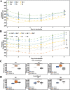
Fig 2. Characterization of embryoid body (EB) quality and daily growth.
(A) The graphs depict EBs diameter in U-bottom wells coated with anti-adherence solution. (B) EB growth progression cultured in V-bottom wells coated with anti-adherence solution. Graphs shows different color-coded seeding concentrations given in thousands (“K”) and if centrifuged (“C”). *P<0.001 indicates a significant difference for all EBs of the same well plate and centrifugation. †P<0.001 indicates a significant difference for EBs of the same well plate type but different centrifugation condition. ‡P<0.001 indicates a significant difference for EBs with different plate types but the same centrifugation condition. Scale bar: 200 μm. We considered at least 5 EBs (n = 5) for each experimental group at the beginning of the experiment, and we conducted 5 independent experiments (N = 5).
Fig 3
Fig 3. Diameter and survival of EBs fabricated under different conditions.
(A) Box-plot distribution of the final diameter of EBs at different concentrations. Boxes are color coded to indicate the type of well plate and centrifugation treatment used. Box plots include the maximum, minimum, and mean values observed. A single asterisk (*) indicates a significant difference of P<0.001. A double asterisk (**) indicates a significant difference of P<0.05. We evaluated at least 5 EBs (n = 5) for each experimental group at the beginning of the experiment, and we conducted five independent experiments that included all treatments (N = 5). The distributions of the diameters of EBs produced in V-bottom and U-bottom wells. Average diameters were determined based on the number of EBs (n) that survived the entire culture time. (B) Histogram of EBs produced in U-bottom and V-bottom wells that survived until the end of the experiment. EBs formed in U-bottom wells are shown in blue; EBs formed in V-bottom wells are in orange. The initial number of EBs seeded = 25 in all experiments. (C) Percentages of the numbers of EBs that survived under the different fabrication conditions. (D,E) Brightfield images of spheroids formed at different fabrication conditions after 6 days of culture.
Fig 4
Fig 4. Cell viability in EBs fabricated using different methods.
(A) Live/Dead assay conducted on EBs fabricated in U-bottom plates treated with anti-adherence solution, in V-bottom plates treated with anti-adherence solution, and in ULA plates. Calcein AM and ethidium homodimer-1 were used to stain live (green) and dead (red) cells, respectively. EBs were stained at day 6, before embedding in Matrigel for differentiation. Scale bar: 200 μm. (B) EBs fabricated in ULA plates (control) and conventional U-bottom or V-bottom wells treated with anti-adherence solution exhibited statistically similar cell viabilities (>80%).
Fig 5
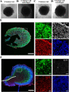
Fig 5. Examples of successful organoid differentiation from EBs.
Brain organoids were formed using guided and unguided differentiation protocols to induce telencephalic differentiation. Regardless of the use of U- or V-bottom wells, the EBs demonstrated budding neuroepithelia at the periphery, hinting at successful organoid formation. Examples of EBs produced in a (A) a U-bottom well without centrifugation, (B) a U-bottom well with centrifugation, (C) a V-bottom well without centrifugation, and (D) a V-bottom well with centrifugation. Scale bar: 500 μm. (E-F) Successful organoid differentiation at 30 daysof culture. Dotted lines indicate the rosette formations in the periphery of the organoid that show the undifferentiated neuroepithelial marker SOX2 and the neural identity markers of β3-tubulin and MAP2. EBs cultured under (E) guided (F) and unguided differentiation protocols showed similar structural locations of the expressed markers, suggesting the neural predestination of the neuroepithelial tissue. (E) Guided protocols yielded a better-defined rosette and exhibited matured β-tubulin and MAP2 expression, while (F) the unguided protocols showed initial signs of axonal (β-tubulin) growth. Scale bar: 200 μm.
Fig 6
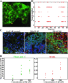
Fig 6. Functionality and possible applications of organoids derived from EBs.
(A) Electrophysiological data were obtained using the voltage-sensitive FluoVolt dye. High resolution (10 ms) imaging of the measured area was used to identify individual neurons. (B) Raster plot of 6 individual neurons shown in (A) reveals frequent neuron firing and suggests synchronous activity at day 60. (C) Disease modeling was tested by exposing the organoids to 200 nM and 500 nM concentrations of soluble amyloid beta (Aβ). (D) Damage was assessed using the Fluor Jade-C fluorescent marker (green) and (E) cell death with the BrdU TUNEL assay (red). Fluorescence images showed the location of the damage. Scale bar: 100 μm. Quantification of the fluorescent image area normalized against the DAPI (blue) signal.

Similar articles

Cited by

References

    1. Eiraku M, Takata N, Ishibashi H, Kawada M, Sakakura E, Okuda S, et al.. Self-organizing optic-cup morphogenesis in three-dimensional culture. Nature. 2011;472: 51–56. doi: 10.1038/nature09941 - DOI - PubMed
    1. Sato T, Vries RG, Snippert HJ, van de Wetering M, Barker N, Stange DE, et al.. Single Lgr5 stem cells build crypt-villus structures in vitro without a mesenchymal niche. Nature. 2009;459: 262–265. doi: 10.1038/nature07935 - DOI - PubMed
    1. Lancaster MA, Renner M, Martin C-A, Wenzel D, Bicknell LS, Hurles ME, et al.. Cerebral organoids model human brain development and microcephaly. Nature. 2013;501: 373–379. doi: 10.1038/nature12517 - DOI - PMC - PubMed
    1. Lancaster MA, Corsini NS, Wolfinger S, Gustafson EH, Phillips AW, Burkard TR, et al.. Guided self-organization and cortical plate formation in human brain organoids. Nat Biotechnol. 2017;35: 659–666. doi: 10.1038/nbt.3906 - DOI - PMC - PubMed
    1. Arora N, Alsous JI, Guggenheim JW, Mak M, Munera J, Wells JM, et al.. A process engineering approach to increase organoid yield. Development. 2017;144: 1128–1136. doi: 10.1242/dev.142919 - DOI - PMC - PubMed

Publication types

Grants and funding

This work was financially supported by the Netherlands Organ-on-Chip Initiative (NOCI) and By NWO (Dutch Research Council) – Gravity and Alzheimer Nederland grants. DCB thankfully acknowledge the financial support of Consejo Nacional de Ciencia y Tecnología (CONACyT) in the form of a doctoral scholarship. GTdS and MMA gratefully acknowledge the financial support from CONACyT (SNI scholarships 26048 and 256730) and the Biocodex Foundation, México. The funders had no role in study design, data collection and analysis, decision to publish, or preparation of the manuscript.