. 2022 Aug;37(4):569-580.
doi: 10.1016/j.virs.2022.05.001. Epub 2022 May 6.

STUB1 regulates antiviral RNAi through inducing ubiquitination and degradation of Dicer and AGO2 in mammals

Affiliations

STUB1 regulates antiviral RNAi through inducing ubiquitination and degradation of Dicer and AGO2 in mammals

Shumin Zhang et al. Virol Sin. 2022 Aug.

Abstract

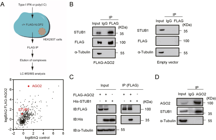
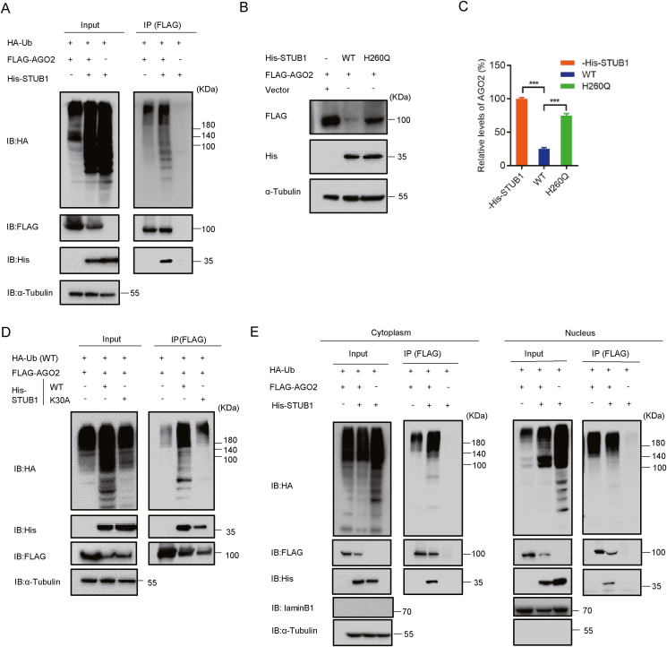
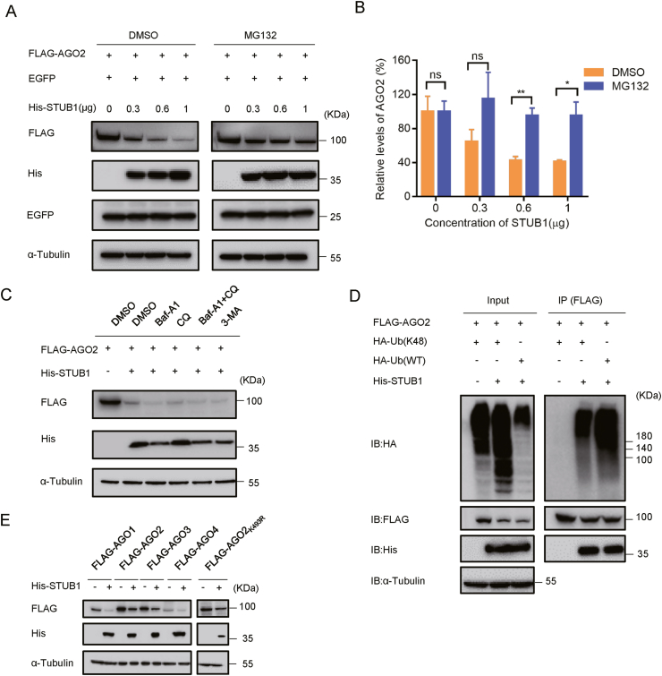
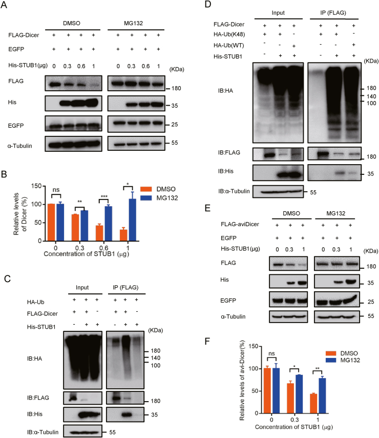
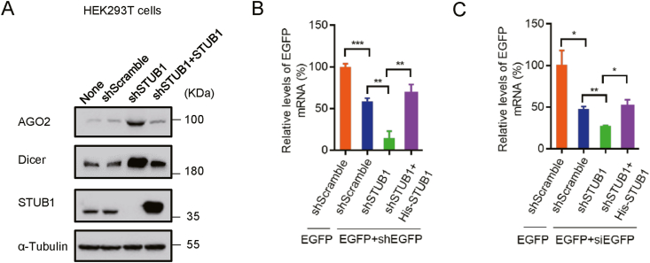
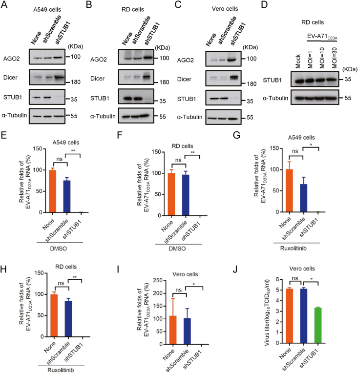
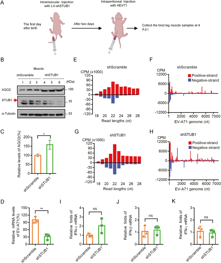
RNA interference (RNAi) is an intrinsic antiviral immune mechanism conserved in diverse eukaryotic organisms. However, the mechanism by which antiviral RNAi in mammals is regulated is poorly understood. In this study, we uncovered that the E3 ubiquitin ligase STIP1 homology and U-box-containing protein 1 (STUB1) was a new regulator of the RNAi machinery in mammals. We found that STUB1 interacted with and ubiquitinated AGO2, and targeted it for degradation in a chaperon-dependent manner. STUB1 promoted the formation of Lys48 (K48)-linked polyubiquitin chains on AGO2, and facilitated AGO2 degradation through ubiquitin-proteasome system. In addition to AGO2, STUB1 also induced the protein degradation of AGO1, AGO3 and AGO4. Further investigation revealed that STUB1 also regulated Dicer's ubiquitination via K48-linked polyubiquitin and induced the degradation of Dicer as well as its specialized form, termed antiviral Dicer (aviDicer) that expresses in mammalian stem cells. Moreover, we found that STUB1 deficiency up-regulated Dicer and AGO2, thereby enhancing the RNAi response and efficiently inhibiting viral replication in mammalian cells. Using the newborn mouse model of Enterovirus A71 (EV-A71), we confirmed that STUB1 deficiency enhanced the virus-derived siRNAs production and antiviral RNAi, which elicited a potent antiviral effect against EV-A71 infection in vivo. In summary, our findings uncovered that the E3 ubiquitin ligase STUB1 was a general regulator of the RNAi machinery by targeting Dicer, aviDicer and AGO1-4. Moreover, STUB1 regulated the RNAi response through mediating the abundance of Dicer and AGO2 during viral infection, thereby providing novel insights into the regulation of antiviral RNAi in mammals.

Keywords: Antiviral RNAi; Argonaute 2 (AGO2); Dicer; STIP1 homology and U-box-containing protein 1 (STUB1).


Figures

Fig. 1
Fig. 1
STUB1 interacts with AGO2 in mammalian cells. A Schematic illustration of the experimental approach used to identify the interactors of AGO2. HEK293T cells were transfected with the FLAG-tagged human AGO2 plasmid or empty vector (control) for 24 ​h, and then lysed and subjected to co-immunoprecipitation by using anti-FLAG antibody. The AGO2-bound complexes were excised and analyzed by LC-MS/MS. Graphs display the intensity-based absolute quantification (iBAQ) score of proteins identified in the FLAG-AGO2-immunoprecipitated complex versus the control. AGO2 and STUB1 were highlighted. B Western blotting analysis of the immunoprecipitated complexes as described in A. C HEK293T cells were co-transfected with FLAG-tagged human AGO2 plasmids and His-tagged human STUB1 plasmids or empty plasmid for 24 ​h, and subjected to co-immunoprecipitation with anti-FLAG antibody, followed by immunoblotting with anti-FLAG, anti-His and anti-α-Tubulin antibodies, respectively. D HEK293T cells were subjected to endogenous co-immunoprecipitation with anti-pan AGO antibody, followed by immunoblotting with anti-AGO2, anti-STUB1 and anti-α-Tubulin antibodies, respectively.
Fig. 2
Fig. 2
The STUB1 TPR domain and AGO2 PIWI domain are indispensable for STUB1-AGO2 interaction. A Schematic illustration of domains for the FLAG-tagged full-length AGO2 and its truncations. B HEK293T cells were co-transfected with His-STUB1 together with FLAG-AGO2 or its truncations for 24 ​h, and then subjected to co-immunoprecipitation with anti-FLAG antibody. C Schematic illustration of domains for the His-tagged full-length STUB1 and its truncations. D HEK293T cells were co-transfected with FLAG-AGO2 together with His-STUB1 or its truncations for 24 ​h, and then subjected to co-immunoprecipitation with anti-FLAG antibody.
Fig. 3
Fig. 3
STUB1 promotes the ubiquitination and degradation of AGO2 dependently on its E3 ligase and chaperon-binding activities. A HEK293T cells were co-transfected with HA-Ub together with FLAG-AGO2 and/or His-STUB1 for 24 ​h, respectively, and subjected to co-immunoprecipitation with anti-FLAG antibody. The ubiquitinated AGO2 was detected with anti-HA antibody, and AGO2-bound STUB1 was detected with anti-His antibody. B–C HEK293T cells were co-transfected with FLAG-AGO2 together with wild-type (WT) STUB1, STUB1H260Q mutant or empty plasmid, respectively, followed by immunoblotting with anti-FLAG antibody to detect AGO2. The experiments were repeated three times, and means ​± ​S.D. were plotted. Student's t-test was used. ∗∗∗, P ​< ​0.001. D HEK293T cells were co-transfected with HA-Ub together with FLAG-AGO2 and His-STUB1 or His-STUB1K30A, respectively, followed by immunoblotting with anti-HA antibody to detect ubiquitinated AGO2. E HEK293T cells were co-transfected with HA-Ub together with FLAG-AGO2 and/or His-STUB1 for 24 ​h, respectively. The Cellular nuclear and cytoplasmic fractions were separated and were immunoprecipitated with anti-FLAG antibody, followed by immunoblotting with anti-HA antibody to detect ubiquitinated AGO2. LaminB1 and tubulin were used as the nuclear and cytoplasmic makers, respectively.
Fig. 4
Fig. 4
STUB1 down-regulates the AGO2 protein level through the ubiquitin-proteasome system. AB HEK293T cells were co-transfected FLAG-AGO2 together with EGFP and different concentrations of His-STUB1, respectively. After 24 ​h transfection, cells were treated with DMSO or MG132 (10 ​μmol/L) for 9 ​h. The resulting cell lysates were immunoblotted with the indicated antibodies. The experiments were repeated three times, and means ​± ​S.D. were plotted. Student's t-test was used. ∗, P ​< ​0.05; ∗∗, P ​< ​0.01; ns, no significant. C HEK293T cells were co-transfected with FLAG-AGO2 together with His-STUB1 or empty vector for 24 ​h, and then treated with DMSO, 20 ​μmol/L chloroquine (CQ), 100 ​nmol/L Bafilomycin A1 (Baf-A1), 10 ​μmol/L 3-methyladenine (3-MA) or 100 ​nmol/L Baf-A1 together with 10 ​μmol/L 3-MA. After 24 ​h treatment, the resulting cell lysates were immunoblotted with the indicated antibodies. D HEK293T cells were co-transfected with FLAG-AGO2 together with His-STUB1 and WT Ub or K48 Ub for 24 ​h, respectively, and subjected to co-immunoprecipitation with anti-FLAG antibody, and followed by immunoblotting with anti-HA antibody to detect ubiquitinated AGO2. E HEK293T cells were co-transfected with FLAG-AGO1, FLAG-AGO2, FLAG-AGO3, FLAG-AGO4 or FLAG-AGO2K493R together with His-STUB1 or empty plasmid for 24 ​h, respectively, and subjected to western blotting with anti-FLAG antibody to detect AGO proteins.
Fig. 5
Fig. 5
STUB1 regulates Dicer ubiquitination for proteasomal degradation. A-B HEK293T cells were co-transfected FLAG-Dicer together with EGFP and different concentrations of His-STUB1, respectively. After 24 ​h transfection, cells were treated with DMSO or MG132 (10 ​μmol/L) for 9 ​h. The resulting cell lysates were immunoblotted with the indicated antibodies. The experiments were repeated three times, and means ​± ​S.D. were plotted. Student's t-test was used. ∗, P ​< ​0.05; ∗∗, P ​< ​0.01; ∗∗∗, P ​< ​0.001; ns, no significant. C HEK293T cells were co-transfected with HA-Ub together with FLAG-Dicer and/or His-STUB1 for 24 ​h, respectively, and subjected to co-immunoprecipitation with anti-FLAG antibody. The ubiquitinated AGO2 was detected with anti-HA antibody, and Dicer-bound STUB1 was detected with anti-His antibody. D HEK293T cells were co-transfected with FLAG-Dicer together with His-STUB1 and WT Ub or K48-Ub for 24 ​h, respectively, and subjected to co-immunoprecipitation with anti-FLAG antibody, and followed by immunoblotting with anti-HA antibody to detect ubiquitinated Dicer. EF HEK293T cells were co-transfected FLAG-aviDicer together with EGFP and different concentrations of His-STUB1, respectively. After 24 ​h transfection, cells were treated with DMSO or MG132 (10 ​μmol/L) for 9 ​h. The resulting cell lysates were immunoblotted with the indicated antibodies. Student's t-test was used. ∗, P ​< ​0.05; ∗∗, P ​< ​0.01; ns, no significant.
Fig. 6
Fig. 6
STUB1 regulates the RNAi response through mediating the abundance of Dicer and AGO2 in mammalian cells. A HEK293T cells were transfected with lentivirus-based shScramble or shSTUB1. The stable STUB1 knocking-down cells were examined via Western blotting with anti- AGO2, anti-Dicer and anti-STUB1 antibodies, respectively. The stable STUB1 knocking-down cells were transfected with His-STUB1 for 24 ​h to generate the STUB1 expression restored cells (shSTUB1+STUB1). BC The shScramble, shSTUB1 and shSTUB1+STUB1 cells were co-transfected with EGFP together with EGFP-specific shRNA (shEGFP) or siEGFP for 24 ​h, and EGFP mRNA levels were examined by qRT-PCR. The experiments were repeated three times, and means ​± ​S.D. were plotted. Student's t-test was used. ∗, P ​< ​0.05; ∗∗, P ​< ​0.01; ∗∗∗, P ​< ​0.001.
Fig. 7
Fig. 7
STUB1 deficiency inhibits the replication of VSR-deficient Enterovirus A71 (EV-A71 D23A) through up-regulating Dicer and AGO2 in mammalian cells. A-C A549, RD and Vero cells were transfected with lentivirus-based shScramble or shSTUB1, respectively. Cells without lentivirus treatment were used as controls (None). The stable STUB1 knocking-down cells were examined via Western blotting with anti- AGO2, anti-Dicer and anti-STUB1 antibodies, respectively. D The protein levels of STUB1 were examined in RD cells infected with different MOI EV-A71D23A at 24 ​h.p.i. EJ The indicated A549, RD and Vero cells were used to infected with EV-A71D23A at an MOI of 1 treated with DMSO or Ruxolitinib (2 ​μmol/L), respectively. At 24 ​h.p.i., the levels of EV-A71D23A genomic RNAs were detected by qRT-PCR. The viral titers in infected Vero cells were examined. TCID50, 50% tissue culture infectious doses per milliliter. The experiments were repeated three times, and means ​± ​S.D. were plotted. Student's t-test was used. ∗, P ​< ​0.05; ∗∗, P ​< ​0.01.
Fig. 8
Fig. 8
STUB1 deficiency enhances antiviral RNAi response in vivo. A The lentivirus-based shScramble or mouse shSTUB1 was intramuscularly injected into the hind leg muscles of 1-day-old ICR mice (n ​= ​3 for each group). Two days later, mice were then i.p. injected with 1 ​× ​106 ​PFU ​EV-A71. At 4 ​d.p.i., mice were euthanized and the hind leg muscles were harvested for subsequent analysis. B The expression levels of mouse AGO2 and STUB1 in shScramble and shSTUB1 mice were tested by Western blotting. The resulting cell lysates were immunoblotted with the indicated antibodies. C The relative expression levels of AGO2 in shScramble and shSTUB1 mice as indicated in B were normalized by tubulin, and the level of AGO2 in shScramble mice was defined as 100%. Student's t-test was used. ∗, P ​< ​0.05. D The levels of EV-A71 genomic RNAs in shScramble and shSTUB1 mice were detected by qRT-PCR. Student's t-test was used. ∗∗, P ​< ​0.01. EH Size distribution and abundance (counts per million of total vsRNA reads, CPM) of total vsRNAs sequenced from shScramble and shSTUB1 mice infected with EV-A71. Red, positive-stranded vsRNAs; blue, negative-stranded vsRNAs. The distribution of 22±1-nt vsRNA reads mapped to the (+)- and (−)-stranded EV-A71 genome and the relative abundances of (+)- and (−)-stranded vsRNAs are indicated. IK The mRNA levels of IFN-α, IFN-β and INF-γ in shScramble and shSTUB1 mice were detected by qRT-PCR. Student's t-test was used. ns, no significant.

Similar articles

Cited by

References

    1. Adiliaghdam F., Basavappa M., Saunders T.L., Harjanto D., Prior J.T., Cronkite D.A., Papavasiliou N., Jeffrey K.L. A requirement for Argonaute 4 in mammalian antiviral defense. Cell Rep. 2020;30:1690–1701. e1694. - PMC - PubMed
    1. Backes S., Langlois R.A., Schmid S., Varble A., Shim J.V., Sachs D., tenOever B.R. The Mammalian response to virus infection is independent of small RNA silencing. Cell Rep. 2014;8:114–125. - PMC - PubMed
    1. Bronevetsky Y., Villarino A.V., Eisley C.J., Barbeau R., Barczak A.J., Heinz G.A., Kremmer E., Heissmeyer V., McManus M.T., Erle D.J., Rao A., Ansel K.M. T cell activation induces proteasomal degradation of Argonaute and rapid remodeling of the microRNA repertoire. J. Exp. Med. 2013;210:417–432. - PMC - PubMed
    1. Chang H.M., Martinez N.J., Thornton J.E., Hagan J.P., Nguyen K.D., Gregory R.I. Trim71 cooperates with microRNAs to repress Cdkn1a expression and promote embryonic stem cell proliferation. Nat. Commun. 2012;3:923. - PMC - PubMed
    1. Chen J., Lai F., Niswander L. The ubiquitin ligase mLin41 temporally promotes neural progenitor cell maintenance through FGF signaling. Genes Dev. 2012;26:803–815. - PMC - PubMed