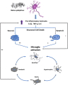
Microglia and status epilepticus in the immature brain
- PMID: 35531942
- PMCID: PMC10173848
- DOI: 10.1002/epi4.12610
Microglia and status epilepticus in the immature brain
Abstract
Microglia are the resident immune cells of the Central Nervous System (CNS), which are activated due to brain damage, as part of the neuroinflammatory response. Microglia undergo morphological and biochemical modifications during activation, adopting a pro-inflammatory or an antiinflammatory state. In the developing brain, status epilepticus (SE) promotes microglia activation that is associated with neuronal injury in some areas of the brain, such as the hippocampus, thalamus, and amygdala. However, the timing of this activation, the anatomical pattern, and the morphological and biochemical characteristics of microglia in the immature brain are age-dependent and have not been fully characterized. Therefore, this review focuses on the response of microglia to SE and its relationship to neurodegeneration.
Keywords: status epilepticus; immature brain; microglia.
© 2022 The Authors. Epilepsia Open published by Wiley Periodicals LLC on behalf of International League Against Epilepsy.
Conflict of interest statement
The authors declare no conflicts of interests. We confirm that we have read the Journal's position on issues involved in ethical publication.
Figures

Similar articles
-
Glia activation and cytokine increase in rat hippocampus by kainic acid-induced status epilepticus during postnatal development.Neurobiol Dis. 2003 Dec;14(3):494-503. doi: 10.1016/j.nbd.2003.08.001. Neurobiol Dis. 2003. PMID: 14678765
-
Status epilepticus induces a particular microglial activation state characterized by enhanced purinergic signaling.J Neurosci. 2008 Sep 10;28(37):9133-44. doi: 10.1523/JNEUROSCI.1820-08.2008. J Neurosci. 2008. PMID: 18784294 Free PMC article.
-
Altered morphological dynamics of activated microglia after induction of status epilepticus.J Neuroinflammation. 2015 Nov 4;12:202. doi: 10.1186/s12974-015-0421-6. J Neuroinflammation. 2015. PMID: 26538404 Free PMC article.
-
Neuronal injury in chronic CNS inflammation.Best Pract Res Clin Anaesthesiol. 2010 Dec;24(4):551-62. doi: 10.1016/j.bpa.2010.11.001. Epub 2010 Nov 29. Best Pract Res Clin Anaesthesiol. 2010. PMID: 21619866 Review.
-
Techniques and Methods of Animal Brain Surgery: Perfusion, Brain Removal, and Histological Techniques.In: Kobeissy FH, editor. Brain Neurotrauma: Molecular, Neuropsychological, and Rehabilitation Aspects. Boca Raton (FL): CRC Press/Taylor & Francis; 2015. Chapter 15. In: Kobeissy FH, editor. Brain Neurotrauma: Molecular, Neuropsychological, and Rehabilitation Aspects. Boca Raton (FL): CRC Press/Taylor & Francis; 2015. Chapter 15. PMID: 26269921 Free Books & Documents. Review.
Cited by
-
Mechanisms of Organophosphate Toxicity and the Role of Acetylcholinesterase Inhibition.Toxics. 2023 Oct 18;11(10):866. doi: 10.3390/toxics11100866. Toxics. 2023. PMID: 37888716 Free PMC article. Review.
-
Investigating the genetic contribution in febrile infection-related epilepsy syndrome and refractory status epilepticus.Front Neurol. 2023 Apr 3;14:1161161. doi: 10.3389/fneur.2023.1161161. eCollection 2023. Front Neurol. 2023. PMID: 37077567 Free PMC article.
-
A Pediatric Rat Model of Organophosphate-Induced Refractory Status Epilepticus: Characterization of Long-Term Epileptic Seizure Activity, Neurologic Dysfunction and Neurodegeneration.J Pharmacol Exp Ther. 2024 Jan 17;388(2):416-431. doi: 10.1124/jpet.123.001794. J Pharmacol Exp Ther. 2024. PMID: 37977810 Free PMC article.
-
Single-Cell Transcriptomic Analyses of Brain Parenchyma in Patients With New-Onset Refractory Status Epilepticus (NORSE).Neurol Neuroimmunol Neuroinflamm. 2024 Jul;11(4):e200259. doi: 10.1212/NXI.0000000000200259. Epub 2024 May 29. Neurol Neuroimmunol Neuroinflamm. 2024. PMID: 38810181 Free PMC article.
-
Protein profiling and assessment of amyloid beta levels in plasma in canine refractory epilepsy.Front Vet Sci. 2023 Dec 21;10:1258244. doi: 10.3389/fvets.2023.1258244. eCollection 2023. Front Vet Sci. 2023. PMID: 38192726 Free PMC article.
References
-
- Brain Injury Association . Brain injury overview. https://www.biausa.org/brain‐injury/about‐brain‐injury/basics/overview. Accessed 23 December 2021.
-
- Thompson K, Wasterlain C. Lithium‐pilocarpine status epilepticus in the immature rabbit. Brain Res Dev Brain Res. 1997;100:1–4. - PubMed
-
- Thompson K, Holm AM, Schousboe A, Popper P, Micevych P, Wasterlain CW. Hippocampal stimulation produces neuronal death in the immature brain. Neuroscience. 1998;82:337–48. - PubMed
-
- Sankar R, Shin DH, Wasterlain CG. Serum neuron‐specific enolase is a marker for neuronal damage following status epilepticus in the rat. Epilepsy Res. 1997;28:129–36. - PubMed
Publication types
MeSH terms
LinkOut - more resources
Full Text Sources

