. 2022 May 5;12(1):7334.
doi: 10.1038/s41598-022-10925-2.

Sophisticated expression responses of ZNT1 and MT in response to changes in the expression of ZIPs

Affiliations

Sophisticated expression responses of ZNT1 and MT in response to changes in the expression of ZIPs

Shino Nagamatsu et al. Sci Rep. .

Abstract

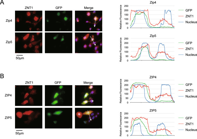
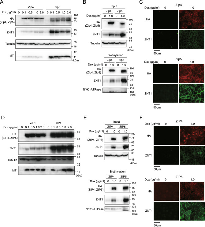
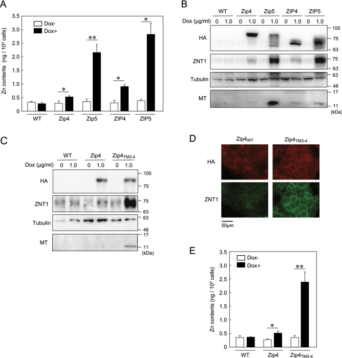
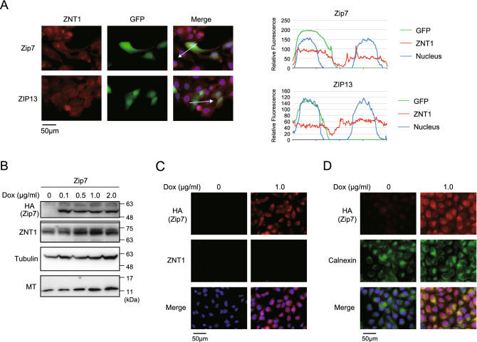
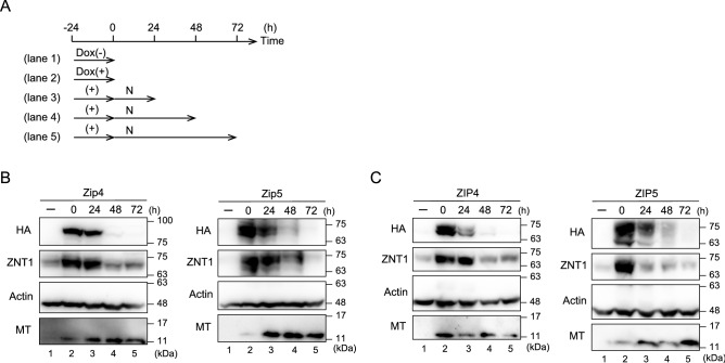
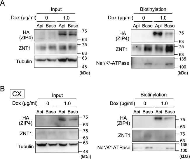
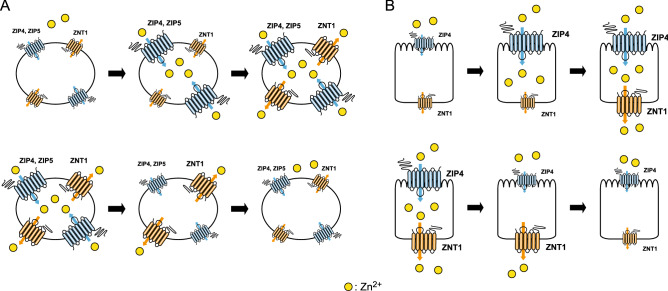
The zinc homeostatic proteins Zn transporter 1 (ZNT1) and metallothionein (MT) function in dampening increases in cytosolic zinc concentrations. Conversely, the expression of ZNT1 and MT is expected to be suppressed during decreases in cytosolic zinc concentrations. Thus, ZNT1/MT homeostatic responses are considered to be essential for maintaining cellular zinc homeostasis because cellular zinc concentrations are readily altered by changes in the expression of several Zrt-/Irt-like proteins (ZIPs) under both physiological and pathological conditions. However, this notion remains to be tested experimentally. Here, we investigated the aforementioned homeostatic process by analyzing ZNT1 and MT protein expression in response to ZIP expression. Overexpression of cell-surface-localized ZIPs, such as ZIP4 and ZIP5, increased the cellular zinc content, which caused an increase in the expression of cell-surface ZNT1 and cytosolic MT in the absence of zinc supplementation in the culture medium. By contrast, elimination of the overexpressed ZIP4 and ZIP5 resulted in decreased expression of ZNT1 but not MT, which suggests that differential regulation of ZNT1 and MT expression at the protein level underlies the homeostatic responses necessary for zinc metabolism under certain conditions. Moreover, increased expression of apically localized ZIP4 facilitated basolateral ZNT1 expression in polarized cells, which indicates that such a coordinated expression mechanism is crucial for vectorial transcellular transport. Our results provide novel insights into the physiological maintenance of cellular zinc homeostasis in response to alterations in cytosolic zinc concentrations caused by changes in the expression of ZIPs.


Conflict of interest statement

The authors declare no competing interests.

Figures

Figure 1
Figure 1
Cell-surface ZNT1 expression is enhanced by Dox-induced expression of mouse Zip4 and Zip5 and human ZIP4 and ZIP5. (A,B) Transiently expressed Zip4/Zip5 and ZIP4/ZIP5 enhanced cell-surface ZNT1 expression. U2OS cells were transiently transfected with an IRES-GFP expression plasmid harboring Zip4-HA or Zip5-HA cDNA (A) or ZIP4-HA or ZIP5-HA cDNA (B), and transfected cells were identified by their GFP fluorescence. DAPI staining is also shown in the merged images, where line-profile analysis was used to semi-quantitatively determine relative fluorescence intensity (white arrows); the graphs on the right show the relative fluorescence intensity along each arrow. Each experiment was performed at least thrice, and representative results from independent experiments are shown.
Figure 2
Figure 2
ZNT1 and MT expression increases in a coordinated manner in response to Dox-induced expression of Zip4/Zip5 or ZIP4/ZIP5. (A) Upregulated expression of ZNT1 and MT accompanied Dox-induced expression of Zip4/Zip5 in MDCK cells. MDCK cells stably expressing Zip4 or Zip5 were cultured with the indicated concentrations of Dox for 24 h, and then ZNT1 and MT expression was examined. Tubulin was used as a loading control. (B,C) ZNT1 accumulated on the cell surface upon Dox-induced Zip4/Zip5 expression. MDCK cells cultured with or without 1.0 μg/mL Dox for 24 h were used in cell-surface biotinylation assays (B) or subject to immunofluorescence staining (C). In (B) tubulin and Na+/K+-ATPase were used as the loading control for input and biotinylation, respectively. (D–F) Same experiments as in (A–C) but performed using ZIP4 and ZIP5. Each experiment was performed at least thrice, and representative results from independent experiments are shown.
Figure 3
Figure 3
Potent induction of ZNT1 expression mediated by Zip5 is associated with Zip5 cytosolic variable loop. (A) Zinc content measured using ICP-MS analysis. Parental MDCK cells and MDCK cells stably expressing Zip4-HA, Zip5-HA, ZIP4-HA, or ZIP5-HA were cultured with 1.0 μg/mL Dox for 24 h and then collected for ICP-MS analyses (n = 3). (B) Immunoblotting performed using cell lysates prepared from the indicated cells cultured as in (A). (C) ZNT1 and MT expression induced by Zip4TM3–4-HA mutant was higher than that induced by Zip4. Parental MDCK cells and MDCK cells stably expressing Zip4-HA or Zip4TM3–4-HA were cultured with 1.0 μg/mL Dox for 24 h. (D) Immunofluorescence labeling for confirming enhanced cell-surface expression of ZNT1 in MDCK cells stably expressing Zip4TM3–4-HA; the cells were cultured as in (C). (E) Zinc content, measured using ICP-MS analysis, in indicated MDCK cells cultured as in (C) (n = 3). In (B,C), tubulin was used as the loading control. Each experiment was performed at least thrice, and representative results from independent experiments are shown.
Figure 4
Figure 4
Expression alteration of intracellularly localized ZIPs does not substantially alter ZNT1 and MT expression. (A) Transiently expressed mouse Zip7 and human ZIP13 did not enhance cell-surface ZNT1 expression. U2OS cells were transiently transfected with an IRES-GFP expression plasmid harboring Zip7-HA or ZIP13-HA cDNA and then examined as described in Fig. 1. Merged images with DAPI staining are also shown, and the right graphs show the relative fluorescence intensity determined semi-quantitatively using line-profile analysis. (B,C) Expression of ZNT1 and MT was only slightly altered by Dox-induced expression of Zip7 in MDCK cells. MDCK cells stably expressing Zip7 were cultured with the indicated concentrations of Dox for 24 h in (B) or with 1.0 μg/mL Dox in C, and ZNT1 and MT expression levels were examined through immunoblotting (B) and immunofluorescence staining (C). In (B), tubulin was used as the loading control. (D) Confirmation of ER localization of induced Zip7. MDCK cells were cultured with or without 1.0 μg/mL Dox for 24 h and then immunofluorescence staining was performed; concurrent immunostaining of the ER marker calnexin was used to assess Zip7 subcellular localization. Each experiment was performed at least thrice, and representative results from independent experiments are shown.
Figure 5
Figure 5
Elimination of the induced expression of ZIPs results in a reduced expression of cell-surface ZNT1. (A) Schematic depicting the assay time course. MDCK cells stably expressing Zip4, Zip5, ZIP4, or ZIP5 were cultured with or without 0.1 μg/mL Dox for 24 h, and then the cells were cultured in normal medium (N) for an additional 24–72 h after washing thrice with PBS. (B,C) ZNT1 expression upregulated by Dox-induced expression of Zip4 or Zip5 (B) or ZIP4 or ZIP5 (C) decreased after Dox removal. However, a marked reduction of MT expression did not accompany the reduction of ZNT1 expression. Actin was used as the loading control. Each experiment was performed at least thrice, and representative results from independent experiments are shown.
Figure 6
Figure 6
Increased expression of apically localized ZIP4 induces the expression of basolaterally localized ZNT1. (A) MDCK cells stably expressing ZIP4-HA and grown in transwell plates were treated with Dox for 24 h. Next, the biotinylation reagent sulfo-NHS-SS-biotin was added to either the apical (Api) or basolateral (Baso) compartment of the transwell plate. Lastly, solubilized proteins captured using streptavidin beads were analyzed by immunoblotting with specific antibodies. (B) Same experiments as in (A), performed similarly but employing a zinc-deficient medium generated using Chelex-100 resin-treated FCS (CX). In (A,B), input refers to aliquots of the biotinylated proteins before avidin capture, and biotinylation refers to avidin-captured proteins. Tubulin and Na+/K+-ATPase were used as the loading control for the input and for monitoring the efficiency of basolateral membrane biotinylation, respectively.
Figure 7
Figure 7
Model depicting ZIP-driven ZNT1 expression for homeostatic control. (A) Model showing ZIP-driven ZNT1 expression in generic cells. Increased expression of cell-surface ZIPs results in increased zinc content in the cytosol, which leads to the enhanced expression of ZNT1 and the subsequent dampening of increases in cytosolic zinc concentration (upper). Conversely, decreased expression of cell-surface ZIPs causes a decrease in the zinc content in the cytosol, which results in the decreased expression of ZNT1 and thus the maintenance around a homeostatic setpoint at a specific concentration in each cell type (lower). (B) Prediction model showing ZIP4-driven ZNT1 expression in the zinc-absorption process in enterocytes. Increased ZIP4 expression on the apical surface results in enhanced zinc content in the cytosol, which, in turn, leads to the upregulation of ZNT1 expression and thereby enables efficient export of zinc on the basolateral side into the blood stream (upper). Conversely, decreased ZIP4 expression on the apical surface causes the opposite responses through a decrease in the zinc content in the cytosol and decreased ZNT1 expression, and thus leads to reduced zinc export (lower).

Similar articles

Cited by

References

    1. Hara T, et al. Physiological roles of zinc transporters: Molecular and genetic importance in zinc homeostasis. J. Physiol. Sci. 2017;67:283–301. doi: 10.1007/s12576-017-0521-4. - DOI - PMC - PubMed
    1. Kambe T, Suzuki E, Komori T. Zinc transporters—A review and a new view from biochemistry. In: Fukada T, Kambe T, editors. Zinc Signaling. 2. Springer; 2019. pp. 23–56.
    1. Kambe T, Taylor KM, Fu D. Zinc transporters and their functional integration in mammalian cells. J. Biol. Chem. 2021;296:100320. doi: 10.1016/j.jbc.2021.100320. - DOI - PMC - PubMed
    1. Drozd A, Wojewska D, Peris-Diaz MD, Jakimowicz P, Krezel A. Crosstalk of the structural and zinc buffering properties of mammalian metallothionein-2. Metallomics. 2018;10:595–613. doi: 10.1039/C7MT00332C. - DOI - PubMed
    1. Krezel A, Maret W. The bioinorganic chemistry of mammalian metallothioneins. Chem. Rev. 2021;121:14594–14648. doi: 10.1021/acs.chemrev.1c00371. - DOI - PubMed