. 2022 May 5;12(1):7322.
doi: 10.1038/s41598-022-10907-4.

Modified lipid metabolism and cytosolic phospholipase A2 activation in mesangial cells under pro-inflammatory conditions

Affiliations

Modified lipid metabolism and cytosolic phospholipase A2 activation in mesangial cells under pro-inflammatory conditions

Roberto Boi et al. Sci Rep. .

Abstract

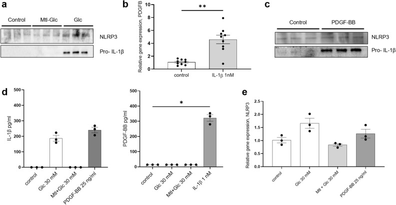
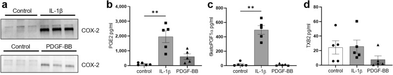
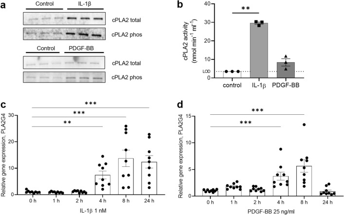
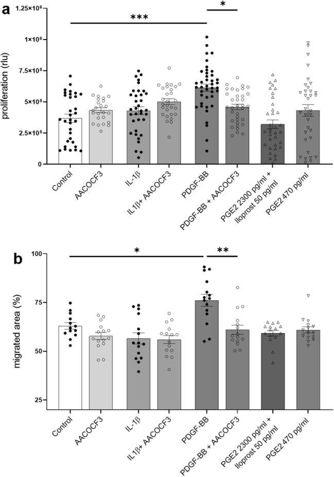
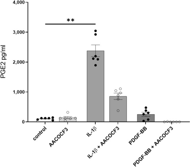
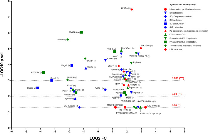
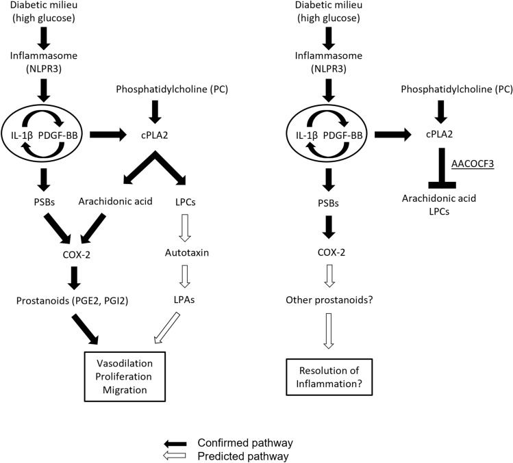
Diabetic kidney disease is a consequence of hyperglycemia and other complex events driven by early glomerular hemodynamic changes and a progressive expansion of the mesangium. The molecular mechanisms behind the pathophysiological alterations of the mesangium are yet to be elucidated. This study aimed at investigating whether lipid signaling might be the missing link. Stimulation of human mesangial cells with high glucose primed the inflammasome-driven interleukin 1 beta (IL-1β) secretion, which in turn stimulated platelet-derived growth factor (PDGF-BB) release. Finally, PDGF-BB increased IL-1β secretion synergistically. Both IL-1β and PDGF-BB stimulation triggered the formation of phosphorylated sphingoid bases, as shown by lipidomics, and activated cytosolic phospholipase cPLA2, sphingosine kinase 1, cyclooxygenase 2, and autotaxin. This led to the release of arachidonic acid and lysophosphatidylcholine, activating the secretion of vasodilatory prostaglandins and proliferative lysophosphatidic acids. Blocking cPLA2 release of arachidonic acid reduced mesangial cells proliferation and prostaglandin secretion. Validation was performed in silico using the Nephroseq database and a glomerular transcriptomic database. In conclusion, hyperglycemia primes glomerular inflammatory and proliferative stimuli triggering lipid metabolism modifications in human mesangial cells. The upregulation of cPLA2 was critical in this setting. Its inhibition reduced mesangial secretion of prostaglandins and proliferation, making it a potential therapeutical target.


Conflict of interest statement

The authors declare no competing interests.

Figures

Figure 1
Figure 1
High glucose priming of IL-1β, stimulation of IL-1β on PDGF-BB production, and vice-versa. (a) Activation of the inflammasome NLRP3 and subsequent increase of pro-IL-1β protein levels were measured after 24 h stimulation of mesangial cells with Glc 30 mM. Osmotic control was Glc plus mannitol (Mtl), final concentration 30 mM. (b) 24 h stimulation of human mesangial cells with IL-1β 1 nM increased the levels of PDGFB mRNA. (c) In turn, a 24 h stimulation of mesangial cells with 25 ng/ml of PDGF-BB activated NLRP3 activity and increased pro-IL-1β protein levels. (d) Bio-Plex analysis of IL-1β, and PDGF-BB in media after high glucose, osmotic control, IL-1β and PDGF-BB 48 h stimulations. *P < 0.05. Data are reported as mean ± SEM. Samples under the LOD were considered equal to the lowest value in the measurable range (0.25 pg/ml for IL-1β and 13.96 pg/ml for PDGF-BB). (e) NLRP3 gene expression levels increased after 24 h stimulation with Glc 30 mM and PDGF-BB 25 ng/ml. Uncropped blots after chemiluminescence development and total protein stain free blots are presented in Supplementary Fig. S4a. Quantification of the bands in the western blot is reported in Supplementary Fig. S5.
Figure 2
Figure 2
Decreased total ceramides and sphingomyelins and increased phosphorylated sphingoid bases after 24 h stimulation of mesangial cells with IL-1β and PDGF-BB. Lipidomics experiments showed a decrease of the whole sphingolipid cellular pool (Cer, LacCer, dhCer, GluCer, SM, dhSG, SG, S1P, dhS1P, PhytoSG) after the treatments (a) and the increase of phosphorylated sphingoid bases (b) compared to total sphingoid bases (dhSG, SG, S1P, dhS1P, PhytoSG). Data were normalized against cell count and divided by the relative cell count normalization factor. Sphk1 (phosphorylation of sphingoid bases) protein levels are increased by both stimulations, while Cerk (phosphorylation of ceramides) is downregulated at protein (c) and gene level (d). SPTLC1 (de novo ceramide synthesis) is not regulated at protein level (c) and CERS2 (dhCer synthesis from dhSG) is downregulated at the gene level (e). *P < 0.05; **P < 0.01; ***P < 0.001. Data are reported as mean ± SEM. Uncropped blots after chemiluminescence development and total protein stain free blots are presented in Supplementary Fig. S4b. Quantification of the bands in the western blot is reported in Supplementary Fig. S5.
Figure 3
Figure 3
Induction of COX-2 by phosphorylated sphingoid bases and prostaglandins secretion. COX-2 protein levels increased after 24 h stimulation of human mesangial cells with IL-1β or PDGF-BB (a). PGE2 is secreted in the medium after both treatments (b), while PGI2 (measured through its metabolite 6-keto PGF1α) is secreted after the IL-1β treatment only (c). Thromboxane A2 (measured through its metabolite TXB2) secretion is not regulated (d). *P < 0.05; **P < 0.01. Data are reported as mean ± SEM. Uncropped blots after chemiluminescence development and total protein stain free blots are presented in Supplementary Fig. S4c. Quantification of the bands in the western blot is reported in Supplementary Fig. S5.
Figure 4
Figure 4
Activation of rate-limiting enzyme cPLA2, upstream of COX-2. Phospholipase cPLA2 is upregulated and phosphorylated in human mesangial cells after 24 h stimulations with IL-1β and PDGF-BB (a). cPLA2 activity is accordingly increased (b). The activity in the controls was below the LOD of 3.5 nmol/min/ml. In this case, the LOD value was plotted in the graph. PLA2G4A gene expression was already upregulated after 4 h in both treatments (c, d). *P < 0.05; ***P < 0.001. Data are reported as mean ± SEM. Uncropped blots after chemiluminescence development and total protein stain free blots are presented in Supplementary Fig. S4d. Quantification of the bands in the western blot is reported in Supplementary Fig. S5.
Figure 5
Figure 5
Inhibition of cPLA2 with AACOCF3 1 μM reduces proliferation, secretion, and prostaglandin secretion. AACOCF3 reduced PDGF-BB driven cell proliferation (a) and migration (b). No effect was observed when PGE2 and iloprost (PGI2) were administered together (mimicking their secretion levels obtained after 24 h IL-1β stimulation of mesangial cells) and at low PGE2 concentrations (mimicking PGE2 secretion levels after 24 h PDGF-BB stimulation) (b). Prostaglandin administration had no migratory effect on mesangial cells (b). *P < 0.05; **P < 0.01; ***P < 0.001, rlu relative light units. Data are reported as mean ± SEM.
Figure 6
Figure 6
Inhibition of cPLA2 with AACOCF3 1 μM reduces IL-1β, PDGF-BB induced PGE2 secretion. **P < 0.01. Data are reported as mean ± SEM.
Figure 7
Figure 7
Volcano plot of the in silico data validation performed using glomerular gene expression data from Nephroseq and glomerular mRNA sequencing data from Levin et al.. Human gene names are reported in uppercase, mouse genes names in lowercase. (a) Hodgin et al. db/db C57BLKS mouse model—DKD vs non-DKD. (b) Hodgin et al., eNOS-deficient C57BLKS db/db mouse model—DKD vs non-DKD. (c) Hodgin et al., DBA/2 mouse model—DKD vs non-DKD. (d) Ju et al. DKD human renal biopsies vs healthy individuals. (e) Levin et al. DKD human renal biopsies vs healthy individuals. (f) Woroniecka et al. DKD human renal biopsies vs healthy individuals. For the outliers, the relative percentiles were reported after the gene names and. COPA values were plotted instead of log2 fold changes.
Figure 8
Figure 8
Signaling pathway overview. Hyperglycemia stimulates mesangial production of IL-1β which activates PDGF-BB secretion. In turn, PDGF-BB promotes IL-1β secretion, sustaining and boosting the pathway activation. IL-1β and PDGF-BB stimulation give rise to the production of phosphorylated sphingoid bases, activating COX-2 transcription. At the same time, IL-1β and PDGF-BB stimulate cPLA2 activation. Arachidonic acid (AA) released by cPLA2 is converted into prostanoids by COX-2 and downstream enzymes. The other product of cPLA2, lysophosphatidylcholine (LPC), is converted into lysophosphatidic acid (LPA) by autotaxin. Thus, hyperglycemia triggered the activation of an interplay between IL-1β and PDGF-BB which stimulates the secretion of lipid hormones (prostanoids PGE2, PGI2, but also lysophosphatidic acid) with hemodynamic, proliferative, and migratory effects at glomerular level. Inhibition of the cPLA2 reaction with AACOCF3 blocks the supply of AA for the COX-2 reaction, thus resolving the inflammatory stimulus. In addition, LPC is not produced, and this blocks the supply for the autotaxin reaction and LPA mediated proliferative response.

Similar articles

Cited by

References

    1. Thomas MC, et al. Diabetic kidney disease. Nat. Rev. Dis. Primers. 2015;1:15018. doi: 10.1038/nrdp.2015.18. - DOI - PMC - PubMed
    1. Tonneijck L, et al. Glomerular hyperfiltration in diabetes: Mechanisms, clinical significance, and treatment. J. Am. Soc. Nephrol. 2017;28:1023–1039. doi: 10.1681/ASN.2016060666. - DOI - PMC - PubMed
    1. Mitrofanova A, Burke G, Merscher S, Fornoni A. New insights into renal lipid dysmetabolism in diabetic kidney disease. World J. Diabetes. 2021;12:524–540. doi: 10.4239/wjd.v12.i5.524. - DOI - PMC - PubMed
    1. Dogan Y, Akarsu S, Ustundag B, Yilmaz E, Gurgoze MK. Serum IL-1beta, IL-2, and IL-6 in insulin-dependent diabetic children. Mediators Inflamm. 2006;2006:59206. doi: 10.1155/mi/2006/59206. - DOI - PMC - PubMed
    1. Gouda W, Mageed L, Abd El Dayem SM, Ashour E, Afify M. Evaluation of pro-inflammatory and anti-inflammatory cytokines in type 1 diabetes mellitus. Bull. Natl. Res. Centre. 2018;42:14. doi: 10.1186/s42269-018-0016-3. - DOI