. 2022 May 5;41(1):164.
doi: 10.1186/s13046-022-02378-2.

EIF4A3-induced circTOLLIP promotes the progression of hepatocellular carcinoma via the miR-516a-5p/PBX3/EMT pathway

Affiliations

EIF4A3-induced circTOLLIP promotes the progression of hepatocellular carcinoma via the miR-516a-5p/PBX3/EMT pathway

Yachong Liu et al. J Exp Clin Cancer Res. .

Abstract

Background: Circular RNAs (circRNAs) function as crucial regulators in multiple cancers, including hepatocellular carcinoma (HCC). However, the roles of circRNAs in HCC remains largely unknown.

Methods: circTOLLIP was identified in HCC by screening of two public circRNA microarray datasets and detected in HCC cells and tissues through quantitative real-time PCR (qRT-PCR) and in situ hybridization (ISH). Gain- and loss-of-function assays were performed to confirm the biological effects of circTOLLIP on HCC in vitro and in vivo. Mechanistically, bioinformatics analysis of online databases, MS2-RNA pulldown, biotin-labeled circTOLLIP/miR-516a-5p RNA pulldown, RNA immunoprecipitation (RIP), luciferase reporter assay, fluorescence in situ hybridization assay (FISH) and RNA sequencing were used to confirm the regulation of Eukaryotic initiation factor 4A3 (EIF4A3) on circTOLLIP and the interaction among circTOLLIP, miR-516a-5p and PBX homeobox 3 (PBX3).

Results: circTOLLIP was significantly upregulated in HCC cells and tissues. High circTOLLIP expression was correlated with poor overall survival (OS) and disease-free survival (DFS) in patients. circTOLLIP promoted the proliferation and metastasis of HCC cells in vitro and in vivo. Mechanistically, EIF4A3 promoted the biogenesis of circTOLLIP without affecting its stability. Moreover, circTOLLIP sponged miR-516a-5p to elevate the expression of PBX3, thereby activating the epithelial-to-mesenchymal transition (EMT) pathway and facilitating tumor progression in HCC.

Conclusions: Our findings indicate that EIF4A3-induced circTOLLIP promotes the progression of HCC through the circTOLLIP/miR-516a-5p/PBX3/EMT axis.

Keywords: EMT; HCC; PBX3; circTOLLIP.


Conflict of interest statement

All authors declare that they have no competing interests.

Figures
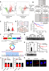
Fig. 1
Fig. 1
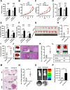
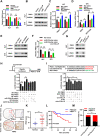
Identification and characterization of circTOLLIP in HCC. a Volcano plot of circRNAs from GEO datasets of GSE97332 and GSE78520. Significantly upregulated and downregulated circRNAs are separately denoted in red and green. b Venn diagram showing the overlap between the two datasets. CircRNAs with P < 0.01 and |log2(foldchange)| ≥ 2 were chosen. c qRT–PCR analysis of the circTOLLIP expression in tissues. T, tumor tissues; N, adjacent nontumor tissues. d Representative ISH images of circTOLLIP in microarrays containing 138 paired HCC specimens. e Kaplan–Meier curve of the correlation between circTOLLIP expression and overall survival (OS). low circTOLLIP group: n = 63, high circTOLLIP group: n = 49. f Schematic display of circTOLLIP formation and the principle of divergent and convergent primer design. The back-splicing junction sites were confirmed by Sanger sequencing. g PCR amplification of circTOLLIP and its linear isoform using divergent and convergent primers from cDNA and genomic DNA (gDNA). GAPDH was used as control. h qRT–PCR analysis of circTOLLIP and TOLLIP mRNA levels with or without RNase R treatment. i Stability of circTOLLIP and TOLLIP RNA with or without actinomycin D treatment at the specific time point measured by qRT–PCR. j The RNA level of circTOLLIP in the nucleus and cytoplasm of HLF and 97H cells. k The subcellular location of circTOLLIP was validated mainly in the cytoplasm by using a FISH assay. Nuclei, blue; circTOLLIP, red. Scale bars = 10 μm
Fig. 2
Fig. 2
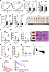
circTOLLIP promotes the proliferation and migration of HCC cells in vitro and in vivo. a-c The colony formation assay and CCK-8 assay were performed in HLF and 97H cells. d-f The statistic graphs of scratch wound healing assay and cell migration and invasion assays. g The subcutaneous xenograft tumors from vector or overexpressed circTOLLIP 97H cells. h-i Tumor weight and tumor volume of subcutaneous xenografts. j-k Liver orthotopic transplantation and metastasis models in nude mice with circTOLLIP-overexpressing and vector HLF cells. Representative pictures are presented that show HE staining of isolated liver tissue and a general image of the whole liver. The number of visible tumor nodules was counted and pointed by red arrows. l-p Lung metastasis assay was performed with 97H-circTOLLIP cells in nude mice by tail vein injection. Representative pictures showing lung metastatic nodules, the incidence of lung metastasis (l), HE staining of lung tissue (m) and lung fluorescence imaging (o, p) were presented. The number of lung metastatic nodules was counted in lung tissues after HE staining(n)
Fig. 3
Fig. 3
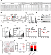
EIF4A3 promotes the biogenesis of circTOLLIP. a Prediction of the putative EIF4A3 binding sites in TOLLIP pre-mRNA by CircInteractome database. b Six positions (a-f) in TOLLIP pre-mRNA were selected to design qPCR primers and 4 plasmids (A1-A4) containing EIF4A3 binding sites were constructed to pull down EIF4A3 protein. c RIP assay using EIF4A3 antibody verified the direct binding of EIF4A3 and TOLLIP pre-mRNA. d Western blot analysis of EIF4A3 protein with MS2-RNA pulldown assay. Vector and GAPDH were used as negative controls. e-f qRT–PCR analysis of circTOLLIP level in HLF and 97H cells with transfection of EIF4A3 siRNA or EIF4A3 overexpression plasmid. g-h Stability of circTOLLIP in EIF4A3-overexpressing or EIF4A3 knockdown 97H cells with actinomycin D treatment. i Representative images of IHC staining in 138 pairs of HCC tissues. j Statistical analysis of the IHC results. k Correlation analysis between circTOLLIP and EIF4A3 expression in HCC tumors
Fig. 4
Fig. 4
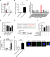
circTOLLIP serves as a sponge for miR-516a-5p. a RIP was performed with an anti-AGO2 antibody in 97H cells. b-c Relative circTOLLIP and microRNA enrichment using biotinylated control probe and specific circTOLLIP probes in 97H cells. d Relative luciferase reporter activity of WT-circTOLLIP after transfection with mimics of the most 4 most enriched microRNAs in HEK-293 T cells. e Schematic view of miR-516a-5p putative binding sites with circTOLLIP and construction of correspondent mutant circTOLLIP reporter plasmid. f Luciferase reporter activity of circTOLLIP in 97H cells co-transfected with miR-516a-5p mimic or NC mimic. g RIP was performed using an anti-AGO2 antibody in 97H cells transfected with miR-516a-5p mimic or NC mimic. h circTOLLIP enrichment using biotinylated NC probe and miR-516a-5p probes in 97H cells. i Colocalization between cicTOLLIP and miR-516a-5p was observed by FISH assay. Scale bars = 50 μm
Fig. 5
Fig. 5
miR-516a-5p inhibits the proliferation and metastasis of HCC cells in vitro and in vivo. a CCK-8, cell colony formation assay (b), scratch wound healing assay (c), and cell migration and invasion assays (d, e) were performed in HLF and 97H cells transfected with miR-516a-5p mimic or NC mimic. f The subcutaneous xenograft tumors from nude mice that transplanted with stablely overexpressing miR-516a-5p and vector 97H cells. g qRT–PCR analysis of miR-516a-5p level in xenograft tumors and quantification of tumor volume (h) and weight (i). j Livers from nude mice orthotopically transplanted with miR-516a-5p-overexpressing and vector 97H cells. Representative images showing the livers from the nude mice and HE staining of isolated liver tissues. The miR-516a-5p expression was analyzed in two groups (k) and the number of visible tumor nodules was counted (l). m-n The relative miR-516a-5p levels in 52 paired HCC and adjacent nontumor tissues. o The correlation between circTOLLIP and miR-516a-5p in HCC tissues. p Kaplan–Meier analysis of the correlation between miR-516a-5p expression and OS in HCC according to data from Kaplan–Meier Plotter database. Low miR-516a-5p group: n = 87, high miR-516a-5p group: n = 79
Fig. 6
Fig. 6
circTOLLIP relieves repression of miR-516a-5p on PBX3. a-b PBX3 expression in HLF and 97H cells with circTOLLIP overexpression or knockdown. c-d qRT–PCR analysis of PBX3 and HIST3H2A in HLF and 97H cells transfected with miR-516a-5p mimic or inhibitor. e PBX3 protein levels in HLF and 97H cells transfected miR-516a-5p mimic or inhibitor. f-g PBX3 expression in HLF and 97H cells transfected with miR-516a-5p mimic alone or in combination with circTOLLIP. h Luciferase reporter activity of PBX3-3’UTR in 97H cells cotransfected with miR-516a-5p mimic or inhibitor. i Luciferase reporter activity of PBX3-3’UTR in 97H cells with circTOLLIP overexpression or knockdown. j Representative images of PBX3 IHC staining in 138 pairs of HCC tissues and statistical analysis of the IHC scores (k). l The correlation between PBX3 expression and OS by Kaplan–Meier analysis. Low PBX3 group: n = 40, high PBX3 group: n = 65. m Correlation analysis between circTOLLIP and PBX3 expression in HCC tumors
Fig. 7
Fig. 7
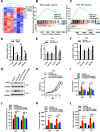
circTOLLIP stimulates EMT by miR-516a-5p/PBX3 axis. a Clustering heatmap showing the RNA-seq profiles in vector and PBX3-overexpressing 97H cells. b-c Gene set enrichment analysis (GSEA) of genes from RNA-seq profiles and TCGA database comparing with EMT_PATHWAY. d Among the EMT-related genes in TCGA, those with the highest positive correlation with PBX3 expression were selected and intersected with the differentially upregulated genes in the RNA-seq profiles. The top four genes (PTX3, COL6A2, CCN2, and MATN3) were verified to be upregulated in 97H cells with PBX3 overexpression compared with vector cells. And these genes were also upregulated in HLF and 97H cells with circTOLLIP overexpression (e, f) though qRT–PCR analysis. g Western blot analysis of PBX3 protein expression in circTOLLIP-overexpressing cells with or without transfection of miR-516a-5p mimic or PBX3 siRNA. h-i CCK-8 and colony formation assays to evaluate the cell proliferation ability respectively in four group cells. j-l Scratch wound healing assay and cell migration and invasion assays to evaluate the cell metastatic ability
Fig. 8
Fig. 8
Schematic diagram of the mechanism by which circTOLLIP promotes HCC progression. EIF4A3-induced circTOLLIP promotes the expression of PBX3 by sponging miR-516a-5p, in turn activating the PBX3/EMT signaling pathway and promoting the progression of HCC

Similar articles

Cited by

References

    1. Sung H, Ferlay J, Siegel RL, Laversanne M, Soerjomataram I, Jemal A, et al. Global cancer statistics 2020: GLOBOCAN estimates of incidence and mortality worldwide for 36 cancers in 185 countries. CA Cancer J Clin. 2021;71(3):209–249. doi: 10.3322/caac.21660. - DOI - PubMed
    1. Llovet JM, Kelley RK, Villanueva A, Singal AG, Pikarsky E, Roayaie S, et al. Hepatocellular carcinoma. Nat Rev Dis Primers. 2021;7(1):6. doi: 10.1038/s41572-020-00240-3. - DOI - PubMed
    1. European Association for the Study of the Liver Electronic address eee, European association for the study of the L. EASL clinical practice guidelines: management of hepatocellular carcinoma. J Hepatol. 2018;69(1):182–236. doi: 10.1016/j.jhep.2018.03.019. - DOI - PubMed
    1. Pinto Marques H, Gomes da Silva S, De Martin E, Agopian VG, Martins PN. Emerging biomarkers in HCC patients: Current status. Int J Surg. 2020;82S:70–76. doi: 10.1016/j.ijsu.2020.04.043. - DOI - PubMed
    1. Llovet JM, De Baere T, Kulik L, Haber PK, Greten TF, Meyer T, et al. Locoregional therapies in the era of molecular and immune treatments for hepatocellular carcinoma. Nat Rev Gastroenterol Hepatol. 2021;18(5):293–313. doi: 10.1038/s41575-020-00395-0. - DOI - PubMed

MeSH terms