

SARM1 Depletion Slows Axon Degeneration in a CNS Model of Neurotropic Viral Infection
- PMID: 35493328
- PMCID: PMC9043327
- DOI: 10.3389/fnmol.2022.860410
SARM1 Depletion Slows Axon Degeneration in a CNS Model of Neurotropic Viral Infection
Abstract
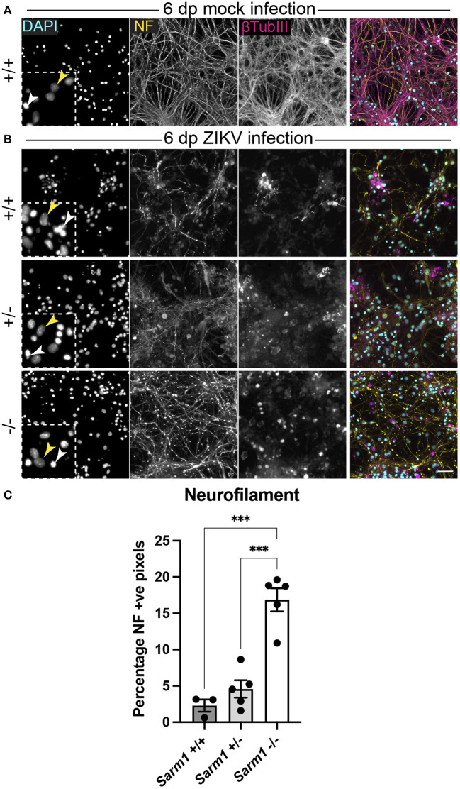
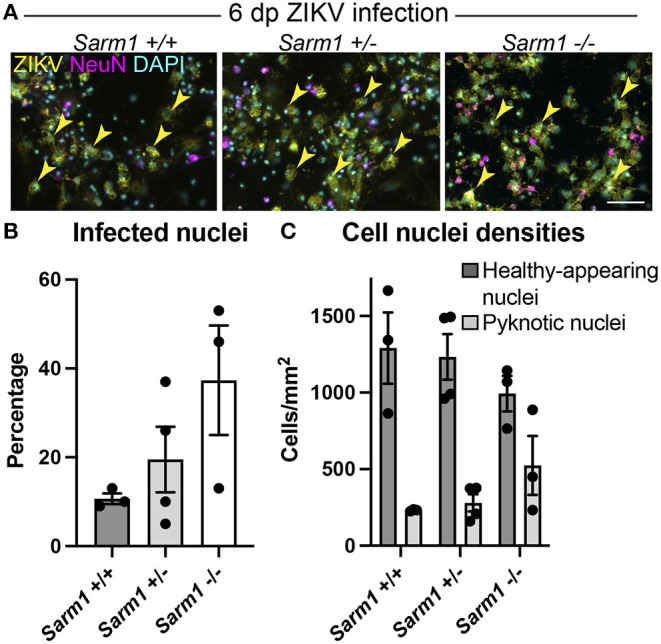
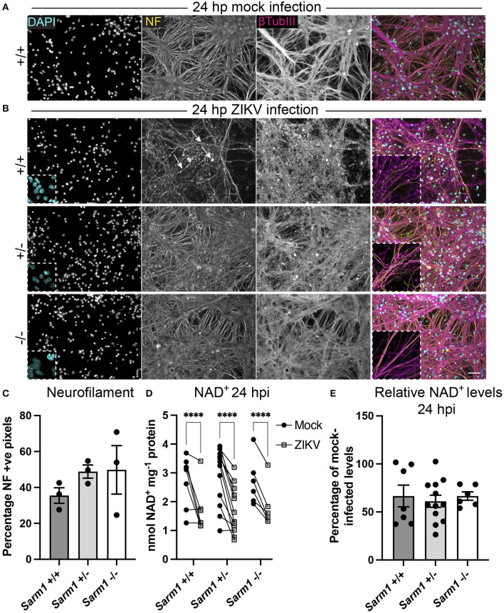
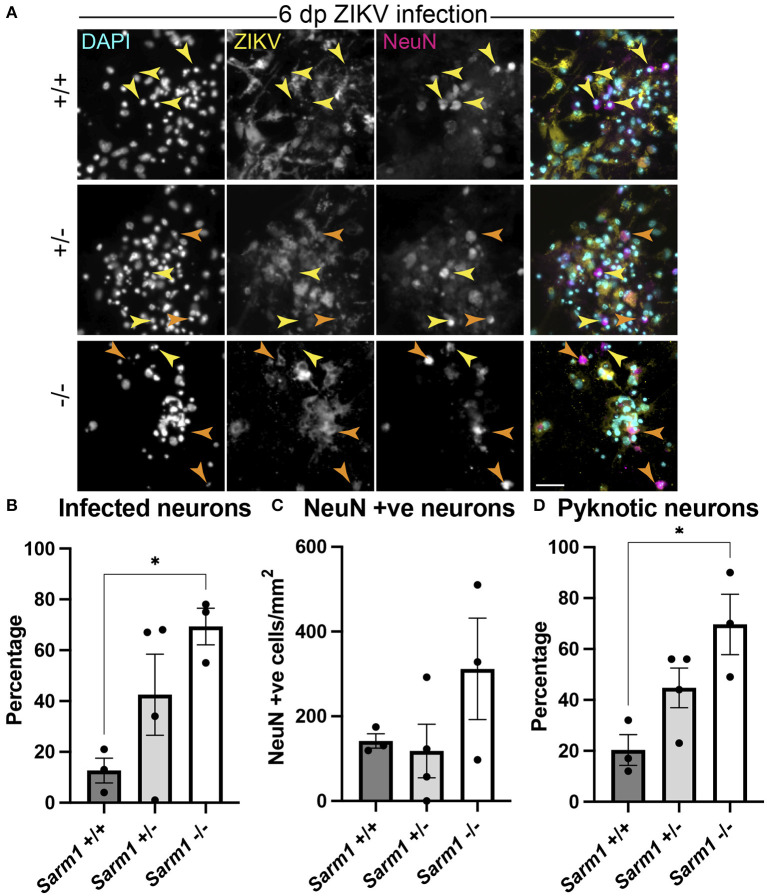
Zika virus (ZIKV) is a neurotropic flavivirus recently linked to congenital ZIKV syndrome in children and encephalitis and Guillain-Barré syndrome in adults. Neurotropic viruses often use axons to traffic to neuronal or glial cell somas where they either remain latent or replicate and proceed to infect new cells. Consequently, it has been suggested that axon degeneration could represent an evolutionarily conserved mechanism to limit viral spread. Whilst it is not known if ZIKV transits in axons, we previously reported that ZIKV infection of glial cells in a murine spinal cord-derived cell culture model of the CNS is associated with a profound loss of neuronal cell processes. This, despite that postmitotic neurons are relatively refractory to infection and death. Here, we tested the hypothesis that ZIKV-associated degeneration of neuronal processes is dependent on activation of Sterile alpha and armadillo motif-containing protein 1 (SARM1), an NADase that acts as a central executioner in a conserved axon degeneration pathway. To test this, we infected wild type and Sarm1 homozygous or heterozygous null cell cultures with ZIKV and examined NAD+ levels as well as the survival of neurons and their processes. Unexpectedly, ZIKV infection led to a rapid SARM1-independent reduction in NAD+. Nonetheless, the subsequent profound loss of neuronal cell processes was SARM1-dependent and was preceded by early changes in the appearance of β-tubulin III staining. Together, these data identify a role for SARM1 in the pathogenesis of ZIKV infection, which may reflect SARM1's conserved prodegenerative function, independent of its NADase activity.
Keywords: Zika virus; glia; neurofilament; nicotinamide adenine dinucleotide; tubulin (microtubules).
Copyright © 2022 Crawford, Antoniou, Komarek, Schultz, Donald, Montague, Barnett, Linington, Willison, Kohl, Coleman and Edgar.
Conflict of interest statement
The authors declare that the research was conducted in the absence of any commercial or financial relationships that could be construed as a potential conflict of interest.
Figures
Similar articles
-
SARM1 is responsible for calpain-dependent dendrite degeneration in mouse hippocampal neurons.J Biol Chem. 2024 Feb;300(2):105630. doi: 10.1016/j.jbc.2024.105630. Epub 2024 Jan 8. J Biol Chem. 2024. PMID: 38199568 Free PMC article.
-
SARM1-specific motifs in the TIR domain enable NAD+ loss and regulate injury-induced SARM1 activation.Proc Natl Acad Sci U S A. 2016 Oct 11;113(41):E6271-E6280. doi: 10.1073/pnas.1601506113. Epub 2016 Sep 26. Proc Natl Acad Sci U S A. 2016. PMID: 27671644 Free PMC article.
-
Natural variants of human SARM1 cause both intrinsic and dominant loss-of-function influencing axon survival.Sci Rep. 2022 Aug 16;12(1):13846. doi: 10.1038/s41598-022-18052-8. Sci Rep. 2022. PMID: 35974060 Free PMC article.
-
Multifaceted roles of SARM1 in axon degeneration and signaling.Front Cell Neurosci. 2022 Aug 25;16:958900. doi: 10.3389/fncel.2022.958900. eCollection 2022. Front Cell Neurosci. 2022. PMID: 36090788 Free PMC article. Review.
-
The curious case of SARM1: Dr. Jekyll and Mr. Hyde in cell death and immunity?FEBS J. 2023 Jan;290(2):340-358. doi: 10.1111/febs.16256. Epub 2021 Nov 12. FEBS J. 2023. PMID: 34710262 Review.
Cited by
-
SARM1 detection in myelinating glia: sarm1/Sarm1 is dispensable for PNS and CNS myelination in zebrafish and mice.Front Cell Neurosci. 2023 Apr 5;17:1158388. doi: 10.3389/fncel.2023.1158388. eCollection 2023. Front Cell Neurosci. 2023. PMID: 37091921 Free PMC article.
-
NMNAT2 is a druggable target to drive neuronal NAD production.Nat Commun. 2024 Jul 24;15(1):6256. doi: 10.1038/s41467-024-50354-5. Nat Commun. 2024. PMID: 39048544 Free PMC article.
-
Axon Biology in ALS: Mechanisms of Axon Degeneration and Prospects for Therapy.Neurotherapeutics. 2022 Jul;19(4):1133-1144. doi: 10.1007/s13311-022-01297-6. Epub 2022 Oct 7. Neurotherapeutics. 2022. PMID: 36207571 Free PMC article. Review.
-
Abnormal intraepidermal nerve fiber density in disease: A scoping review.Front Neurol. 2023 Apr 20;14:1161077. doi: 10.3389/fneur.2023.1161077. eCollection 2023. Front Neurol. 2023. PMID: 37153658 Free PMC article.
-
SARM1-Dependent Axon Degeneration: Nucleotide Signaling, Neurodegenerative Disorders, Toxicity, and Therapeutic Opportunities.Neuroscientist. 2024 Aug;30(4):473-492. doi: 10.1177/10738584231162508. Epub 2023 Mar 31. Neuroscientist. 2024. PMID: 37002660 Free PMC article. Review.
References
-
- Angeletti C., Amici A., Gilley J., Loreto A., Trapanotto A. G., Antoniou C., et al. . (2021). SARM1 is a multi-functional NAD(P)ase with prominent base exchange activity, all regulated by multiple physiologically-relevant NAD metabolites. iScience. 25:103812. 10.1016/j.isci.2022.103812 - DOI - PMC - PubMed
-
- Bijland S., Thomson G., Euston M., Michail K., Thümmler K., Mücklisch S., et al. . (2019). An in vitro model for studying CNS white matter: functional properties and experimental approaches. [version 1; peer review: 2 approved]. F1000Research 8, 117. 10.12688/f1000research.16802.1 - DOI - PMC - PubMed
-
- Brito Ferreira M. L., Militão de Albuquerque M., de F. P., de Brito C. A. A., de Oliveira França R. F., Porto Moreira Á. J., de Morais Machado, et al. . (2020). Neurological disease in adults with Zika and chikungunya virus infection in Northeast Brazil: a prospective observational study. Lancet Neurol. 19, 826–839. 10.1016/S1474-4422(20)30232-5 - DOI - PMC - PubMed
Grants and funding
LinkOut - more resources
Full Text Sources
Other Literature Sources
Molecular Biology Databases
Research Materials