Review
. 2022;19(1):662-677.
doi: 10.1080/15476286.2022.2067712. Epub 2021 Dec 31.

Mechanisms of ribosome recycling in bacteria and mitochondria: a structural perspective

Affiliations
Review

Mechanisms of ribosome recycling in bacteria and mitochondria: a structural perspective

Savannah M Seely et al. RNA Biol. 2022.

Abstract

In all living cells, the ribosome translates the genetic information carried by messenger RNAs (mRNAs) into proteins. The process of ribosome recycling, a key step during protein synthesis that ensures ribosomal subunits remain available for new rounds of translation, has been largely overlooked. Despite being essential to the survival of the cell, several mechanistic aspects of ribosome recycling remain unclear. In eubacteria and mitochondria, recycling of the ribosome into subunits requires the concerted action of the ribosome recycling factor (RRF) and elongation factor G (EF-G). Recently, the conserved protein HflX was identified in bacteria as an alternative factor that recycles the ribosome under stress growth conditions. The homologue of HflX, the GTP-binding protein 6 (GTPBP6), has a dual role in mitochondrial translation by facilitating ribosome recycling and biogenesis. In this review, mechanisms of ribosome recycling in eubacteria and mitochondria are described based on structural studies of ribosome complexes.

Keywords: GTPBP6; HflX; Ribosome; elongation factor G; protein synthesis; ribosome recycling; ribosome recycling factor; tRNA; translation factors.


Conflict of interest statement

No potential conflict of interest was reported by the author(s).

Figures

Figure 1.
Figure 1.
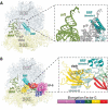
RRF is a tRNA mimic with a flexibly disposed domain II. (A) Ribbon diagram of the E. coli RRF crystal structure (PDB 1EK8) [37]. Domains I and II are distinctly colored and connected by flexible linkers. (B) L-shaped structure of tRNA. (C) Crystal structures of RRF aligned by domain I show that domain II rotates about the axis of domain I (PDBs: 1EK8, teal; 1DD5, gold; 1EH1, green; 1GE9, magenta) [37–40].
Figure 2.
Figure 2.
Domain II of RRF is flexibly disposed on the 70S ribosome. Crystal structures of RRF on the 70S ribosome aligned by 23S rRNA. In the absence of EF-G, domain II of RRF occupies different positions relative to the 23S rRNA helix H69 (PDBs: 4V5Y, E. coli 70S-paromomycin-RRF; 4V54, E. coli 70S-RRF; 4V5A, T. thermophilus 70S-RRF) [32,33].
Figure 3.
Figure 3.
RRF in the post-termination complex (PoTC) is not compatible with tRNA in the p/P state of binding and EF-G on the 70S ribosome. (A) RRF (teal/light blue) bound to the 70S ribosome with p/E-tRNA (olive) (PDB 4V9D [56]). The p/P-tRNA (gray) is shown. Inset: Domain I of RRF clashes with tRNA bound in the p/P state. (B) Structure of EF-G-GDPCP in the extended state bound to the 70S ribosome (PDB 4V5F [4]). EF-G is colored by domain according to the bar chart. Inset: Domain IV of EF-G in the extended conformation is not compatible with RRF without further rotation of RRF domain II about the long-axis of domain I.
Figure 4.
Figure 4.
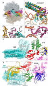
Pre-recycling complex with p/R- and E-site tRNAs. (A) Overview of pre-recycling complex (PDB 6UCQ [62]) with E-site tRNA (orange), p/R-tRNA (pink), RRF (teal and light blue), and EF-G in the compact state (colored by domain). (B) RRF domain II positioned in a ‘ready-to-attack’ state. Domain II (teal) locates in the niche created by H69 (orange), h44 (cerium), and uS12 (brown). RRF from crystal structures in the absence of EF-G superimposed through domain I of RRF (PDBs 4V5A, gold; 4V55, magenta) [32,33]. (C) Close-up view of the tRNA interaction with RRF domain I wherein the p/R-tRNA CCA-end is crunched and displaced by ~22 Å toward the E site and exhibits shape complementarity with RRF. The classical p/P-tRNA is not be compatible with RRF on the 70S ribosome. (D) The CCA-end of the p/R-tRNA is squeezed between 23S rRNA helices H74 and H80 (orange). (E) Interactions between compact EF-G and RRF. Domain II of RRF interacts favorably with EF-G domains III and V. (F) Interactions between RRFmt and EF-G2mt in the post-recycling complex (PDB 7L20 [115]) wherein EF-G2mt has undergone rearrangements of domains III, IV and V. Domain IV of EF-G2mt forms favorable interactions with the surface of RRFmt domain II, which has rotated to avoid a steric collision with EF-G.
Figure 5.
Figure 5.
Cryo-EM structure of E. coli 50S subunit bound to HflX. (A) Overview of E. coli HflX bound to the 50S subunit (PDB 5ADY [91]). (B) Close-up view of the HflX N-terminal domain with superimposed p/P- and p/E-tRNAs showing that the p/P-tRNA is not compatible with HflX. (C) The GDPCP nucleotide in the G-domain of HflX locates ~45 Å away from the 23S rRNA sarcin-ricin loop (SRL). (D) In EF-G-GDPCP bound to the 70S ribosome, the GDPCP nucleotide in the G-domain is closer (~20 Å) to the SRL (PDB 4V9H [8]).
Figure 6.
Figure 6.
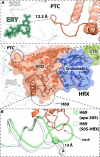
Interactions of HflX with the 50S ribosomal subunit. (A) Close-up view of the PTC loop within the HflX N-terminal domain (orange) (PDB 5ADY [91]) with the antibiotic erythromycin (ERY) (green) bound in the nascent peptide exit tunnel (NPET) (PDB 6ND6 [130]). The nearest distance between HflX and ERY is more than 12 Å. (B) The N-terminal domain of HflX (orange) displaces 23S rRNA helix H69 by ~13 Å (white) relative to the apo form (green) of the 50S subunit.
Figure 7.
Figure 7.
Binding of RRFmt on the post-termination mitoribosome is stabilized by large subunit interactions and is not compatible with tRNA in the p/P state. (A) RRFmt (teal/light blue/light green) bound to the 55S ribosome (PDB 6NU2) with p/E-tRNA (olive) and p/P-tRNA (gray) shown (PDB 7NSJ and 7NSI respectively) [112,116]. (B) Domain I of RRFmt is not compatible with tRNA bound in the p/P state. (C) The N-terminal extension (NTE) of RRFmt exhibits stabilizing interactions with large subunit mitoribosomal proteins uL16 and bL27 (tan and brown respectively).
Figure 8.
Figure 8.
Cryo-EM structure of mitochondrial RRFmt and EF-G2mt bound to the 39S mitoribosomal subunit. (A) Overview of RRFmt and EF-G2mt bound to the 39S mitoribosomal subunit post-recycling (related to Fig. 4F) (PDB 7L20 [115]). (B) Close-up view of RRFmt domain I interactions with EF-G2mt domain IV (gold). The structure shows that the C-terminal extension (CTE) of EF-G1mt (PDB 6VLZ, purple [117]) would collide with domain I of RRF, preventing EF-G1mt from participating in the recycling step. (C) Superposition of RRFmt from the 55S class I structure (dark turquoise, PDB: 7L08) [115] aligned by domain I on the class three complex with RRFmt (teal) and EF-G2mt (omitted for clarity) on the 39S, wherein RRFmt domain II is rotated approximately 45° toward the SSU and exhibits a collision with the superimposed h44.
Figure 9.
Figure 9.
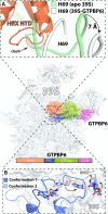
Cryo-EM structure of GTPBP6 bound to the 39S mitoribosome subunit. (A) Close-up view of the interactions between the GTPBP6 N-terminal domain (PDB 7OF4, orange [129]) and H69. Helix H69 in the apo 39S subunit is green (PDB 6NU3 [112]), showing that it is not compatible with the binding of GTPBP6, shifting by ~7 Å (PDB 7OF4, white [129]). (B) Two conformations of the PTC region upon GTPBP6 binding to the 39S mitoribosome subunit. Superimposition of the 39S subunit with PTC conformation 1 (PDB 7OF4, dark blue [129]) with that of the 39S subunit with PTC conformation 2 (PDB 7OF6, light blue [129]) reveals rearrangements of PTC residues A3089 (E. coli A2602), U3072 (E. coli U2585), and U2993 (E. coli U2506).

Similar articles

Cited by

References

    1. Hussain T, Llacer JL, Wimberly BT, et al. Large-scale movements of IF3 and tRNA during bacterial translation initiation. Cell. 2016;167:133–144 e113. - PMC - PubMed
    1. Sprink T, Ramrath DJ, Yamamoto H, et al. Structures of ribosome-bound initiation factor 2 reveal the mechanism of subunit association. Sci Adv. 2016;2:e1501502. - PMC - PubMed
    1. Kaledhonkar S, Fu Z, Caban K, et al. Late steps in bacterial translation initiation visualized using time-resolved cryo-EM. Nature. 2019;570(7761):400–404. DOI:10.1038/s41586-019-1249-5. - DOI - PMC - PubMed
    1. Gao YG, Selmer M, Dunham CM, et al. The structure of the ribosome with elongation factor G trapped in the posttranslocational state. Science. 2009;326:694–699. - PMC - PubMed
    1. Schmeing TM, Voorhees RM, Kelley AC, et al. The crystal structure of the ribosome bound to EF-Tu and aminoacyl-tRNA. Science. 2009;326:688–694. - PMC - PubMed

Publication types

MeSH terms

LinkOut - more resources