Review
. 2022 May 25;122(10):9331-9356.
doi: 10.1021/acs.chemrev.1c01023. Epub 2022 Apr 21.

NMR Provides Unique Insight into the Functional Dynamics and Interactions of Intrinsically Disordered Proteins

Affiliations
Review

NMR Provides Unique Insight into the Functional Dynamics and Interactions of Intrinsically Disordered Proteins

Aldo R Camacho-Zarco et al. Chem Rev. .

Abstract

Intrinsically disordered proteins are ubiquitous throughout all known proteomes, playing essential roles in all aspects of cellular and extracellular biochemistry. To understand their function, it is necessary to determine their structural and dynamic behavior and to describe the physical chemistry of their interaction trajectories. Nuclear magnetic resonance is perfectly adapted to this task, providing ensemble averaged structural and dynamic parameters that report on each assigned resonance in the molecule, unveiling otherwise inaccessible insight into the reaction kinetics and thermodynamics that are essential for function. In this review, we describe recent applications of NMR-based approaches to understanding the conformational energy landscape, the nature and time scales of local and long-range dynamics and how they depend on the environment, even in the cell. Finally, we illustrate the ability of NMR to uncover the mechanistic basis of functional disordered molecular assemblies that are important for human health.


Conflict of interest statement

The authors declare no competing financial interest.

Figures

Figure 1
Figure 1
NMR probes biomolecular conformational changes on a vast range of time scales. NMR spin relaxation provides accurate information on the reorientational properties of relaxation-active interactions, normally interatomic bonds, up to tens of nanoseconds. In the fast exchange limit, a single NMR peak represents a population weighted average over the chemical shifts of each populated substate. When the exchange rate is in the same range as the difference in chemical shifts of the distinct states, on time scales from tens of microseconds to hundreds of milliseconds in proteins, line-broadening is observed, and 1H, 13C, and 15N NMR exchange approaches can be used to characterize interconversion between the different conformational states. Exchange that is significantly slower than the difference in chemical shifts of the distinct states gives rise to slow exchange, allowing all states to be individually investigated.
Figure 2
Figure 2
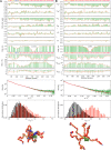
Experimental comparison of conformational behavior of the intrinsically disordered δ subunit of bacterial RNA polymerase. (A) Experimental parameters measured on wild-type protein (green bars) compared to ensemble-averaged values calculated from 10 ensembles comprising 200-strong ASTEROIDS ensembles (red lines). From top to bottom: secondary chemical shifts, paramagnetic relaxation enhancements (labeled at residue 132), residual dipolar couplings, and SAXS. Bottom: comparison of distribution of radii of gyration from a statistical coil pool (black) and the ASTEROIDS ensemble (red). Structural models of five conformations are displayed below the plots (ordered domain in green, IDR in yellow with positively and negatively charged residues highlighted in blue and red, respectively. (B) Same parameters for the mutated protein in which a lysine-rich tract 96KAKKKKAKK104 are replaced by 96EAEEEEAEE104. This results in a clear abrogation of long-range contacts with the C-terminal half of the domain that collapse the protein. This collapse, and its abrogation, are visible not only in SAXS and PRE data but also in the residual dipolar coupling data. (Reproduced with permission from Kuban et al. 2019 Copyright 2019 ACS).
Figure 3
Figure 3
Temperature-dependent 15N relaxation maps three modes of intrinsically disordered protein dynamics. (A) 15N auto- and cross-relaxation rates of NT measured at different magnetic field strengths (green, 600 MHz 1H frequency; blue, 700 MHz; red, 850 MHz; orange, 950 MHz) and at different temperatures (top: 298 K, second row 288 K, third row 278 K, bottom 274 K). (B–F) Analysis of all relaxation data in (A), using a three-component model-free approach, with characteristic correlation times related via an Arrhenius expression. (B) Slow (τ3) and intermediate (τ2) correlation times at 274 K (red), 278 K (orange), 288 K (green), and 298 K (blue). (C) Activation energies for slow (red) and intermediate (blue) time scales for each residue. (D–F) Amplitude of slow (D), intermediate (E), and fast (F) time scale contributions (Reproduced with permission from Abyzov et al. JACS 2016 Copyright 2016 ACS).
Figure 4
Figure 4
Viscosity-dependent 15N relaxation maps distinct response of local and longer-range dynamics in intrinsically disordered proteins. (A) Transverse (R2) and longitudinal (R1) relaxation, transverse cross-correlated DD/CSA (ηxy) and heteronuclear {1H}-15N nuclear Overhauser enhancement (NOE) recorded at 600, 700, and 850 MHz as a function of concentration of Dextran 40. (B) Longitudinal water relaxation (solid red line, normalized to the value in free solution; ρ0) shows a similar dependence on concentration of viscogen to the intermediate time scale motion (green points). The slow motional component (purple) resembles approximately 3* ρ0 (dotted line). (C) Friction coefficients (ε) for intermediate backbone (blue) and slower, segmental (red) motions. (D) Cartoon representation of the length scales of intermediate and slower motions (Reproduced with permission from Adamski et al. JACS 2019 Copyright 2019 ACS).
Figure 5
Figure 5
Residue-specific friction coefficients are transferable between different in vitro crowding environments and even predict values measured in cellulo. (A) Experimental 15N relaxation rates recorded on Sendai virus NT in the presence of 135g/L PEG (gray bars) compared to values calculated using sequence-specific friction coefficients (eq 11) (red lines) determined as a function of Dextran concentrations and water relaxation in the sample of interest. For comparison, relaxation rates predicted under dilute conditions are shown in blue. (B) Relaxation rates measured at 600 MHz 1H frequency at a concentration of 90 g/L PEG (colors as in (A)). (C) 15N relaxation rates recorded in-cell (red points) compared to values calculated on the basis of dynamic parameters determined in vitro (green bars and line). Orange bars and lines show rates predicted for dilute conditions. Experimentally determined friction coefficients and the experimental measurement of the water R1,0in cellulo were used in the prediction. (Reproduced with permission from Adamski et al. JACS 2019 Copyright 2019 ACS).
Figure 6
Figure 6
NMR relaxation allows for the identification of ensembles of time-dependent trajectories that represent fast motions in interconverting substates. (A) Experimental 15N relaxation rates recorded on Sendai virus NT at 298 K in dilute conditions (gray bars) compared to values calculated from 4 μs of MD simulation, (blue line). The red line shows values calculated from the ABSURD procedure targetting only transverse relaxation measured at 850 MHz (orange box). (B) The ABSURD procedure results in average time-dependent correlation functions that can be decomposed into local and segmental motions of the peptide chain. (Reproduced with permission from Salvi et al. JPCL 2016 Copyright 2016 ACS and Salvi et al. Angewandte Chemie 2017 Copyright Wiley 2017).
Figure 7
Figure 7
Temperature-dependent NMR relaxation identifies accurate and transferable molecular force fields for IDPs. Experimental 15N{1H} steady-state nOes (gray bars) measured on Sendai virus NT at different magnetic fields (left 600 MHz, middle 700 MHz, and right 850 MHz) and temperatures. ABSURD-selected ensembles of trajectories using Charmm36m combined with the TIP4P/2005 water model (red) reproduces experimental values better than when combined with TIP3P (blue), at all temperatures. (Reproduced with permission from Salvi et al. Sci. Adv. 2019 Copyright 2019 AAAS).
Figure 8
Figure 8
Multinuclear CPMG relaxation dispersion maps the molecular recognition trajectory of an intrinsically disordered protein as it binds its physiological partner. (A) 1H, 13C, and 15N CPMG were used to map the interaction trajectory of Sendai virus NT with the C-terminal domain of the phosphoprotein (PX). The combination of multinuclear CPMG, measured at multiple substoichiometric admixtures (2, 3.5, 5, and 8% of PX compared to NT) provides the necessary information to reconstruct a complex interaction trajectory involving both folding and binding. (B) The first step involves funnelling of one of the helical elements present in the equilibrium of rapidly exchanging substates, in an encounter complex on the surface of PX. (C) The second step involves binding of the stabilized helix into a groove between two helices on the surface of PX. (D) Relaxation dispersion measured on NT confirms that the second step coincides with events occurring on the surface of NT. (E) Representation of the most likely interaction trajectory derived from the ensemble of the experimental data. (Reproduced with permission from Schneider et al. JACS 2015 Copyright 2015 American Chemical Society).
Figure 9
Figure 9
NMR detects essential, ultraweak interactions in the dynamic assembly of Measles virus nucleo/phosphoprotein complex. (A) 15N–1H HSQC spectrum of the complex formed between PTAIL and the nucleoprotein. The complex comprises more than 450 intrinsically disordered amino acids. (B) Representation of the two interaction sites involved in the complex. The phosphoprotein of Measles virus (yellow) is known to bind the nucleoprotein (gray) in a tight complex at its N-terminal end. NMR reveals a second binding site (δα4) that is 150 amino acids away from the first binding site, in the middle of a long intrinsically disordered domain that binds a distal site of the nucleoprotein. NMR exchange (C) 15N CPMG and (D) rotating frame relaxation in the free and bound forms of the region 140–304 of PTAIL, reveals that the intrinsic affinity of this second site is 5 orders of magnitude lower than the known binding site. (E) Normalized peak intensities (I/I0) of P1–304 (50 μM) with P1–50N1–525 (gray, 25; red, 50; green, 100; and blue, 150 μM concentrations of P1–304. (F) Interaction profile of P1–304,HELL → AAAA mutation (concentrations as in E). Mutation of these four residues in the binding site knocks out the second interaction and replication. (Reproduced with permission from Milles et al. Sci. Adv. 2018 Copyright 2018 AAAS).
Figure 10
Figure 10
Influenza polymerase forms a highly dynamic assembly with the intrinsically disordered host transcription factor ANP32a in a species specific-way. (A) PREs measured on hANP32A (orange, experimental; and blue, representative ensembles selected using ASTEROIDS) in the presence of paramagnetically labeled human adapted 627-NLS. (B) Same information for avANP32A in the presence of paramagnetically labeled avian adapted 627-NLS. (C, D) Representation of the dynamic complexes determined from the data shown in A and B, respectively. Multivalent interactions between ANP32a (yellow/red) and the 627 domain (gray) are localized to the basic patch on the surface of 627. In the case of avANP32A and avian adapted 627-NLS(E), ANP32A disordered domain is in general closer to the NLS domain (yellow) mediated by the hydrophobic hexapeptide (green). (E) Position of the cysteine residues used to label 627-NLS. (F) Representation of the ensemble of conformers of the hANP32A:627-NLS complex. (G) Average distance difference matrix (in Å) between ANP32A (x-axis) and the 627-NLS domains (y-axis) over the two ensembles. (Reproduced with permission from Camacho-Zarco et al. Nat. Commun. 2020).

Similar articles

Cited by

References

    1. Uversky V. N. Natively Unfolded Proteins: A Point Where Biology Waits for Physics. Protein Sci. 2002, 11, 739–756. 10.1110/ps.4210102. - DOI - PMC - PubMed
    1. Tompa P. Intrinsically Unstructured Proteins. Trends Biochem. Sci. 2002, 27, 527–533. 10.1016/S0968-0004(02)02169-2. - DOI - PubMed
    1. Dyson H. J.; Wright P. E. Intrinsically Unstructured Proteins and Their Functions. Nat. Rev. Mol. Cell Biol. 2005, 6, 197–208. 10.1038/nrm1589. - DOI - PubMed
    1. Uversky V. N.; Dunker A. K. Understanding Protein Non-Folding. Biochim. Biophys. Acta 2010, 1804, 1231–1264. 10.1016/j.bbapap.2010.01.017. - DOI - PMC - PubMed
    1. Tompa P.; Davey N. E.; Gibson T. J.; Babu M. M. A Million Peptide Motifs for the Molecular Biologist. Mol. Cell 2014, 55, 161–169. 10.1016/j.molcel.2014.05.032. - DOI - PubMed

Publication types

MeSH terms

Substances