Review
. 2022 Apr;28(4):678-689.
doi: 10.1038/s41591-022-01765-8. Epub 2022 Apr 19.

Engineered cellular immunotherapies in cancer and beyond

Affiliations
Review

Engineered cellular immunotherapies in cancer and beyond

Amanda V Finck et al. Nat Med. 2022 Apr.

Abstract

This year marks the tenth anniversary of cell therapy with chimeric antigen receptor (CAR)-modified T cells for refractory leukemia. The widespread commercial approval of genetically engineered T cells for a variety of blood cancers offers hope for patients with other types of cancer, and the convergence of human genome engineering and cell therapy technology holds great potential for generation of a new class of cellular therapeutics. In this Review, we discuss the goals of cellular immunotherapy in cancer, key challenges facing the field and exciting strategies that are emerging to overcome these obstacles. Finally, we outline how developments in the cancer field are paving the way for cellular immunotherapeutics in other diseases.


Figures

Fig. 1 ∣
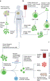
Fig. 1 ∣. Autologous and allogeneic engineered cell manufacturing.
a, Tumor mass is excised and TILs are isolated. Once collected, autologous TILs are activated, engineered and expanded ex vivo under optimized culture conditions, prior to being reinfused into the patient. b, Peripheral blood is collected and T cells are isolated through leukapheresis. Upon isolation, T cells are activated ex vivo and virally transduced to express a synthetic CAR receptor or an engineered TCR. Following expansion, genetically modified autologous T cells are infused into the patient. c, Cells from various sources are either directly collected from peripheral blood mononuclear cells or are differentiated in vivo. To prevent alloreactivity, human genome engineering, such as removal of the endogenous TCRs in allogeneic T cells, is conducted, and synthetic receptors are inserted. Cells are cryopreserved and banked following manufacturing. Figure created using BioRender.
Fig. 2 ∣
Fig. 2 ∣. Mechanisms of tolerance.
Central (ac) and peripheral (dj) tolerance mechanisms a, Clonal deletion. A developing T cell (in the thymus) or B cell (in the bone marrow) recognizes a self-antigen and is deleted by apoptosis. b, Clonal diversion. A developing T cell receives a medium-strength signal through its receptor in the thymus, which induces FOXP3 expression and differentiation into a natural Treg cell (nTreg). c, Receptor editing. A developing B cell recognizes a self-antigen in the bone marrow and undergoes further genetic recombination events to produce a new antigen receptor on its surface that no longer responds to self-antigen. d, Regulation. nTreg cells (from the thymus), inducible Treg (iTreg) cells (generated in the periphery), regulatory B cells (Breg) and CD8+ T suppressor cells work through various contact-dependent and contact-independent modes to suppress immune responses in the periphery. e, Anergy. A state of unresponsiveness induced when a T cell receives a signal through its cognate antigen receptor (TCR-Ag-MHC) in the absence of costimulation (CD28-B7 or CD40-CD40L). f, Deletion. Strong signals through the cognate antigen receptors on lymphocytes can bring about activation-induced cell death. g, Exhaustion. The persistence of antigen during an ongoing immune response can lead to a state of hyporesponsiveness. h, Immunologic ignorance. Some organs (such as the anterior chamber of the eyes) are immune-privileged, and lymphocytes have diminished access to these tissues. i, Accommodation. B cells produce antibodies that fix complement and damage a transplanted organ; but in the presence of a persistent antigen, the B-cell and antibody repertoires change, and produce antibodies that protect from complement fixation, thereby protecting the transplanted organ from damage. j, Organ-specific tolerance. Some organs are more tolerogenic than others, such as the liver (adapted from Ezekian at al. with permission).
Fig. 3 ∣
Fig. 3 ∣. Innate and adaptive mechanisms of cell recognition and rejection.
The contribution of each pathway in allogeneic cell rejection can vary depending on donor cell source, transplant location, and host levels of immunogenicity. Innate mechanisms of rejection are shown in parts ac. a, Complement-dependent cellular cytotoxicity is initiated after pre-existing donor-specific anti-HLA antibodies (DSAs) recognize non-self MHC-I. Complement component C1q then recognizes these DSA–pMHC I complexes and initiates the complement cascade, leading to the formation of membrane attack complexes, which induce donor cell apoptosis (as seen in HSCT and solid organ transplant). b, NK cell ‘missing self’ cytotoxicity: NK cells have activating and inhibiting receptors (that is, KIR in humans and Ly49 in mice). In the presence of an activating signal, KIR detection of self MHC-I will prevent killing by NK cells. However, if self MHC-I is not detected on the target cell, NK cytotoxicity will not be inhibited. c, Antibody-dependent cellular cytotoxicity can be mediated by DSAs and effector cells bearing Fcγ receptors (that is, monocytes, NK cells and macrophages). d, Adaptive mechanisms of rejection. Donor antigen presentation and priming can be mediated by direct and indirect mechanisms. CD4+ T cells can be primed indirectly by donor peptide loaded on host MHC-II (i) or semi-directly on recycled donor MHC-II (ii). Another form of semi-direct antigen recognition is when MHC-I molecules are recycled from donor cells, which results in CD8+ T cell recognition of donor antigen in the presence of indirectly activated CD4+ T cells (iii). Finally, host CD4+ T cells can also become activated directly on donor antigen-presenting cells (iv). e, Host CD4+ T cells promote activation of CD8+ T cells, NK cells, macrophages and B cells. Activated CD4+ T cells (called type 1 helper T cells, TH1 cells) can provide proinflammatory cytokines to improve direct cytotoxicity of T cells and other effector-mediated cellular cytotoxicity mechanisms. TH1-type cytokines can also activate macrophages, which enhances IL-12 secretion and helps maintain the TH1 subset. In presensitized models, CD4+ T cells can also bolster a macrophage-driven phenotype through CD40–CD40L interactions. TH2-skewed CD4+ T cells also enhance antibody-dependent cellular cytotoxicity through antigen-specific activation of B cells and secretion of cytokines that are important for class switching (IL-4/IL-5) and proliferation (IL-2). pMHC, peptide-bound MHC; APC, antigen-presenting cell; IFNγ, interferon γ; GMCSF, granulocyte–macrophage colony-stimulating factor; CTL, cytotoxic T lymphocytes. Figure created using BioRender.
Fig. 4 ∣
Fig. 4 ∣. Permutations of cell therapies for cancer treatment.
a, The availability of numerous types of cell therapies creates the possibility for a wide range of combinatorial therapeutic approaches. Within each cell type, there is a wide range of cellular modifications that can be made, ranging from addition of synthetic receptors,- and secreted payloads,- to genetic deletions,,. This diverse toolbox of cell therapies can be used in numerous combinations, which may be more effective than each one as monotherapy. Note that only a subset of examples is listed for each category of cellular modification. b, There are additional therapeutic modalities that have the potential to synergize with cell therapies-, thus further increasing the number of potential therapeutic combinations. c, Two examples of potential therapeutic combinations are shown that could improve the ability to directly target tumor cells and increase the endogenous immune response. Figure created using BioRender.

Similar articles

Cited by

References

    1. Combination Products (FDA, accessed 1 March 2022); https://www.fda.gov/combination-products
    1. Grupp SA et al. Chimeric antigen receptor-modified T cells for acute lymphoid leukemia. N. Engl. J. Med 368, 1509–1518 (2013). - PMC - PubMed
    1. Fischbach MA, Bluestone JA & Lim WA Cell-based therapeutics: the next pillar of medicine. Sci. Transl. Med 5, 179ps177 (2013). - PMC - PubMed
    1. Frangoul H et al. CRISPR–Cas9 gene editing for sickle cell disease and β-thalassemia. N. Engl. J. Med 384, 252–260 (2021). - PubMed
    1. Approved Cellular and Gene Therapy (FDA, 2022); https://www.fda.gov/vaccines-blood-biologics/cellular-gene-therapy-produ...

Publication types

Substances