. 2022 Apr 15;13(4):350.
doi: 10.1038/s41419-022-04798-6.

Targeted CUL4A inhibition synergizes with cisplatin to yield long-term survival in models of head and neck squamous cell carcinoma through a DDB2-mediated mechanism

Affiliations

Targeted CUL4A inhibition synergizes with cisplatin to yield long-term survival in models of head and neck squamous cell carcinoma through a DDB2-mediated mechanism

Trace M Jones et al. Cell Death Dis. .

Abstract

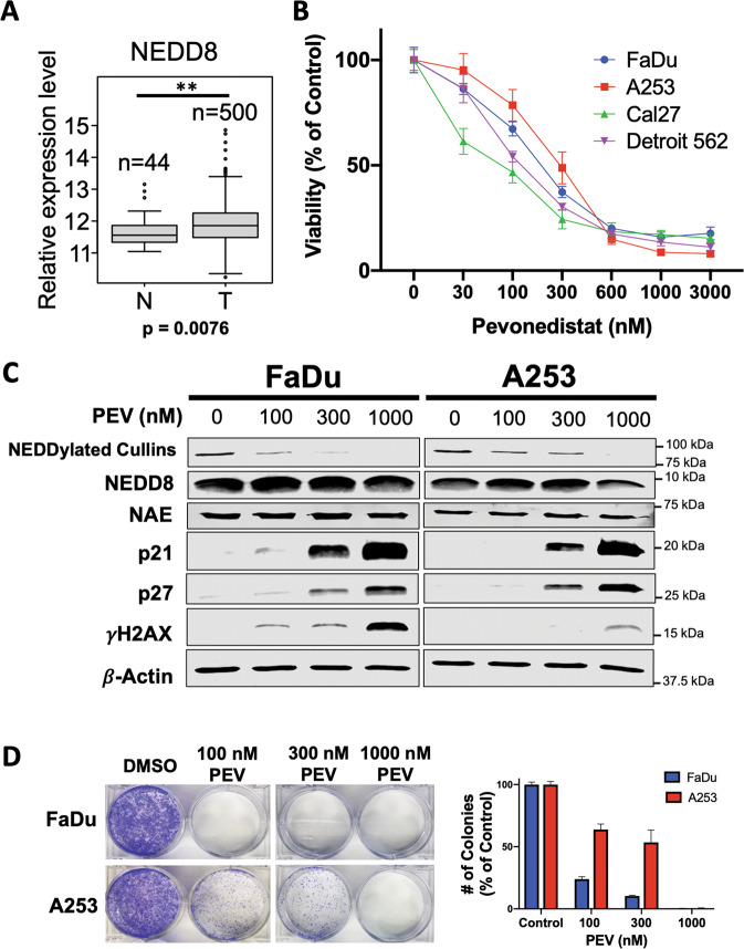
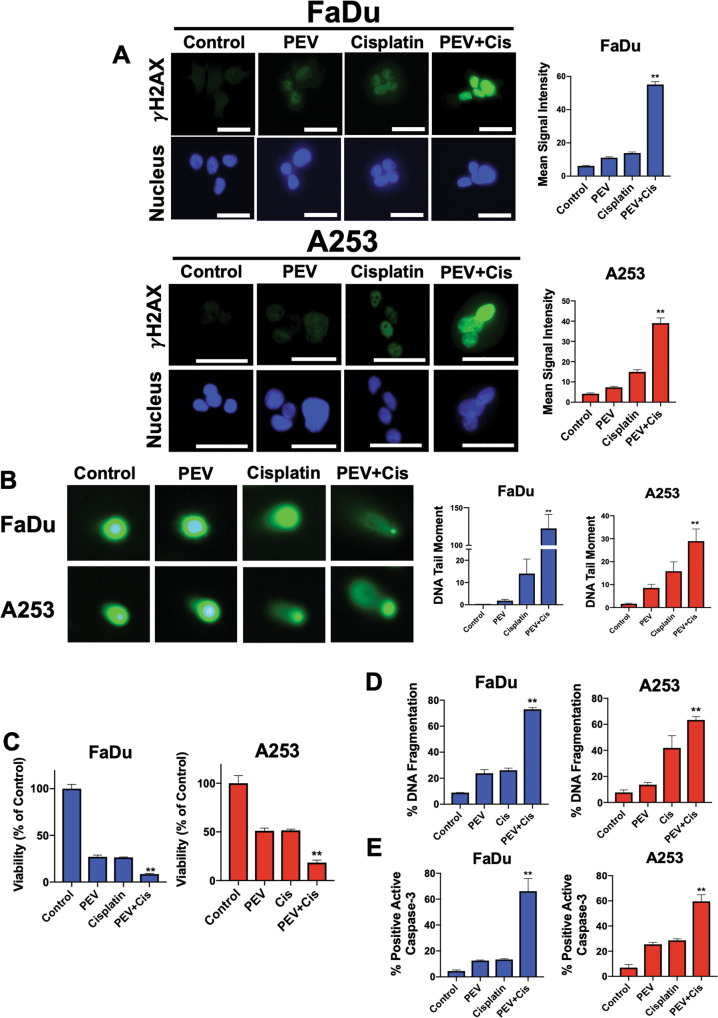
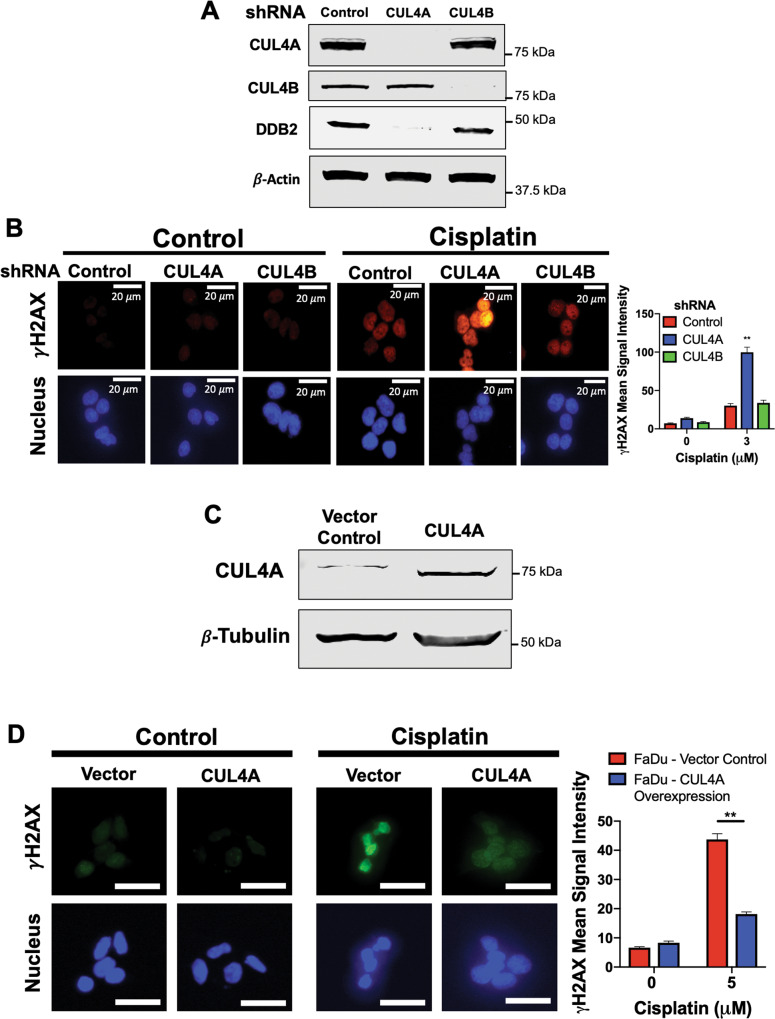
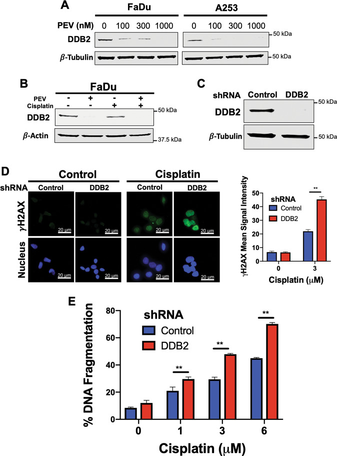
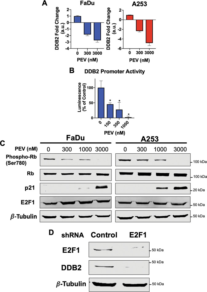
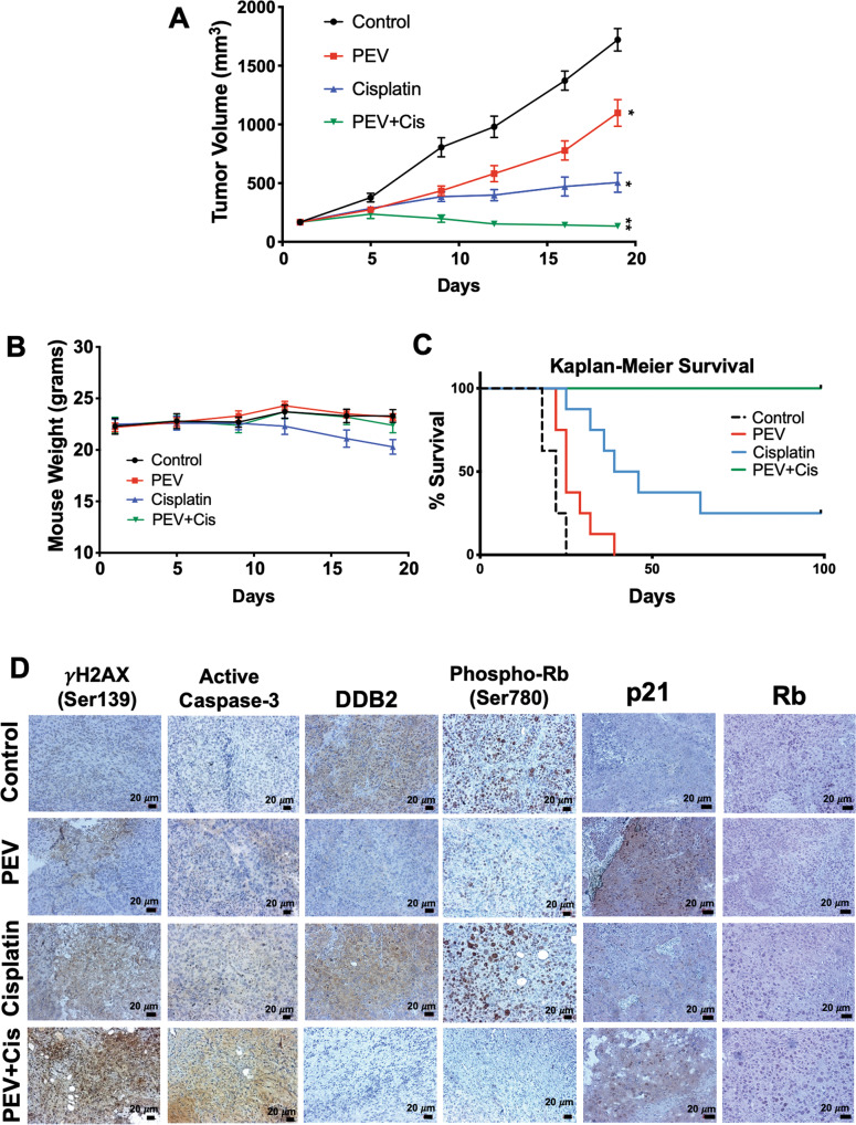
Patients with late-stage and human papillomavirus (HPV)-negative head and neck squamous cell carcinoma (HNSCC) continue to have a very poor prognosis. The development of more effective novel therapies that improve overall survival and overcome drug resistance is an urgent priority. Here we report that HNSCC tumors significantly overexpress NEDD8 and exhibit high sensitivity to the first-in-class NEDD8-activating enzyme (NAE) inhibitor pevonedistat. Additional studies established that disruption of NEDD8-mediated protein turnover with pevonedistat dramatically augmented cisplatin-induced DNA damage and apoptosis in HNSCC models. Further analysis revealed that the specific pevonedistat target CUL4A played an essential role in driving the synergy of the pevonedistat and cisplatin combination. Targeted inhibition of CUL4A resulted in significant downregulation in Damage Specific DNA binding protein 2 (DDB2), a DNA-damage recognition protein that promotes nucleotide excision repair and resistance to cisplatin. Silencing of CUL4A or DDB2 enhanced cisplatin-induced DNA damage and apoptosis in a manner similar to that of pevonedistat demonstrating that targeted inhibition of CUL4A may be a novel approach to augment cisplatin therapy. Administration of pevonedistat to mice bearing HNSCC tumors significantly decreased DDB2 expression in tumor cells, increased DNA damage and potently enhanced the activity of cisplatin to yield tumor regression and long-term survival of all animals. Our findings provide strong rationale for clinical investigation of CUL4A inhibition with pevonedistat as a novel strategy to augment the efficacy of cisplatin therapy for patients with HNSCC and identify loss of DDB2 as a key pharmacodynamic mediator controlling sensitivity to this regimen.


Conflict of interest statement

The authors declare no competing interests.

Figures

Fig. 1
Fig. 1. The NEDD8 pathway is a rational target for HNSCC therapy.
A NEDD8 is significantly elevated in clinical cases of HNSCC. Gene expression data from the GDC data portal were analyzed for levels of NEDD8 in 500 HNSCC tumor (T) and 44 adjacent normal (N) tissue samples. Mean ± SD. **Indicates a significant difference at p = 0.0076. B Pevonedistat decreases HNSCC cell viability in a dose-dependent manner. FaDu, A253, Cal27, and Detroit-562 cell lines were treated with varying concentrations of pevonedistat for 72 h. Viability was assessed by MTT assay. Mean ± SD, n = 8. C Pevonedistat stabilizes CRL targets in HNSCC cell lines. FaDu and A253 HNSCC cells were treated with the indicated concentrations of pevonedistat for 24 h. NEDD8, NAE, p21, p27, and γH2AX were measured by immunoblotting. β-Actin was used as a loading control. D Pevonedistat inhibits colony formation in HNSCC cell lines. FaDu and A253 cell lines were plated and treated with various concentrations of pevonedistat for 24 h. The cells were then incubated in fresh media for 10 days and colonies were quantified. Mean ± SD, n = 3.
Fig. 2
Fig. 2. Pevonedistat significantly augments cisplatin-mediated DNA damage.
A Pevonedistat and cisplatin treatment significantly increase γH2AX expression. FaDu and A253 cells were treated with 600 nM pevonedistat, 3 μM cisplatin, or the combination for 24 h. γH2AX levels were measured by immunocytochemistry and quantified using ImageJ software. Representative images are shown. DAPI was used as a counterstain. Mean signal intensity ± SEM, n = 54 cells per condition. Scale bar = 20 μm. B Pevonedistat significantly augments cisplatin-induced DNA damage. Cells were treated with 600 nM pevonedistat, 3 μM cisplatin, or the combination for 24 h. DNA damage was determined by comet assay and DNA tail moment was determined. Mean ± SEM, n = 27. C Pevonedistat significantly enhances cisplatin-mediated reductions in cell viability. FaDu and A253 cells were treated with 0.3 μM pevonedistat, 1–3 μM cisplatin, or the combination for 72 h. Cell viability was determined by MTT assay. Mean ± SD, n = 4. D, E Pevonedistat significantly augments cisplatin-induced apoptosis. FaDu and A253 cells were treated with 1 μM pevonedistat, 10 μM cisplatin, or the combination for 24 h. Apoptosis was measured by PI-FACS analysis (D) and active caspase-3 assay (E). Mean ± SD, n = 3. **Indicates a significant difference from either monotherapy, p < 0.05.
Fig. 3
Fig. 3. CUL4A regulates cisplatin-induced DNA damage.
A Lentiviral shRNA knockdown of CUL4A decreases DDB2 expression. FaDu cells were transfected with lentiviral shRNA particles targeting CUL4A, CUL4B or a scramble control. The levels of CUL4A, CUL4B, and DDB2 were determined by immunoblotting. B Knockdown of CUL4A leads to increased γH2AX expression. FaDu cells were transfected with lentiviral shRNA targeting CUL4A, CUL4B, or a scramble control. Cells were then treated with 3 μM cisplatin for 24 h. γH2AX levels were determined and quantified by ImageJ software. DAPI was used as a counterstain. Representative images are shown. Mean signal intensity ±SEM, n = 20 cells per condition. **Indicates a significant difference from other treated groups, p < 0.01. C FaDu cells were transfected with lentiviral particles containing a functional CUL4A expression vector. Overexpression of CUL4A was confirmed by immunoblotting following puromycin selection. D Overexpression of CUL4A decreases cisplatin-mediated DNA damage. FaDu cells transfected with CUL4A overexpression and control plasmid were incubated with 5 μM cisplatin for 24 h. γH2AX expression was observed and quantified using ImageJ software. DAPI was used as a nuclear counterstain. Representative images are shown. Mean signal intensity ±SEM, n = 66 cells per condition. Scale bar = 20 μm. **Indicates a significant difference between samples, p < 0.01.
Fig. 4
Fig. 4. Pevonedistat-mediated downregulation of DDB2 drives its synergy with cisplatin.
A, B Pevonedistat decreases the expression of DDB2. FaDu and A253 cells were treated with the indicated concentrations of pevonedistat (A) and were treated with 600 nM pevonedistat, 3 μM cisplatin, and the combination (B) for 24 h. DDB2 expression was determined by immunoblotting. C DDB2 was silenced in FaDu cells using lentiviral shRNA. Cells were infected with DDB2 and scramble control shRNA and placed under puromycin selection. DDB2 levels were determined by immunoblotting. D Knockdown of DDB2 increases γH2AX levels following cisplatin treatment. FaDu cells transfected with lentiviral shDDB2 or control particles were treated with 3 μM cisplatin for 24 h. γH2AX signal was observed by fluorescent imaging and quantified using ImageJ software. DAPI was used as a counterstain. Mean signal intensity ±SEM, n = 58 cells per condition. E Knockdown of DDB2 increases cisplatin-induced apoptosis. FaDu cells transfected with lentiviral DDB2 or control shRNA were treated with the indicated concentrations of cisplatin for 48 h. Apoptosis was determined by PI-FACS analysis. Mean ± SD, n = 3. **Indicates a significant difference between indicated groups, p < 0.01.
Fig. 5
Fig. 5. Inhibition of E2F1 underlies pevonedistat-driven suppression of DDB2 levels.
A Pevonedistat treatment results in the transcriptional downregulation of DDB2. FaDu and A253 cells were treated with the indicated concentrations of pevonedistat for 24 h. qRT-PCR was used to quantify changes in DDB2 expression. Mean ± SD, n = 3. B Pevonedistat decreases DDB2 promoter activity. FaDu cells were transfected with lentiviral particles containing a DDB2 promoter flanking a luciferase gene. Cells stably expressing this construct were treated with the indicated concentrations of pevonedistat for 24 h and DDB2 promoter activity was quantified. Mean ± SD, n = 3. *Indicates a significant difference from control, p < 0.05. C Pevonedistat increases p21 expression, which is associated with a reduction in Rb phosphorylation. FaDu and A253 cells were treated with the indicated concentrations of pevonedistat for 24 h. Phospho-Rb, total Rb, E2F1, and p21 levels were determined by immunoblotting. D E2F1 knockdown results in decreased DDB2 expression. E2F1 was silenced by lentiviral shRNA. The expression levels of E2F1 and DDB2 were measured by immunoblotting.
Fig. 6
Fig. 6. Pevonedistat cooperates with cisplatin to yield long-term survival.
A The pevonedistat and cisplatin combination promotes tumor regression. FaDu tumors were established in mice. When tumors reach ~150 mm3 in size, mice were treated with vehicle control, 60 mg/kg pevonedistat SC QDx5, 3 mg/kg cisplatin IP twice a week, or the combination for approximately 3 weeks. Tumors were measured twice weekly during treatment. Mean ± SEM, n = 10 per group. B The combination of pevonedistat and cisplatin is well-tolerated in mice. Weight measurements were taken twice a week. Mean ± SEM, n = 10 per group. C The pevonedistat and cisplatin combination promotes long-term survival in tumor-bearing mice. FaDu tumors were established in mice and treated as described in A. Mice were monitored until Day 100 of the experiment. No signs of toxicity or disease were observed. Overall survival was determined by Kaplan–Meier survival analysis, n = 8 per group. D Pevonedistat decreases DDB2 expression in vivo and enhances cisplatin-induced γH2AX and cleaved caspase-3 levels in tumor specimens. γH2AX, cleaved caspase-3, DDB2, p21, phospho-Rb, and Rb expression was determined by immunohistochemistry.

Similar articles

Cited by

References

    1. Bose P, Brockton NT, Dort JC. Head and neck cancer: from anatomy to biology. Int J Cancer. 2013;133:2013–23. doi: 10.1002/ijc.28112. - DOI - PubMed
    1. Bray F, Ferlay J, Soerjomataram I, Siegel RL, Torre LA, Jemal A. Global cancer statistics 2018: GLOBOCAN estimates of incidence and mortality worldwide for 36 cancers in 185 countries. CA Cancer J Clin. 2018;68:394–424. doi: 10.3322/caac.21492. - DOI - PubMed
    1. Chow LQM. Head and neck cancer. New Engl J Med. 2020;382:60–72. doi: 10.1056/NEJMra1715715. - DOI - PubMed
    1. Zhou L, Zhang W, Sun Y, Jia L. Protein neddylation and its alterations in human cancers for targeted therapy. Cell Signal. 2018;44:92–102. doi: 10.1016/j.cellsig.2018.01.009. - DOI - PMC - PubMed
    1. Hori T, Osaka F, Chiba T, Miyamoto C, Okabayashi K, Shimbara N, et al. Covalent modification of all members of human cullin family proteins by NEDD8. Oncogene. 1999;18:6829–34. doi: 10.1038/sj.onc.1203093. - DOI - PubMed

Publication types

MeSH terms