. 2022 Apr;29(4):403-413.
doi: 10.1038/s41594-022-00758-y. Epub 2022 Apr 14.

CENP-N promotes the compaction of centromeric chromatin

Affiliations

CENP-N promotes the compaction of centromeric chromatin

Keda Zhou et al. Nat Struct Mol Biol. 2022 Apr.

Abstract

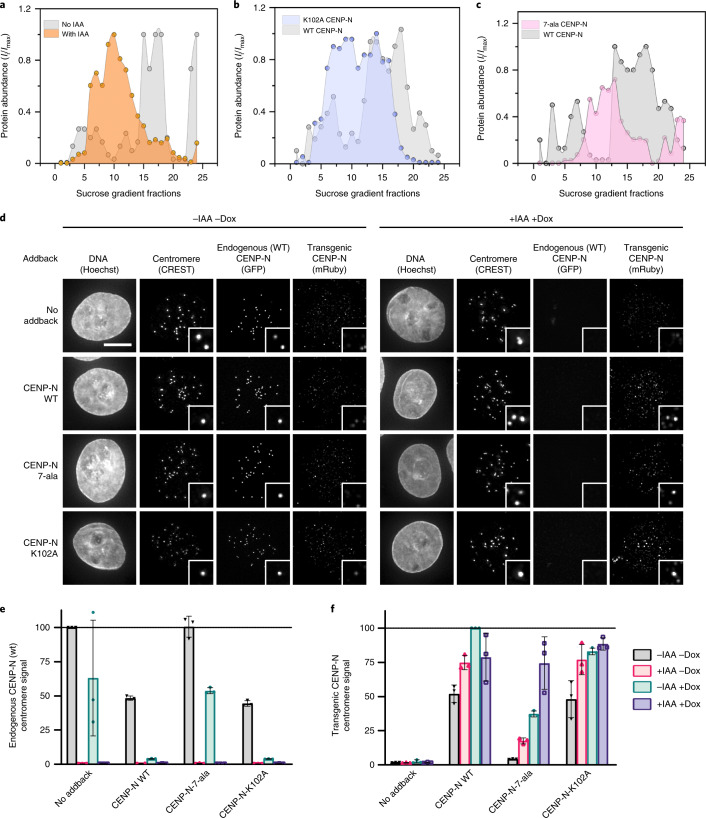
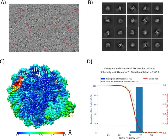
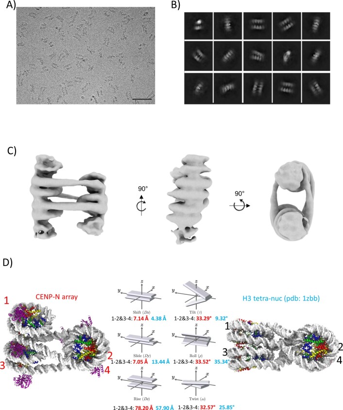
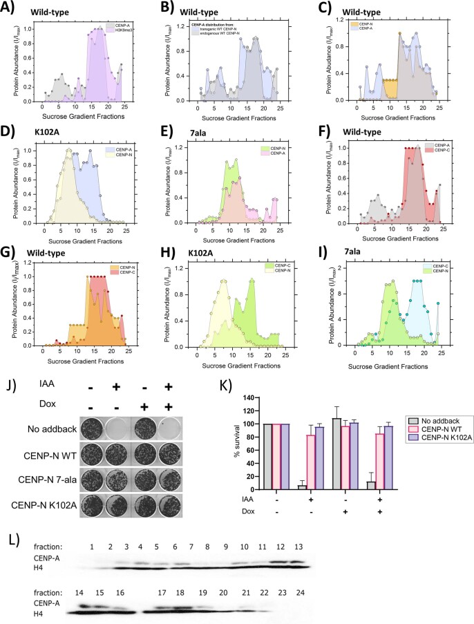
The histone variant CENP-A is the epigenetic determinant for the centromere, where it is interspersed with canonical H3 to form a specialized chromatin structure that nucleates the kinetochore. How nucleosomes at the centromere arrange into higher order structures is unknown. Here we demonstrate that the human CENP-A-interacting protein CENP-N promotes the stacking of CENP-A-containing mononucleosomes and nucleosomal arrays through a previously undefined interaction between the α6 helix of CENP-N with the DNA of a neighboring nucleosome. We describe the cryo-EM structures and biophysical characterization of such CENP-N-mediated nucleosome stacks and nucleosomal arrays and demonstrate that this interaction is responsible for the formation of densely packed chromatin at the centromere in the cell. Our results provide first evidence that CENP-A, together with CENP-N, promotes specific chromatin higher order structure at the centromere.


Conflict of interest statement

The authors declare no competing interests

Figures

Fig. 1
Fig. 1. CENP-N mediates CENP-A nucleosome stacking in vitro.
a, A model of two CENP-A nucleosomes (α satellite DNA) connected by two copies of CENP-N was fit into the density map. Protein identity is indicated by color codes. Arrows highlight the weak density attributed to a second CENP-N on the other side of the CENP-A nucleosomes. b, Simulation plots of nucleosome stacks in the absence or presence of CENP-N. This plot used two coordinate points from each nucleosome, namely the geometric center of the C1′ atoms from nucleotides located at the dyad and opposite of the dyad (Extended Data Fig. 4 for illustration). The C1’ atoms of every nucleotide in one nucleosome, pictured as the bottom nucleosome of each graph in Fig. 1b, were used to align each frame of the trajectory. The dyad and opposing points in the other nucleosome, pictured as the top nucleosome of each graph in Fig. 1b, were plotted to depict the relative sampling of each stacked-nucleosome system. c, SV-AUC (enhanced van Holde–Weischet plots) for CENP-A (CA) or H3 mononucleosomes (MN) in complex with CENP-N1–289 (CN) or full-length CENP-N/CENP-L (CN + CL). d, FRET analysis of CENP-A mononucleosome (CA MN) interactions in the absence or presence of CENP-N. The donor is CENP-A mononucleosomes containing Alexa 488-labeled H2B; the acceptor is a CENP-A mononucleosome containing Atto N 647-labeled H2B (250 nM donor and acceptor nucleosome concentrations were used). FRET intensity changes in dependence of [CENP-N]. The final NaCl concentration was 70 mM. Error bars are from four independent measurements of two biological replicates. Data are presented as mean values ± s.d. Source data
Fig. 2
Fig. 2. Structural basis for CENP-N-dependent nucleosome-nucleosome interaction.
a, The CENP-N α6 helix interacts with nucleosomal DNA of a second nucleosome without contacting histones (nonspecific nucleosome). Top, overview of interactions. Lower left, the interface between CENP-N α6 and DNA is highlighted. Lower middle, a surface charge representation (unit, kT e−1) in the same orientation. Lower right, the specific interaction on the other side of CENP-N with the CENP-A RG loop. b, Single mutations on CENP-N α6 affect nucleosome-nucleosome interactions, as shown by AUC. c, FRET analysis of the interaction between CENP-A mononucleosome (CA MN) and H3 mononucleosome (H3 MN) in the absence or presence of CENP-N. The donor is CENP-A mononucleosomes containing Alexa 488-labeled H2B; the acceptor is a H3 mononucleosome containing Atto N 647-labeled H2B (250 nM donor and acceptor nucleosome were used); FRET intensity changes in dependence of [CENP-N]. Error bars are from five independent measurements of two biological replicates. Data are presented as mean values ± s.d. d, The H4 N-terminal tail is essential for CENP-N-mediated nucleosome stacking (van Holde–Weischet plots of sedimentation). ∆19 indicates H4 tail deletion (amino acids 1–18). e, Comparison of different modes of nucleosome stacking. ‘Nuc1’ represents the reference nucleosome that interacts specifically with the indicated factor. ‘Nuc2’ is the neighboring nucleosome which interacts with Nuc1 or its binding factors non-specifically. Top, models for stacked mononucleosomes. 1AOI is the PDB code for a previously published nucleosome structure. Bottom, ‘superhelix locations’ (SHLs) (1-6) and the nucleosomal dyad axis (SHL 0; ɸ) of nuc2 (brown color, DNA only), are indicated, with nuc1 shown in a dotted circle (gray color), depicts the relative orientations of nuc1 and nuc2 and CENP-N or cGAS, respectively. Only half of the nucleosomal DNA is shown for clarity. Source data
Fig. 3
Fig. 3. CENP-N folds CENP-A chromatin into a regular fiber.
a, A nucleosomal array consisting of six CENP-A nucleosomes with CENP-N was fit into the density map. Dashed circles indicate weak densities representing more disordered nucleosomes at both ends. b, CENP-N interacts with nucleosomal DNA of a neighboring nucleosome (CENP-N α6 binds between SHL6 and 7 (left)). This is in contrast to mononucleosome stacks, where CENP-N α6 binds between SHL4 to 5 (right). c, The relative arrangement of nucleosomes in the arrays was expressed in analogy to the arrangement of DNA bases in a double helix. Parameters for CENP-N fibers (left, EMD-26333) and H1 fibers are shown. The vectors in the middle show the parameters of the analysis (in red for CENP-A arrays, and cyan for H1 arrays).
Fig. 4
Fig. 4. CENP-N promotes the compaction of centromeric chromatin in vivo.
a, CENP-A distribution in sucrose gradient in the presence of WT CENP-N (gray) and after IAA-induced degradation of CENP-N (orange). An equal amount of chromatin was loaded per well on an SDS gel, resolved by blotting against H4 histone (Extended Data Fig. 9l). b,c, Comparison of CENP-A distribution in the presence of transiently expressed CENP-N variants, WT CENP-N (gray), K102A CENP-N (lilac), and 7-ala CENP-N (pink, 7 positively charged amino acids on α6 are mutated to alanine). Dots in ac represent a mean of two biological replicates. Imax and Ii in ac represent a maximum signal intensity from all fractions and the signal intensity in a given fraction, respectively. Solid lines represent interpolation between experimental data points (using Akima spline). d, Representative images of nuclei (Hoechst stain) showing endogenous WT CENP-N (GFP) or transgenic CENP-N variant (anti-Ruby antibody) localization at centromeres (CREST signal). Scale bar, 5 µm. Insets show magnified images of example centromeres (CREST foci). Cells were treated for 24 hours with doxycycline (dox) to induce expression of mRuby2-3×FLAG-tagged CENP-N (WT or mutant) and/or with indole acetic acid (IAA) to deplete endogenous auxin-inducible degron (AID)-tagged CENP-N-GFP as indicated. e,f, Normalized centromeric fluorescence signal corresponding to endogenous WT CENP-N (GFP) (e) or transgenic WT or CENP-N variant (anti-mRuby antibody) (f). Each dot represents the median centromeric CENP-N signal from >100 cells from 1 biological replicate. Line and error bars are mean of three biological replicates ± s.d. Source data
Extended Data Fig. 1
Extended Data Fig. 1. CryoEM analysis of CENP-N in complex with CENP-A mono-nucleosomes (reconstituted with 147 bp palindromic α satellite DNA).
a) Raw cryoEM micrograph. Stacks are indicated by red lines. Size bar is 50 nm. b) 2D classifications of particles at the mono-nucleosome level. c) Local resolution of the 3D map for CENP-N in complex with CENP-A mono-nucleosomes. The color key indicates the resolution. d) 3DFSC analysis of reconstructed 3D map.
Extended Data Fig. 2
Extended Data Fig. 2. CryoEM analysis of CENP-N mediated stacks of CENP-A mono-nucleosomes reconstituted with palindromic α-satellite DNA.
a) Flow-chart of the analysis of di-nucleosomes in the stacks. b) 2D classification of di-nucleosomes with CENP-N. c) Local resolution of the 3D map for stacked CENP-A nucleosomes in complex with CENP-N. d) 3DFSC analysis of the reconstructed 3D map. The plot reveals significant orientation bias in the map reconstruction. Therefore, the estimated global resolution of 3.54 Å does not represent the overall map quality. In some orientations, the resolution is closer to 10 Å.
Extended Data Fig. 3
Extended Data Fig. 3. CryoEM analysis of CENP-N mediated stacks of CENP-A mono-nucleosomes reconstituted with 601 DNA.
a) Representative cryoEM micrograph. Stacks are highlighted by red lines. Size bar is 50 nm. b) 2D classification of the di-nucleosome with CENP-N in the stacks. c) Flow-chart on the analysis of di-nucleosomes in the stacks. d) Comparison of di-nucleosome maps derived for α satellite and 601 CENP-A nucleosomes.
Extended Data Fig. 4
Extended Data Fig. 4. MD simulations of stacked nucleosomes with 0, 1, or 2 CENP-N.
a) Diagram of the points used to construct stacked-nucleosome sampling graphs as depicted in Fig. 1c, in addition to nucleosome parameters calculated for stacked-nucleosome simulations. Nucleosomes (blue) are shown in face-on (left) and profile (right) viewpoints, with DNA represented in a darker blue. Dyad points and their opposing points are represented as small red and green circles, respectively. CENP-N is shown in purple to provide a point-of-reference. b) From left to right, six histograms of stacking parameters for di-nucleosome systems: Shift, Slide, Rise (top), and Tilt, Roll, Twist (bottom), in analogy to the parameters used to describe the geometry of the DNA double helix. Histograms do not contain the first 100 ns of simulation time which was allotted for each system to achieve equilibration.
Extended Data Fig. 5
Extended Data Fig. 5. Solution assays for CENP-N induced CENP-A nucleosome stacking.
a) FRET analysis of homotypic CENP-A mono-nucleosome (CA) or H3 mono-nucleosome (H3) interactions in the absence or presence of CENP-N. Donor is a mono-nucleosomes containing Alexa 488 labeled H2B; Acceptor is a mono-nucleosome containing Atto N 647 labeled H2B. 250 nM donor and acceptor nucleosome; FRET intensity in dependence of [CENP-N]. Final concentration for NaCl is around 100 mM. Error bars from three independent measurements. Data are presented as mean values + /- SD. b) Salt concentration affects nucleosome stack formation. AUC analysis (van Holde-Weischet plots) of CENP-N in complex with CENP-A mono-nucleosomes at 60 and 200 mM NaCl). CN: CENP-N1–289 in complex with CENP-A mono-nucleosome. CA_MN: CENP-A mono-nucleosome. c) CENP-N mutant (K102A) binds to CENP-A nucleosomes as well as wild-type CENP-N (5% native PAGE). 250 nM CENP-A nucleosome was combined with CENP-N at ratios of 2:1, 4:1 and 8:1 in buffer containing 50 mM NaCl, 20 mM Tris-HCl (pH 7.8), 1 mM EDTA, 1 mM DTT. d) deletion of the H4 N-terminal tail (∆19) does not affect the specific interaction between CENP-N and CENP-A nucleosomes. CENP-N was mixed with CENP-A nucleosome containing full length H4 or (∆19) H4 at a 2:1 ratio in the same buffer as in A). Source data
Extended Data Fig. 6
Extended Data Fig. 6. Residue contacts of the CENP-N α6-helix with DNA of the DNA-directed nucleosome from simulations containing one (A) and two CENP-N (B).
CENP-N 1 and CENP-N 2 are distinguished by the binding orientation of the α6-helix with the DNA grooves. CENP-N 1 (blue) binds directly into the DNA minor groove while CENP-2 (red) does not. Protein-DNA contacts were defined between the heavy atoms of residues within 4.0 Å of one another. Standard errors were derived using n = 15, where n is the number of statistically independent data points in each window as was determined by calculating the statistical inefficiency.
Extended Data Fig. 7
Extended Data Fig. 7. CryoEM analysis of 12mer nucleosomal arrays in presence of CENP-N.
a) raw cryoEM image of CENP-N in complex with 12-207mer 601 array. Size bar is 50 nm. b) raw cryoEM image of CENP-N in complex with 12-167mer array. Size bar is 50 nm. c) 2D classification of CENP-N in complex with 12-167mer array. d) Local resolution map of chromatin fiber. Color key indicates the resolution. e) 3DFSC analysis of cryoEM electron map.
Extended Data Fig. 8
Extended Data Fig. 8. CENP-N affects the folding of chromatin arrays.
a) Comparison between tetra-nucleosomes in a chromatin fiber with CENP-N, and of a canonical tetra-nucleosome. Analysis of the relative orientation of the nucleosomes, in analogy to DNA base pair analysis, is shown in the middle panel, with numbers for the CENP-N array in red, and tetranucleosome array in black. b) CryoEM analysis of crosslinked chromatin arrays without CENP-N, raw cryoEM image. Size bar is 100 nm c) 2D classification of CENP-N in complex with 12-167mer array. d) Low resolution 3D cryoEM map illustrates ladder-like arrangement of nucleosomes.
Extended Data Fig. 9
Extended Data Fig. 9. Western blot detection of centromeric proteins across 5-40% sucrose gradients and clonogenic survival assay.
a) Comparison of CENP-A (gray) and H3K9me3 (lilac) distribution in the presence of endogenous WT-CENP-N. b) CENP-A distribution in the presence of endogenous (gray) and transgenic (blue) WT-CENP-N. c) Comparison of CENP-A (blue) and CENP-N (orange) in the presence of WT CENP-N. d) Comparison of CENP-A (blue) and CENP-N (yellow) in the presence of K102A CENP-N. e). Comparison of CENP-A (pink) and CENP-N (green) distribution in the presence of 7ala CENP-N. f) Comparison of CENP-A (gray) and CENP-C (red) distribution in the presence of endogenous WT-CENP-N. g) CENP-N (orange) and CENP-C (red) distribution in the presence of endogenous WT-CENP-N h) CENP-N (yellow) and CENP-C (green) distribution in the presence of K102A CENP-N. i) CENP-N (yellow) and CENP-C (green) distribution in the presence of 7ala CENP-N. j) Representative crystal violet-stained colonies from clonogenic survival assay showing viable CENP-N AID cells with the indicated transgenic CENP-N variant treated with or without 0.1 mM IAA and doxycycline for 14 days. After treatment of 1000 seeded cells/well, surviving colonies were fixed and stained with crystal violet stain. k) Quantification of average percentage survival (average crystal violet stain intensity) of CENP-N AID cells (except CENP-N 7-ala) from 3 biological replicates normalized to untreated cells. (CENP-N 7-ala survival data is from single replicate). Data are presented as mean values± SD. l) Western blot of CENP-A and H4 histone distribution across 5–40 % sucrose gradient. Presence of CENP-A signal indicates fractions containing the centromeric chromatin whereas H4 signal represents the overall amount of chromatin loaded onto a SDS gel. Source data
Extended Data Fig. 10
Extended Data Fig. 10. The structure of CENP-A α satellite nucleosome in complex with CENP-NNT.
a) 2.65 Å cryoEM map of CENP-A α satellite nucleosome in complex with CENP-NNT. The components are colored as indicated. b) Density (with model) of the interface between CENP-N and histones. c) Density (with model) of the interface between CENP-N and nearby DNA. d) The model to density of DNA. e) α satellite DNA is compressed by one base pair in the crystal structure of the nucleosome (pdb 1KX5), compared to our cryoEM structure where DNA ends are unconstrained.

Similar articles

Cited by

References

    1. Luger K, Mader AW, Richmond RK, Sargent DF, Richmond TJ. Crystal structure of the nucleosome core particle at 2.8 A resolution. Nature. 1997;389:251–260. - PubMed
    1. Dekker J, Mirny L. The 3D genome as moderator of chromosomal communication. Cell. 2016;164:1110–1121. - PMC - PubMed
    1. Maeshima K, Ide S, Babokhov M. Dynamic chromatin organization without the 30-nm fiber. Curr. Opin. Cell Biol. 2019;58:95–104. - PubMed
    1. Boettiger A, Murphy S. Advances in chromatin imaging at kilobase-scale resolution. Trends Genet. 2020;36:273–287. - PMC - PubMed
    1. Ding X, Lin X, Zhang B. Stability and folding pathways of tetra-nucleosome from six-dimensional free energy surface. Nat. Commun. 2021;12:1091. - PMC - PubMed

Publication types

LinkOut - more resources