

Glycine Receptor Subtypes and Their Roles in Nociception and Chronic Pain
- PMID: 35401105
- PMCID: PMC8984470
- DOI: 10.3389/fnmol.2022.848642
Glycine Receptor Subtypes and Their Roles in Nociception and Chronic Pain
Abstract
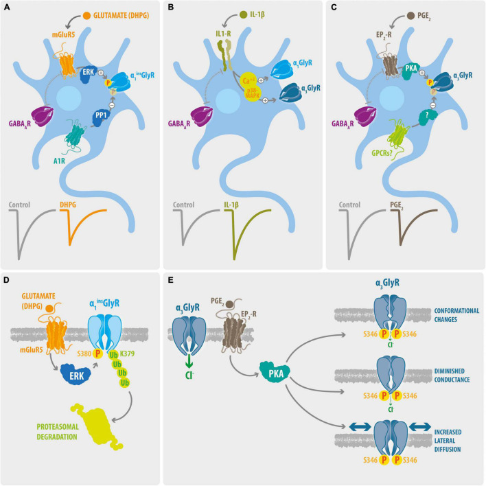
Disruption of the inhibitory control provided by the glycinergic system is one of the major mechanisms underlying chronic pain. In line with this concept, recent studies have provided robust proof that pharmacological intervention of glycine receptors (GlyRs) restores the inhibitory function and exerts anti-nociceptive effects on preclinical models of chronic pain. A targeted regulation of the glycinergic system requires the identification of the GlyR subtypes involved in chronic pain states. Nevertheless, the roles of individual GlyR subunits in nociception and in chronic pain are yet not well defined. This review aims to provide a systematic outline on the contribution of GlyR subtypes in chronic pain mechanisms, with a particular focus on molecular pathways of spinal glycinergic dis-inhibition mediated by post-translational modifications at the receptor level. The current experimental evidence has shown that phosphorylation of synaptic α1β and α3β GlyRs are involved in processes of spinal glycinergic dis-inhibition triggered by chronic inflammatory pain. On the other hand, the participation of α2-containing GlyRs and of β subunits in pain signaling have been less studied and remain undefined. Although many questions in the field are still unresolved, future progress in GlyR research may soon open new exciting avenues into understanding and controlling chronic pain.
Keywords: chronic pain; glycine receptor (GlyR); nociception; phosphorylation; synaptic plasticity.
Copyright © 2022 San Martín, Sazo, Utreras, Moraga-Cid and Yévenes.
Conflict of interest statement
The authors declare that the research was conducted in the absence of any commercial or financial relationships that could be construed as a potential conflict of interest.
Figures
Similar articles
-
Native glycine receptor subtypes and their physiological roles.Neuropharmacology. 2009 Jan;56(1):303-9. doi: 10.1016/j.neuropharm.2008.07.034. Epub 2008 Aug 3. Neuropharmacology. 2009. PMID: 18721822 Review.
-
Glycine Receptor Drug Discovery.Adv Pharmacol. 2017;79:225-253. doi: 10.1016/bs.apha.2017.01.003. Epub 2017 Mar 21. Adv Pharmacol. 2017. PMID: 28528670 Review.
-
Plasticity of synaptic inhibition in mouse spinal cord lamina II neurons during early postnatal development and after inactivation of the glycine receptor alpha3 subunit gene.Eur J Neurosci. 2009 Dec;30(12):2284-92. doi: 10.1111/j.1460-9568.2009.07018.x. Epub 2009 Dec 10. Eur J Neurosci. 2009. PMID: 20092571
-
Functional reconstitution of glycinergic synapses incorporating defined glycine receptor subunit combinations.Neuropharmacology. 2015 Feb;89:391-7. doi: 10.1016/j.neuropharm.2014.10.026. Neuropharmacology. 2015. PMID: 25445488
-
Molecular pharmacology of the glycine receptor chloride channel.Curr Pharm Des. 2007;13(23):2350-67. doi: 10.2174/138161207781368693. Curr Pharm Des. 2007. PMID: 17692006 Review.
Cited by
-
Pain modulation in the spinal cord.Front Pain Res (Lausanne). 2022 Sep 13;3:984042. doi: 10.3389/fpain.2022.984042. eCollection 2022. Front Pain Res (Lausanne). 2022. PMID: 36176710 Free PMC article. Review.
-
Mechanism of human α3β GlyR modulation in inflammatory pain and 2, 6-DTBP interaction.Res Sq [Preprint]. 2024 Aug 7:rs.3.rs-4402878. doi: 10.21203/rs.3.rs-4402878/v1. Res Sq. 2024. PMID: 39149480 Free PMC article. Preprint.
-
Positive Allosteric Modulators of Glycine Receptors and Their Potential Use in Pain Therapies.Pharmacol Rev. 2022 Oct;74(4):933-961. doi: 10.1124/pharmrev.122.000583. Pharmacol Rev. 2022. PMID: 36779343 Free PMC article. Review.
-
Metabolic Biomarkers Differentiate a Surgical Intervertebral Disc from a Nonsurgical Intervertebral Disc.Int J Mol Sci. 2023 Jun 24;24(13):10572. doi: 10.3390/ijms241310572. Int J Mol Sci. 2023. PMID: 37445750 Free PMC article.
-
Gating mechanism of the human α1β GlyR by glycine.Structure. 2024 Oct 3;32(10):1621-1631.e3. doi: 10.1016/j.str.2024.07.012. Epub 2024 Aug 14. Structure. 2024. PMID: 39146932
References
Publication types
LinkOut - more resources
Full Text Sources