. 2022 Apr 8;12(1):5938.
doi: 10.1038/s41598-022-09757-x.

p38 MAPK priming boosts VSMC proliferation and arteriogenesis by promoting PGC1α-dependent mitochondrial dynamics

Affiliations

p38 MAPK priming boosts VSMC proliferation and arteriogenesis by promoting PGC1α-dependent mitochondrial dynamics

Álvaro Sahún-Español et al. Sci Rep. .

Abstract

Vascular smooth muscle cell (VSMC) proliferation is essential for arteriogenesis to restore blood flow after artery occlusion, but the mechanisms underlying this response remain unclear. Based on our previous findings showing increased VSMC proliferation in the neonatal aorta of mice lacking the protease MT4-MMP, we aimed at discovering new players in this process. We demonstrate that MT4-MMP absence boosted VSMC proliferation in vitro in response to PDGF-BB in a cell-autonomous manner through enhanced p38 MAPK activity. Increased phospho-p38 in basal MT4-MMP-null VSMCs augmented the rate of mitochondrial degradation by promoting mitochondrial morphological changes through the co-activator PGC1α as demonstrated in PGC1α-/- VSMCs. We tested the in vivo implications of this pathway in a novel conditional mouse line for selective MT4-MMP deletion in VSMCs and in mice pre-treated with the p38 MAPK activator anisomycin. Priming of p38 MAPK activity in vivo by the absence of the protease MT4-MMP or by anisomycin treatment led to enhanced arteriogenesis and improved flow recovery after femoral artery occlusion. These findings may open new therapeutic opportunities for peripheral vascular diseases.


Conflict of interest statement

The authors declare no competing interests.

Figures
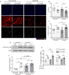
Figure 1
Figure 1
p38 MAPK signaling is increased in MT4-MMP-null VSMCs in vitro leading to boosted VSMC proliferation upon PDGF-BB treatment. (A) Representative confocal microscopy images showing immunostaining for SMA, Ki67 and DAPI in MT4-MMP+/+ and MT4-MMP−/− aortic VSMCs treated or not with PDGF-BB. Scale bar: 50 µm. (B,C) Quantification of the percentage of VSMCs (% SMA+ in DAPI+ cells) and proliferative VSMCs (% Ki67+ in SMA+ cells). n = 9 VSMC cultures in 9 independent experiments. (D) Representative Western blot of phospho-p38 and p38 in lysates of MT4-MMP+/+ and MT4-MMP−/− aortic VSMCs treated or not with PDGF-BB. (E) Quantification of phospho-p38 normalized to total p38. n = 4 VSMC cultures in 4 independent experiments. (F) Quantification of the percentage of proliferative VSMCs (% Ki67+ in SMA+ cells) in MT4-MMP+/+ and MT4-MMP−/− aortic isolated VSMCs treated or not with PDGF-BB in presence or absence of p38 inhibitor (SB203580). n = 5 VSMC cultures in five independent experiments. In (B,C,E,F) data are means ± s.e.m. analysed by two-way ANOVA followed by Benjamini and Hochberg post-test; *, # p < 0.05.
Figure 2
Figure 2
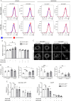
MT4-MMP deletion boosts mitochondrial degradation rate and increases mitochondrial fission in cultured VSMCs through p38 MAPK signaling. (A) Representative flow cytometry histogram plots of MTDR signal in MT4-MMP+/+ and MT4-MMP−/− aortic isolated VSMCs treated or not with PDGF-BB in presence or absence of lysosome inhibitors and p38 inhibitor (SB203580). (B) Quantification of the mitochondrial degradation rate (MTDR MFI in the presence of lysosomal inhibitors divided by MTDR MFI in the absence of lysosomal inhibitors) normalized to MT4-MMP+/+ without PDGF-BB or p38 inhibitor. n = 4 VSMC cultures in 4 independent experiments. (C) Representative confocal microscopy images showing immunostaining for TOMM20 and its thresholded binary image of MT4-MMP+/+ and MT4-MMP−/− VSMCs treated or not with PDGF-BB. Scale bar: 10 µm. (D) Quantification of number of individuals, inverse aspect ratio, mean area and number of branch junctions. n = 4 VSMC cultures in 4 independent experiments. (E) Quantification of relative levels of glycolytic and mitochondrial ATP in technical triplicates of MT4-MMP+/+ and MT4-MMP−/− VSMCs treated or not with PDGF-BB and in absence or presence of SB203580. n = 3 VSMC cultures in 3 independent experiments. In (B,D,E) data are means ± s.e.m. analysed by two-way ANOVA with Benjamini and Hochberg post-test; *, # p < 0.05, ## p < 0.01, ***, ### p < 0.001.
Figure 3
Figure 3
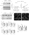
p38 MAPK priming by anisomycin treatment leads to increased mitochondrial fragmentation and PDGF-BB-induced proliferation in VSMCs in a PGC1α-dependent manner. (A) Representative western blot of PGC1α, phospho-p38, p38 and Tubulin in MT4-MMP+/+ and MT4-MMP−/− VSMCs treated or not with PDGF-BB. (B) Correlation graphs between phospho-p38/p38 and PGC1α/Tubulin in MT4-MMP+/+ and MT4-MMP−/− VSMCs treated or not with PDGF-BB. n = 6 VSMC cultures in 6 independent experiments. Correlation was analysed by a simple linear regression test. (C) Western blot of phospho-p38, p38 and Tubulin in two independent batches of PGC1α+/+ and PGC1α−/− VSMCs pre-treated with vehicle (DMSO) or anisomycin. (D) Representative confocal microscopy images showing immunostaining for TOMM20 and its thresholded binary image of PGC1α+/+ and PGC1α−/− VSMCs pre-treated or not with vehicle or anisomycin. Cells were also treated or not afterwards with PDGF-BB (images not shown). Scale bar: 10 µm. (E) Quantification of number of individuals, inverse aspect ratio, mean area and number of branch junctions. n = 5 VSMC cultures in 5 independent experiments. (F) Quantification of the percentage of proliferative VSMCs (DAPI+/SMA+/Ki67+) in PGC1α+/+ and PGC1α−/− VSMCs pre-treated with vehicle (DMSO) or anisomycin and in presence or absence of PDGF-BB. n = 5 VSMC cultures in five 5 independent experiments. In (E,F) data are means ± s.e.m. analysed by two-way ANOVA with Benjamini and Hochberg post-test; *, # p < 0.05, ** p < 0.01, ***, ### p < 0.001, ****, #### p < 0.0001.
Figure 4
Figure 4
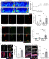
Enhanced blood flow restoration and increased VSMC proliferation and p38 activity in remodeled arterioles after femoral ligation in MT4 MMPΔVSMC mice. (A) Representative laser Doppler images showing Non Ligated and Ligated adductor blood perfusion 1 and 7 days after ischemia in MT4-MMPf/f and MT4-MMPΔVSMC mice. (B) Quantification of the adductor blood flow perfusion (Non Ligated versus Ligated). n = 12 mice per genotype in 2 independent experiments. (C) Representative confocal microscopy images showing immunofluorescence for SMA from the superficial Non Ligated and Ligated MT4-MMPf/f and MT4-MMPΔVSMC adductor muscles. Scale bar: 100 µm. (D) Quantification of the density of remodeled arterioles (#Arterioles > 40 µm of diameter/mm2 tissue) in the superficial Non Ligated and Ligated MT4-MMPf/f and MT4-MMPΔVSMC adductor muscles. n = 6 mice per genotype in 2 independent experiments. (E) Representative confocal microscopy images showing SMA, Erg and EdU from the superficial Ligated MT4-MMPf/f and MT4-MMPΔVSMC adductor muscles. Scale bar: 50 µm. (F) Quantification of the percentage of proliferative VSMCs (Erg-/SMA+/EdU+) within the remodeled arterioles. n = 6 mice per genotype in 2 independent experiments. (G) Representative confocal microscopy images showing immunostaining for SMA, phospho-p38 MAPK and DAPI within the remodeled arterioles. Scale bar: 25 µm. Yellow arrowheads mark VSMCs positive for phospho-p38. (H) Quantification of the percentage of phospho-p38+ VSMCs within the remodeled arterioles. n = 4 MT4-MMPf/f and 5 MT4-MMPΔVSMC in two independent experiments. (I) Representative confocal microscopy images showing immunostaining for SMA, PGC1α and Hoechst in VSMCs within the remodeled arterioles from MT4-MMPf/f and MT4-MMPΔVSMC ligated adductor muscles. Scale bar: 25 µm. (J) Quantification of PGC1α mean signal intensity in VSMCs within the remodeled arterioles from MT4-MMPf/f and MT4-MMPΔVSMC ligated adductor muscles. In (B,D,F) data are means ± s.e.m. analysed by two-way ANOVA with Benjamini and Hochberg post-test. In (H, J) data are means ± s.e.m. analyzed by unpaired t-test. *, # p < 0.05, **, ## p < 0.01, **** p < 0.0001.
Figure 5
Figure 5
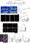
p38 MAPK priming by anisomycin pre-treatment increases blood flow restoration, arteriolar remodeling and VSMC proliferation after femoral ligation. (A) Scheme of the experimental design for anisomycin preconditioning in vivo and its analysis. (B) Representative laser Doppler images showing Non Ligated and Ligated adductor blood 1 and 7 days after ischemia in MT4-MMP+/+ mice pre-treated with vehicle or anisomycin. (C) Quantification of the adductor blood flow perfusion (Non Ligated versus Ligated ratio). n = 9 vehicle mice and 8 anisomycin mice in 3 independent experiments. (D) Representative confocal microscopy images showing immunofluorescence for SMA from the superficial Non Ligated and Ligated adductor muscles of MT4-MMP+/+ mice pre-treated or not with anisomycin. Scale bar: 200 µm. (E) Quantification of the density of remodeled arterioles (#Arterioles > 40 µm of diameter/mm2 tissue) within the superficial Non Ligated and Ligated adductor muscles of wild-type mice pre-treated or not with anisomycin. n = 9 vehicle mice and 8 anisomycin mice in 3 independent experiments. (F) Immunofluorescence of SMA and EdU from the superficial Ligated adductor muscle of wild-type mice pre-treated or not with anisomycin. Scale bar: 50 µm. (G) Quantification of the percentage of proliferative VSMCs (SMA+/EdU+) within the remodeled arterioles. n = 6 vehicle mice and 5 anisomycin mice in 3 independent experiments. (H) Representative confocal microscopy images showing immunostaining for SMA, PGC1α and Hoechst in VSMCs within the remodeled arterioles of the ligated superficial adductors from vehicle- or anisomycin-treated mice. Scale bar: 25 µm. (I) Quantification of PGC1α mean signal intensity in VSMCs within the remodeled arterioles from vehicle- or anisomycin-treated mice. (J) Quantification of the density of remodeled arterioles (#Arterioles > 40 µm of diameter/mm2 tissue) (left) and the percentage of proliferative VSMCs (SMA+/EdU+) (right) within the remodeled arterioles of PGC1α-null mice pre-treated or not with anisomycin 7 days post-ligation. n = 6 vehicle mice and 5 anisomycin mice in one experiment. In (C,E,G,J) data are means ± s.e.m. analyzed by two-way ANOVA with Benjamini and Hochberg post-test. In (I) data are means ± s.e.m. analyzed by unpaired t-test.; *, # p < 0.05, **, ## p < 0.01, ***, ### p < 0.001, **** p < 0.0001.
Figure 6
Figure 6
Graphical abstract: An increase in p38 MAPK phosphorylation in VSMCs (caused by external preconditioning with anisomycin or by knockdown of MT4-MMP) leads to a basal primed state in which mitochondrial dynamics is enhanced via PGC1α. This cellular context allows for a better proliferative response of VSMCs to in vitro treatment with PDGF-BB. Furthermore, in vivo, after femoral artery ligation, such VSMC priming allows for better (PGC1α-dependent) VSMC proliferation and arteriogenesis, and thus more efficient recovery of blood flow.

Similar articles

Cited by

References

    1. Cai W, Schaper W. Mechanisms of arteriogenesis. Acta Biochim. Biophys. Sin. (Shanghai) 2008;40:681–692. doi: 10.1093/abbs/40.8.681. - DOI - PubMed
    1. Schirmer SH, van Nooijen FC, Piek JJ, van Royen N. Stimulation of collateral artery growth: Travelling further down the road to clinical application. Heart. 2008;95:191–197. doi: 10.1136/hrt.2007.136119. - DOI - PubMed
    1. Meier P, et al. Beneficial effect of recruitable collaterals: A 10-year follow-up study in patients with stable coronary artery disease undergoing quantitative collateral measurements. Circulation. 2007;116:975–983. doi: 10.1161/CIRCULATIONAHA.107.703959. - DOI - PubMed
    1. Hellström M, Kalén M, Lindahl P, Abramsson A, Betsholtz C. Role of PDGF-B and PDGFR-β in recruitment of vascular smooth muscle cells and pericytes during embryonic blood vessel formation in the mouse. Development. 1999;126:3047–3055. doi: 10.1242/dev.126.14.3047. - DOI - PubMed
    1. Martins RN, Chleboun JO, Sellers P, Sleigh M, Muir J. The role of PDGF-BB on the development of the collateral circulation after acute arterial occlusion. Growth Factors. 1994;10:299–306. doi: 10.3109/08977199409010996. - DOI - PubMed

Publication types

MeSH terms

Substances