

Mucosal-associated invariant T cells are activated in an interleukin-18-dependent manner in Epstein-Barr virus-associated T/natural killer cell lymphoproliferative diseases
- PMID: 35380609
- PMCID: PMC8982962
- DOI: 10.1093/cei/uxab004
Mucosal-associated invariant T cells are activated in an interleukin-18-dependent manner in Epstein-Barr virus-associated T/natural killer cell lymphoproliferative diseases
Abstract
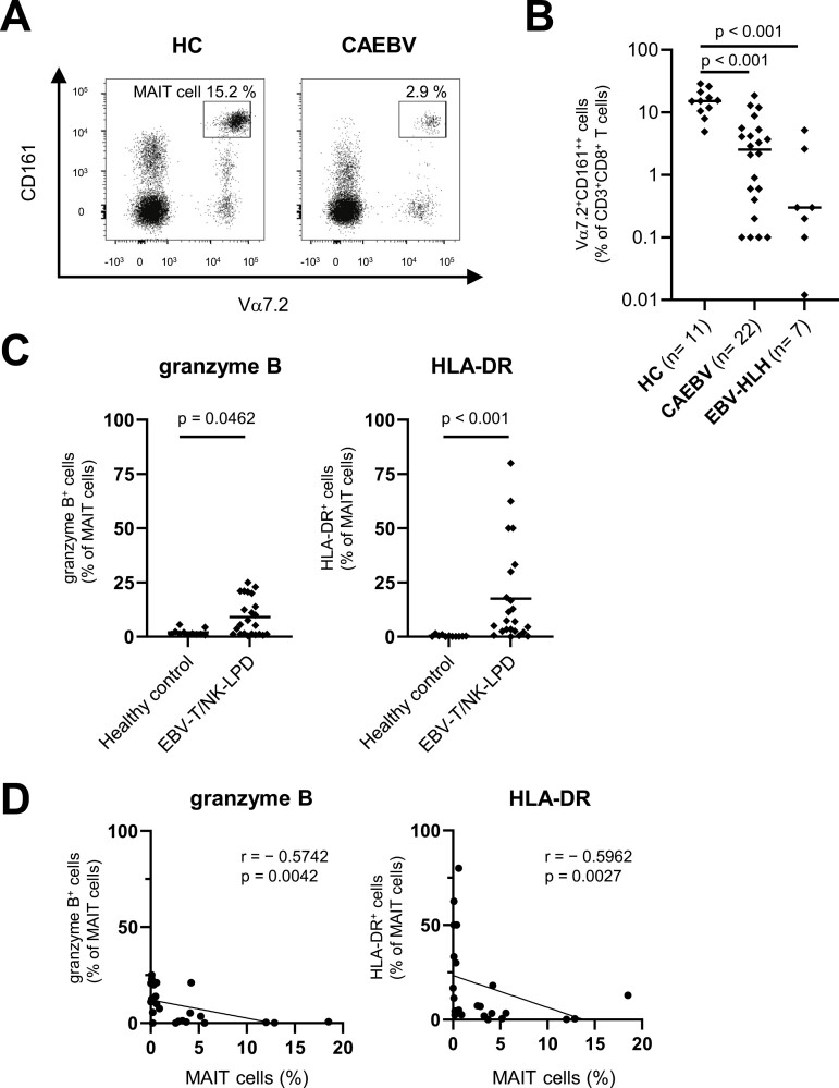
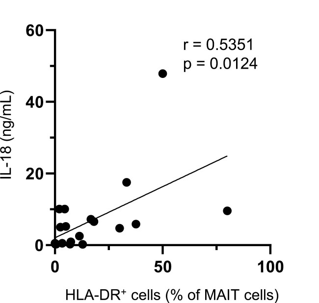
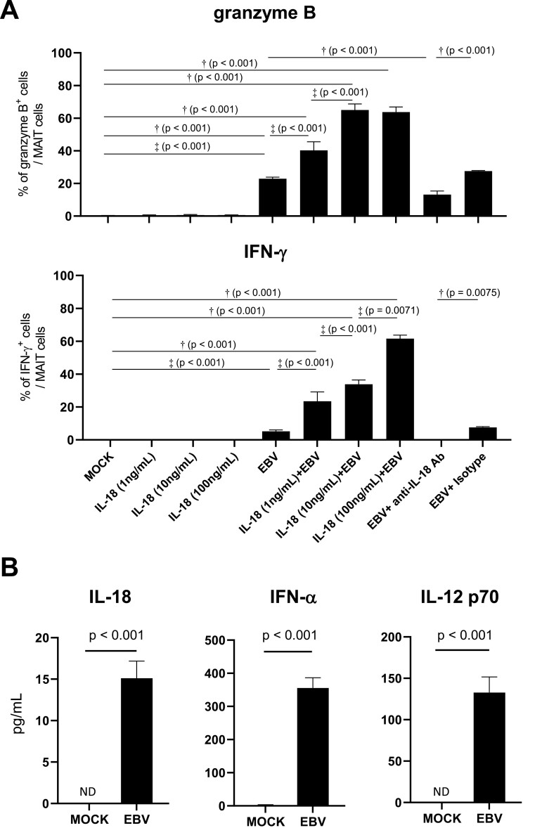
Mucosal-associated invariant T (MAIT) cells are a type of innate immune cells that protect against some infections. However, the involvement of MAIT cells in Epstein-Barr virus-associated T/natural killer cell lymphoproliferative diseases (EBV-T/NK-LPD) is unclear. In this study, we found that MAIT cells were highly activated in the blood of patients with EBV-T/NK-LPD. MAIT cell activation levels correlated with disease severity and plasma IL-18 levels. Stimulation of healthy peripheral blood mononuclear cells with EBV resulted in activation of MAIT cells, and this activation level was enhanced by exogenous IL-18. MAIT cells stimulated by IL-18 might thus be involved in the immunopathogenesis of EBV-T/NK-LPD.
Keywords: Epstein-Barr virus; interleukin-18; mucosal-associated innate T cell.
© The Author(s) 2022. Published by Oxford University Press on behalf of the British Society for Immunology. All rights reserved. For permissions, please e-mail: journals.permissions@oup.com.
Figures


Similar articles
-
Acquired hemophilia A associated with Epstein-Barr-virus-associated T/natural killer-cell lymphoproliferative disease: A case report.Medicine (Baltimore). 2021 Apr 23;100(16):e25518. doi: 10.1097/MD.0000000000025518. Medicine (Baltimore). 2021. PMID: 33879690 Free PMC article.
-
Somatic mutations in KMT2D and TET2 associated with worse prognosis in Epstein-Barr virus-associated T or natural killer-cell lymphoproliferative disorders.Cancer Biol Ther. 2019;20(10):1319-1327. doi: 10.1080/15384047.2019.1638670. Epub 2019 Jul 16. Cancer Biol Ther. 2019. PMID: 31311407 Free PMC article.
-
Central nervous system vasculitis from Epstein-Barr virus-associated T/natural killer-cell lymphoproliferative disorder in children: A case report.Brain Dev. 2019 Oct;41(9):820-825. doi: 10.1016/j.braindev.2019.05.009. Epub 2019 Jun 14. Brain Dev. 2019. PMID: 31208818
-
Epstein-Barr virus-associated T/natural killer-cell lymphoproliferative disorders.J Dermatol. 2014 Jan;41(1):29-39. doi: 10.1111/1346-8138.12322. J Dermatol. 2014. PMID: 24438142 Review.
-
Epstein-Barr virus-positive T/NK-cell lymphoproliferative disorders.Exp Mol Med. 2015 Jan 23;47(1):e133. doi: 10.1038/emm.2014.105. Exp Mol Med. 2015. PMID: 25613730 Free PMC article. Review.
Cited by
-
Interleukin-18 cytokine in immunity, inflammation, and autoimmunity: Biological role in induction, regulation, and treatment.Front Immunol. 2022 Aug 11;13:919973. doi: 10.3389/fimmu.2022.919973. eCollection 2022. Front Immunol. 2022. PMID: 36032110 Free PMC article. Review.
References
-
- Longnecker RM, Kieff E, Cohen JI.. Fields Virology. Philadelphia: Lippincott Williams & Wilkins, 2013.
Publication types
MeSH terms
Substances
LinkOut - more resources
Full Text Sources
Miscellaneous
