Review
. 2022 Apr 4;8(1):159.
doi: 10.1038/s41420-022-00982-x.

Targeting lncRNAs in programmed cell death as a therapeutic strategy for non-small cell lung cancer

Affiliations
Review

Targeting lncRNAs in programmed cell death as a therapeutic strategy for non-small cell lung cancer

Yanqin Luo et al. Cell Death Discov. .

Abstract

Lung cancer is a leading cause of cancer-related mortality worldwide, with non-small cell lung cancer (NSCLC) being the most common histological type. Owing to the limited therapeutic efficacy and side effects of currently available therapies for NSCLC, it is necessary to identify novel therapeutic targets for NSCLC. Long non-coding RNAs (lncRNAs) are non-protein-coding RNAs with a transcript length of more than 200 nucleotides, which play a vital role in the tumorigenesis and progression of multiple cancers, including NSCLC. Induction of programmed cell death (PCD) is the main mechanism leading to tumour cell death in most cancer treatments. Recent studies have demonstrated that lncRNAs are closely correlated with PCD including apoptosis, pyroptosis, autophagy and ferroptosis, which can regulate PCD and relevant death pathways to affect NSCLC progression and the efficacy of clinical therapy. Therefore, in this review, we focused on the function of lncRNAs in PCD of NSCLC and summarized the therapeutic role of targeting lncRNAs in PCD for NSCLC treatment, aiming to provide new sights into the underlying pathogenic mechanisms and propose a potential new strategy for NSCLC therapy so as to improve therapeutic outcomes with the ultimate goal to benefit the patients.


Conflict of interest statement

The authors declare no competing interests.

Figures

Fig. 1
Fig. 1. Relationship between lncRNAs and PCD in NSCLC.
PCD plays a critical role in the pathogenesis of NSCLC. LncRNAs are strongly associated with PCD, which can regulate different types of PCD and related pathways to influence the tumorigenesis and cancer progression of NSCLC.
Fig. 2
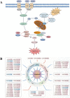
Fig. 2. LncRNAs participate in the regulation of the apoptotic pathways in NSCLC.
A Apoptosis can be initiated through two fundamental pathways: the death-receptor–mediated extrinsic pathway or the intracellular organelle-based intrinsic pathway. The extrinsic pathway can be activated by multiple extra-cellular death ligands, such as Fas ligand (FasL), TNF-α and TNF-related apoptosis-inducing ligand (TRAIL). The binding of ligands and their corresponding membrane death receptors then recruits death adaptor proteins, such as Fas-associated death domain (FADD) and TNF receptor-associated death domain (TRADD), to the death receptors. Subsequently, the oligomerized receptors and recruited adaptor proteins form the death-inducing signalling complex (DISC), which binds to pro-caspase-8 and triggers its activation. Active caspase-8 further proteolytically cleaves and promotes the activation of downstream apoptotic effector proteins caspase-3/6/7, and ultimately apoptosis. Various cytotoxic stimuli and cellular stresses activate the intrinsic pathway by inducing increased expression of BH3-only proteins (BIM, BAD, etc.) and inhibiting anti-apoptotic Bcl-2 proteins (Bcl-2, Bcl-xl, etc.). Activation of the pro-apoptotic effectors Bax and Bak ultimately causes mitochondrial outer membrane permeabilization (MOMP), which induces the release of cytochrome C. Cytochrome C engages apoptotic peptidase-activating factor 1 (APAF-1), subsequently triggers the activation of caspase-9 and in turn downstream effectors caspase-3/6/7, eventually contributing to apoptosis. In addition, active caspase-8 also engages the intrinsic apoptotic pathway indirectly through proteolytic activation of the pro-apoptotic protein BID into tBID. Moreover, the inhibitors of apoptosis proteins (IAPs), such as X-linked inhibitor of apoptosis protein (XIAP) and survivin, play a negative role in modulating both the extrinsic pathway and the intrinsic pathway by potently inhibiting key executioner caspase-3/7 activation and suppressing the initiator caspase-9 activity. B Various lncRNAs, including oncogenic lncRNAs (red box, high expression in NSCLC) and tumour-suppressive lncRNAs (blue box, low expression in NSCLC), participate in lung cancer-associated apoptosis by regulating apoptosis-related proteins.
Fig. 3
Fig. 3. LncRNAs regulate the autophagy pathways in NSCLC.
Autophagy starts with the formation of phagophores. The formation of unc-51-like kinase (ULK) complex from ULK1/2 kinase, ATG13, ATG101 and family-interacting protein FIP200, directly modulated by integrated signals from the mechanistic target of rapamycin complex 1 (mTORC1) and AMP-activated protein kinase (AMPK) signalling, triggers autophagic activity. Activated ULK1 can lead to the phosphorylation of Beclin1, thus activating class III PI3-kinase (VPS34) complex, which consists of VPS34, Beclin-1, VPS15 and ATG14L, primarily responsible for the nucleation of autophagosomal membrane. Two principal ubiquitination systems, ATG5-ATG12 and LC3 conjugation systems participate in autophagophore elongation and conversion into intact autophagosome. Respectively, ATG5 and ATG12 assemble into a complex, which then interacts with ATG16L1 to form a multimeric ATG12-ATG5-ATG16L1 conjugate that is on the outer surface of the autophagosomal membrane. Membrane-bound LC3-II is generated by the conjugation of microtubule-associated protein 1-light chain 3-I (LC3-I) to the lipid phosphatidylethanolamine (PE), providing a docking site for mounting autophagy cargo receptors that enable cargo loading into the autophagic vesicles. Various oncogenic lncRNAs (red font, high expression in NSCLC) participate in lung cancer-associated autophagy by regulating autophagy-related proteins.
Fig. 4
Fig. 4. Roles of lncRNAs in the regulation of pyroptosis in NSCLC.
Pyroptosis can be induced via the caspase-1-dependent canonical pathway and caspase-4,5 (for human)- or caspase-11 (for mouse)-mediated non-canonical pathway. In terms of the canonical pathway, damage-associated molecular patterns (DAMPs) or pathogen-associated molecular patterns (PAMPs) activate the inflammasome sensors NLRP3, which then recruit the effector pro-caspase-1 with the aid of the adaptor protein ASC to assemble inflammasomes, leading to the conversion of pro-caspase-1 into active caspase-1. Activation of NLRP3 inflammasome requires the involvement of the TLR4-mediated NF-κB pathway, which promotes the expression of NLRP3, pro-caspase-1, pro-IL-1β and pro-IL-18 proteins. Activated caspase-1 cleaves gasdermin D (GSDMD) and ultimately mediates pyroptosis. Besides, caspase-1 promotes the maturation of pro-inflammatory cytokines IL-1β and IL-18 by cleaving their precursor proteins, which trigger wide-ranging inflammatory responses. In the non-canonical pathway, human caspase-4 and -5 or their murine homologue caspase-11 can be directly activated by binding to lipopolysaccharide (LPS) and cleave GSDMD with efficiency similar to that of caspase-1, thereby inducing cell pyroptosis. Oncogenic lncRNAs (red font, high expression in NSCLC) participate in lung cancer-associated pyroptosis by regulating pyroptosis-related proteins.
Fig. 5
Fig. 5. LncRNAs participate in the regulation of ferroptosis in NSCLC.
Ferroptosis is initiated by blockade of the cellular antioxidant defences depending on glutathione (GSH). Cystine/glutamate antiporter system Xc- is responsible for transporting intracellular glutamate to the outside of the cell and transferring extra-cellular cystine into the cell. Subsequently, cystine is converted into cysteine for the biosynthesis of GSH. GPX4 converts GSH into oxidized glutathione (GSSG), concurrent with cytotoxic lipid peroxide (L-OOH) reduced to the corresponding alcohol (L-OH), thus reducing ROS accumulation. Cellular uptake of circulating iron (Fe3+) is mediated by TFR1 and intracellular iron can also be exported by ferroportin (FPN). The iron oxidoreductase six-transmembrane epithelial antigen of the prostate 3 (STEAP3) reduces Fe3+ to Fe2+, the latter is released from the endosome via divalent metal transporter 1 (DMT1) and then delivered into the unstable iron pool, thereby leading to ROS generation. Aberrant accumulation of ROS ultimately results in lipid peroxidation and ferroptosis. Oncogenic lncRNAs (red font, high expression in NSCLC) and tumour-suppressive lncRNAs (blue font, low expression in NSCLC) participate in lung cancer-associated ferroptosis by regulating ferroptosis-related proteins.

Similar articles

Cited by

References

    1. Imyanitov EN, Iyevleva AG, Levchenko EV. Molecular testing and targeted therapy for non-small cell lung cancer: current status and perspectives. Crit Rev Oncol Hematol. 2021;157:103194. - PubMed
    1. Zappa C, Mousa SA. Non-small cell lung cancer: current treatment and future advances. Transl Lung Cancer Res. 2016;5:288–300. - PMC - PubMed
    1. Herbst RS, Morgensztern D, Boshoff C. The biology and management of non-small cell lung cancer. Nature. 2018;553:446–54. doi: 10.1038/nature25183. - DOI - PubMed
    1. Duma N, Santana-Davila R, Molina JR. Non-small cell lung cancer: epidemiology, screening, diagnosis, and treatment. Mayo Clin Proc. 2019;94:1623–40. - PubMed
    1. Schmitt AM, Chang HY. Long noncoding RNAs in cancer pathways. Cancer Cell. 2016;29:452–63. - PMC - PubMed