. 2022 Mar 16:10:838612.
doi: 10.3389/fcell.2022.838612. eCollection 2022.

Expression and Roles of Lynx1, a Modulator of Cholinergic Transmission, in Skeletal Muscles and Neuromuscular Junctions in Mice

Affiliations

Expression and Roles of Lynx1, a Modulator of Cholinergic Transmission, in Skeletal Muscles and Neuromuscular Junctions in Mice

Sydney V Doss et al. Front Cell Dev Biol. .

Abstract

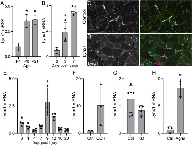
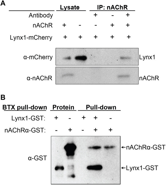
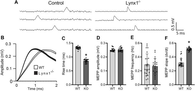
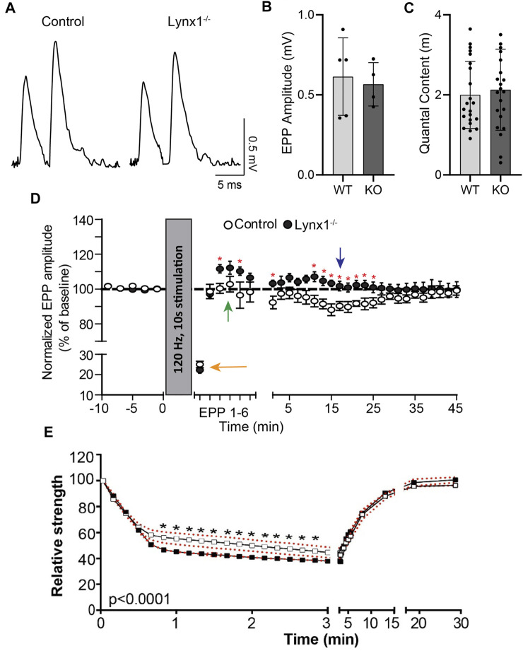
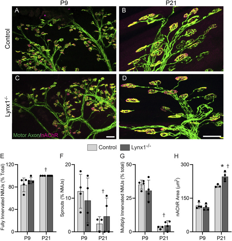
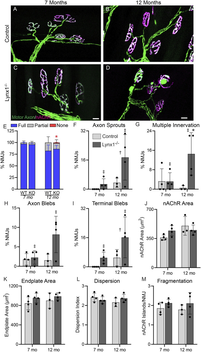
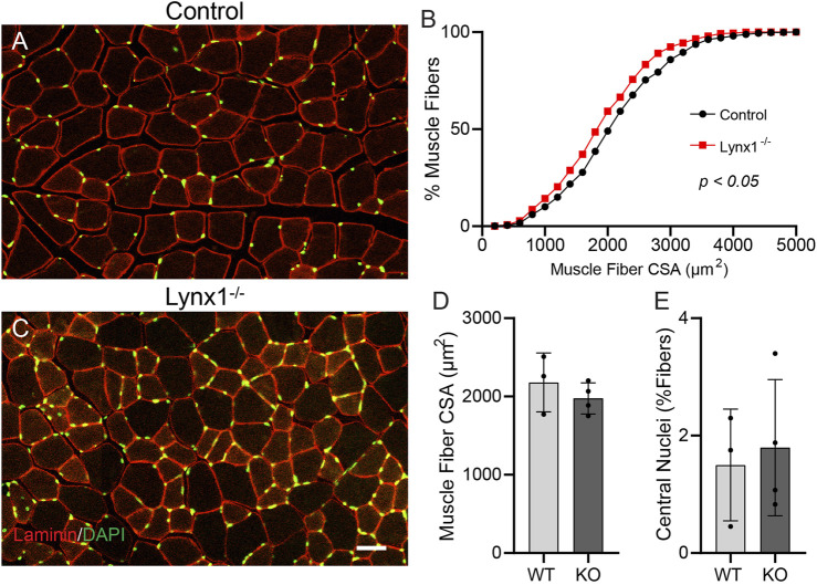
Lynx1 is a glycosylphosphatidylinositol (GPI)-linked protein shown to affect synaptic plasticity through modulation of nicotinic acetylcholine receptor (nAChR) subtypes in the brain. Because of this function and structural similarity to α-bungarotoxin, which binds muscle-specific nAChRs with high affinity, Lynx1 is a promising candidate for modulating nAChRs in skeletal muscles. However, little is known about the expression and roles of Lynx1 in skeletal muscles and neuromuscular junctions (NMJs). Here, we show that Lynx1 is expressed in skeletal muscles, increases during development, and concentrates at NMJs. We also demonstrate that Lynx1 interacts with muscle-specific nAChR subunits. Additionally, we present data indicating that Lynx1 deletion alters the response of skeletal muscles to cholinergic transmission and their contractile properties. Based on these findings, we asked if Lynx1 deletion affects developing and adult NMJs. Loss of Lynx1 had no effect on NMJs at postnatal day 9 (P9) and moderately increased their size at P21. Thus, Lynx1 plays a minor role in the structural development of NMJs. In 7- and 12-month-old mice lacking Lynx1, there is a marked increase in the incidence of NMJs with age- and disease-associated morphological alterations. The loss of Lynx1 also reduced the size of adult muscle fibers. Despite these effects, Lynx1 deletion did not alter the rate of NMJ reinnervation and stability following motor axon injury. These findings suggest that Lynx1 is not required during fast remodeling of the NMJ, as is the case during reformation following crushing of motor axons and development. Instead, these data indicate that the primary role of Lynx1 may be to maintain the structure and function of adult and aging NMJs.

Keywords: Lynx1; acetylcholine receptor; aging; cholinergic transmission; neuromuscular junction; skeletal muscle; synaptic plasticity.


Conflict of interest statement

The authors declare that the research was conducted in the absence of any commercial or financial relationships that could be construed as a potential conflict of interest.

Figures

FIGURE 1
FIGURE 1
Lynx1 levels track structural and functional changes at neuromuscular junctions (NMJs). (A,B) qPCR analysis of Lynx1 mRNA levels in (A) developing tibialis anterior (TA) and extensor digitorum longus (EDL) muscles and in (B) C2C12 myotubes at 3 and 7 days post-fusion compared to unfused myoblasts; *p < 0.05 versus P1 or control. p < 0.05 versus 3 days post-fusion, one-way ANOVA with Bonferroni post hoc. (C,D) Representative images of Lynx1 (green) and fluorescently conjugated α-bungarotoxin (fBTX)-labeled nicotinic acetylcholine receptors (magenta) in control and Lynx1−/− TA muscle cross-sections. (E,F) qPCR analysis of Lynx1 mRNA levels in (E) the TA muscle following fibular nerve crush injury, (F) C2C12 myotubes following 24-h carbachol (CCH) treatment, (G) the TA muscle of vesicular acetylcholine transporter (VAChT) knockdown (KD) mice, and (H) C2C12 myotubes following 24-h z-agrin treatment. *p < 0.05 versus P1 or control, one-way ANOVA with Bonferroni post hoc. All values are mean ± SD. Scale bar = 25 µm.
FIGURE 2
FIGURE 2
Lynx1 interacts with nicotinic acetylcholine receptors (nAChRs). (A) nAChR co-immunoprecipitation (co-IP) of HEK293 cells co-transfected with Lynx1-mCherry and the α1, β1, δ, and ε nAChR subunits. Western blot was performed with anti-mCherry or anti-nAChR antibodies. Lane 1, cell lysate transfected with Lynx1-mCherry and nAChR subunits. Lane 2, cell lysate transfected with Lynx1-mCherry only. Lane 3, nAChR co-IP of cell lysate transfected with Lynx1-mCherry only. Lane 4, no antibody co-IP of cell lysate transfected with Lynx1-mCherry and nAChR subunits. Lane 5, nAChR co-IP of cell lysate transfected with Lynx1-mCherry and nAChR subunits. (B) α-bungarotoxin (BTX) pull-down of purified recombinant glutathione S-transferase (GST)-tagged truncated α1 nAChR and GST-tagged Lynx1. Western blot was performed with a GST antibody. Lanes 1 and 2 show GST-tagged Lynx1 and GST-tagged truncated α1 nAChR at their predicted sizes. Lanes 3–5 show BTX pull-down products of samples with and without recombinant GST-tagged truncated α1 nAChR or GST-tagged Lynx1. n ≥ 3.
FIGURE 3
FIGURE 3
Lynx1 deletion increases nAChR sensitivity. (A) Example traces of spontaneous miniature endplate potential (MEPP) recordings from control and Lynx1−/− extensor digitorum longus (EDL). (B) The average MEPP amplitude of control and Lynx1−/− muscle, where the line width represents the SEM of 100 recordings. (C) The average rise time to peak amplitude of MEPPs represented in (B). (D) The mean amplitude of MEPPs in control and Lynx1−/− muscle. (E) The frequency of MEPPs in control and Lynx1−/− muscle. (F) The average slope of MEPPs to peak amplitude in (B). Control n ≥ 5, Lynx1−/− n ≥ 8. All values are mean ± SD. *p < 0.05, unpaired, two-tailed Student’s t-test.
FIGURE 4
FIGURE 4
Lynx1 reduces synaptic plasticity. (A) Example recordings of endplate potentials (EPPs) elicited by paired-pulse stimulation (0.2 Hz, 10-m interval) from control and Lynx1−/− extensor digitorum longus (EDL). (B) The average EPP amplitude (measured of the first EPP of the pair) and (C) the average quantal content following paired-pulse stimulation. (D) The amplitude of EPPs at baseline and following tetanic stimulation (120 Hz, 10 s). The orange arrow denotes rapid depolarization following initial stimulation. The green arrow denotes post-tetanic potentiation in Lynx1−/− but not control muscle. The blue arrow denotes the absence of long-lasting depression in Lynx1−/− muscle. (E) Neuromuscular fatigue represented as relative strength, as a percent of baseline, following super-imposed muscle stimulations after fatigue protocol in 4-month-old control and Lynx1−/− EDL (red dotted line represents SEM). Values in (B) and (C) are mean ± SD, and values in (D) and (E) are mean ± SEM. *p < 0.05, unpaired, two-tailed Student’s t-test.
FIGURE 5
FIGURE 5
Loss of Lynx1 has no discernable impact on NMJ development. (A–D) Representative images of NMJs in the extensor digitorum longus (EDL) muscles of P9 (A,C) and P21 (B,D) control and Lynx1−/− mice. Motor axons were labeled with YFP (green) and nicotinic acetylcholine receptors (nAChRs) were labeled with fluorescently conjugated α-bungarotoxin (fBTX, magenta). (E–H) Morphological analysis of neuromuscular junctions (NMJs), including (E) the degree of NMJ innervation, (F) the percentage of NMJs with axonal sprouts, (G) the percentage of NMJs innervated by more than one axon, and (H) NMJ area, as determined by the area of nAChR clusters. All values are mean ± SD. Age effect, p < 0.05, two-way ANOVA. *p < 0.05 versus age-matched control, two-way ANOVA with Šídák’s multiple comparisons test. n ≥ 3. Scale bar = 20 µm.
FIGURE 6
FIGURE 6
Lynx1 deletion increases the incidence of age-related changes at adult NMJs. (A,B) Representative images of neuromuscular junctions (NMJs) in the extensor digitorum longus (EDL) of 7-month-old (A,C) and 12-month-old (B,D) control and Lynx1−/− mice. Motor axons were labeled with YFP (green), and nicotinic acetylcholine receptors (nAChRs) were labeled with fluorescently conjugated α-bungarotoxin (fBTX, magenta). (E) The percentage of innervated NMJs, either fully, partially, or not at all. (F–M) Morphological analysis of NMJs, including (F) the presence of axon sprouts, (G) multiple innervations, (H) the presence of preterminal axon blebs, (I) the presence of blebs in the axon terminal, (J) nAChR area, (K) endplate area, (L) nAChR cluster dispersion, and (M) nAChR fragmentation. n = 3–4. *p < 0.05 versus age-matched control; age effect, p < 0.05; genotype effect, p < 0.05; two-way ANOVA with Šídák’s multiple comparisons test. All values are mean ± SD. Scale bar = 20 µm.
FIGURE 7
FIGURE 7
Muscle fibers are smaller but do not exhibit signs of degeneration in Lynx1−/− mice. (A,C) Representative images of tibialis anterior (TA) cross sections from 12-month-old control and Lynx1−/− mice in which muscle fibers are identified by laminin (red) and nuclei are labeled with DAPI (green). (B) Cumulative frequency of muscle fiber cross-sectional area (CSA). p < 0.05, Kolmogorov–Smirnov test. (D) The average muscle fiber CSA. (E) The percentage of muscle fibers with a centrally located nucleus. All values are mean ± SD. Scale bar = 50 µm.
FIGURE 8
FIGURE 8
Loss of Lynx1 does not affect the stability nor repair of neuromuscular junctions (NMJs) following denervation. (A,B) Representative images of NMJs in the extensor digitorum longus (EDL) at 8 (A,C) and 16 days post-nerve crush injury (DPI). Motor axons were labeled with YFP (green), and nicotinic acetylcholine receptors (nAChRs) were labeled with fluorescently conjugated α-bungarotoxin (fBTX, red). (E) The percentage of innervated NMJs, either fully, partially, or not at all. (F–H) Morphological analysis of NMJs, including (F) the presence of axon sprouts, (G) multiple innervations, and (H) nAChR fragmentation. *p < 0.05 versus injury-matched control; genotype effect, p < 0.05; two-way ANOVA with Šídák’s multiple comparisons test. n = 3. All values are mean ± SD. Scale bar = 25 µm.

Similar articles

Cited by

References

    1. Balice-Gordon R., Lichtman J. (1990). In Vivo visualization of the Growth of Pre- and Postsynaptic Elements of Neuromuscular Junctions in the Mouse. J. Neurosci. 10, 894–908. 10.1523/jneurosci.10-03-00894.1990 - DOI - PMC - PubMed
    1. Dalkin W., Taetzsch T., Valdez G. (2016). The Fibular Nerve Injury Method: A Reliable Assay to Identify and Test Factors that Repair Neuromuscular Junctions. JoVE 114, 54186. 10.3791/54186 - DOI - PMC - PubMed
    1. Darabid H., Perez-Gonzalez A. P., Robitaille R. (2014). Neuromuscular Synaptogenesis: Coordinating Partners with Multiple Functions. Nat. Rev. Neurosci. 15, 703–718. 10.1038/nrn3821 - DOI - PubMed
    1. Dellisanti C. D., Yao Y., Stroud J. C., Wang Z.-Z., Chen L. (2007). Crystal Structure of the Extracellular Domain of nAChR α1 Bound to α-bungarotoxin at 1.94 Å Resolution. Nat. Neurosci. 10, 953–962. 10.1038/nn1942 - DOI - PubMed
    1. Feng G., Mellor R. H., Bernstein M., Keller-Peck C., Nguyen Q. T., Wallace M., et al. (2000). Imaging Neuronal Subsets in Transgenic Mice Expressing Multiple Spectral Variants of GFP. Neuron 28, 41–51. 10.1016/S0896-6273(00)00084-2 - DOI - PubMed

LinkOut - more resources