

Case report of mesenteric solitary fibrous tumour and review of the literature: 'once in a blue moon'
- PMID: 35355574
- PMCID: PMC8963141
- DOI: 10.1093/jscr/rjac097
Case report of mesenteric solitary fibrous tumour and review of the literature: 'once in a blue moon'
Abstract
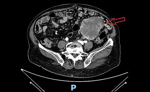
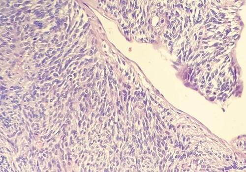
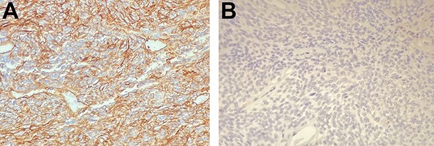
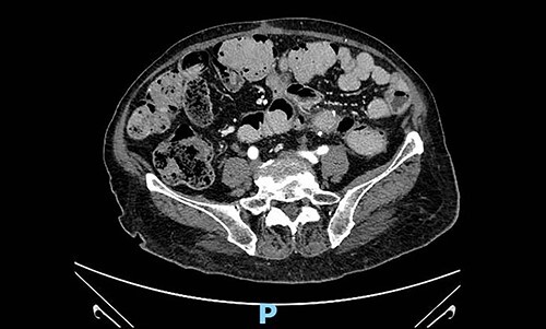
Solitary fibrous tumour (SFT) is a rare mesenchymal tumour, usually originating from the serous surfaces, typically of the pleura and pericardium. However, it can also have localizations in soft tissues and visceral organs. We report the case of a 79-year-old woman affected by mesenteric SFT, localized in the sigmoid colon. We performed open excision of the abovementioned mass en-bloc with the sigmoid colon and left adnexal tissues. Pathological examination of surgical specimen revealed a diagnosis of SFT CD34+, high-risk of metastases. Mesenteric SFTs are even rarer forms of SFT and may be asymptomatic or cause intestinal occlusion. There is no consensus on the management of this tumour. Radical surgical excision is the only curative treatment, while adjuvant therapies are indicated in case of advanced disease. Due to the high metastatic risk of malignant forms, a long follow-up is mandatory in these cases.
Keywords: case-report; mesenchymal neoplasm; oncological surgery; rare neoplasm; solitary fibrous tumour.
Published by Oxford University Press and JSCR Publishing Ltd. All rights reserved. © The Author(s) 2022.
Figures
Similar articles
-
Solitary Fibrous Tumour of the Mesentery: An Uncommon Site for a Rare Tumour.Cureus. 2024 Sep 9;16(9):e69011. doi: 10.7759/cureus.69011. eCollection 2024 Sep. Cureus. 2024. PMID: 39385851 Free PMC article.
-
A malignant solitary fibrous tumour arising from the first lumbar vertebra and mimicking an osteosarcoma: a case report.World J Surg Oncol. 2017 May 11;15(1):100. doi: 10.1186/s12957-017-1161-0. World J Surg Oncol. 2017. PMID: 28494796 Free PMC article.
-
Solitary fibrous tumour of the sigmoid colon mesentery.BMJ Case Rep. 2019 May 8;12(5):e228774. doi: 10.1136/bcr-2018-228774. BMJ Case Rep. 2019. PMID: 31068346 Free PMC article.
-
Solitary fibrous tumour with intramedullary component: case report and review of the literature.Neurol Neurochir Pol. 2014;48(2):144-9. doi: 10.1016/j.pjnns.2013.09.006. Epub 2014 Jan 23. Neurol Neurochir Pol. 2014. PMID: 24821642 Review.
-
Solitary fibrous tumour of the liver-report on metastasis and local recurrence of a malignant case and review of literature.World J Surg Oncol. 2017 Jan 18;15(1):27. doi: 10.1186/s12957-017-1102-y. World J Surg Oncol. 2017. PMID: 28100235 Free PMC article. Review.
Cited by
-
Solitary Fibrous Tumour of the Mesentery: An Uncommon Site for a Rare Tumour.Cureus. 2024 Sep 9;16(9):e69011. doi: 10.7759/cureus.69011. eCollection 2024 Sep. Cureus. 2024. PMID: 39385851 Free PMC article.
References
-
- Goodlad JR, Fletcher CD. Solitary fibrous tumor arising at unusual sites: analysis of a series. Histopathology 1991;19:51222. - PubMed
-
- Gronchi A, Miah AB, Dei Tos AP, Casali PG, Stacchiotti S, on behalf of the ESMO Guidelines Committee, EURACAN and GENTURIS. Soft tissue and visceral sarcomas: ESMO–EURACAN–GENTURIS clinical practice guidelines for diagnosis, treatment and follow-up. Ann Oncol 2021;32:1348–65. - PubMed
-
- Stout AP. Hemangiopericytoma. A study of twenty-five new cases. Cancer 1949;2:1027–54. - PubMed
-
- Fletcher CD, Bridge JA, Pancras CW (eds). World Health Organization Classification of Tumours of Soft Tissue and Bone. Lyon, France: IARC Press, 2013.
-
- Louis DN, Ohgaki H, Wiestler OD, Cavenee WK. WHO Classification of Tumours of the Central Nervous System. World Health Organization Classification of Tumours, 4th edn. Lyon, France: International Agency for Research on Cancer, 2007.
Publication types
LinkOut - more resources
Full Text Sources
Research Materials
