. 2022 Apr 27;96(8):e0027922.
doi: 10.1128/jvi.00279-22. Epub 2022 Mar 30.

PML Body Component Sp100A Restricts Wild-Type Herpes Simplex Virus 1 Infection

Affiliations

PML Body Component Sp100A Restricts Wild-Type Herpes Simplex Virus 1 Infection

Yilei Ma et al. J Virol. .

Abstract

Sp100 (speckled protein 100 kDa) is a constituent component of nuclear structure PML (promyelocytic leukemia) bodies, playing important roles in mediating intrinsic and innate immunity. The Sp100 gene encodes four isoforms with distinct roles in the transcriptional regulation of both cellular and viral genes. Since Sp100 is a primary intranuclear target of infected-cell protein 0 (ICP0), an immediate early E3 ligase encoded by herpes simplex virus 1 (HSV-1), previous investigations attempting to analyze the functions of individual Sp100 variants during HSV-1 infection mostly avoided using a wild-type virus. Therefore, the role of Sp100 under natural infection by HSV-1 remains to be clarified. Here, we reappraised the antiviral capacity of four Sp100 isoforms during infection by a nonmutated HSV-1, examined the molecular behavior of the Sp100 protein in detail, and revealed the following intriguing observations. First, Sp100 isoform A (Sp100A) inhibited wild-type HSV-1 propagation in HEp-2, Sp100-/-, and PML-/- cells. Second, endogenous Sp100 is located in both the nucleus and the cytoplasm. During HSV-1 infection, the nuclear Sp100 level decreased drastically upon the detection of ICP0 in the same subcellular compartment, but cytosolic Sp100 remained stable. Third, transfected Sp100A showed subcellular localizations similar to those of endogenous Sp100 and matched the protein size of endogenous cytosolic Sp100. Fourth, HSV-1 infection induced increased secretion of endogenous Sp100 and ectopically expressed Sp100A, which copurified with extracellular vesicles (EVs) but not infectious virions. Fifth, the Sp100A level in secreting cells positively correlated with its level in EVs, and EV-associated Sp100A restricted HSV-1 in recipient cells. IMPORTANCE Previous studies show that the PML body component Sp100 protein is immediately targeted by ICP0 of HSV-1 in the nucleus during productive infection. Therefore, extensive studies investigating the interplay of Sp100 isoforms with HSV-1 were conducted using a mutant virus lacking ICP0 or in the absence of infection. The role of Sp100 variants during natural HSV-1 infection remains blurry. Here, we report that Sp100A potently and independently inhibited wild-type HSV-1 and that during HSV-1 infection, cytosolic Sp100 remained stable and was increasingly secreted into the extracellular space, in association with EVs. Furthermore, the Sp100A level in secreting cells positively correlated with its level in EVs and the anti-HSV-1 potency of these EVs in recipient cells. In summary, this study implies an active antiviral role of Sp100A during wild-type HSV-1 infection and reveals a novel mechanism of Sp100A to restrict HSV-1 through extracellular communications.

Keywords: EVs; HSV-1; PML; Sp100A; cytosolic Sp100; extracellular vesicle; subcellular localization.


Conflict of interest statement

The authors declare no conflict of interest.

Figures
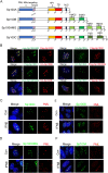
FIG 1
FIG 1
Distinct intranuclear localizations of ectopically expressed Sp100 isoforms. (A) Schematics of protein domains of SP100 splice variants. Destruction-box (D-box), trans-activating region (TR), high mobility group (HMG), bromodomain (BRD) and plant homeodomain (PHD). (B) HEp-2 cells were transfected with plasmids coding for tagged Sp100 isoforms A, B, C, and HMG in pairs. At 24 h posttransfection, cells were fixed, permeabilized, and reacted with antibodies labeled with fluorophores, as indicated. White arrowheads indicate differential intranuclear localizations of Sp100A from the other three Sp100 isoforms. (C to F) HEp-2 cells were transfected with individual plasmids encoding Sp100C (C), Sp100HMG (D), Sp100B (E), or Sp100A (F). At 24 h posttransfection, cells were mock treated (control [Con]) or treated with IFN-β at 1,000 U/mL for 2 h, fixed, permeabilized, and reacted with anti-Flag and anti-PML antibodies. Representative images are shown.
FIG 2
FIG 2
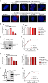
Sp100A inhibited HSV-1 lytic infection at a low multiplicity of infection. (A) Overexpression cell lines of individual Sp100 isoforms (Flag tagged) were established on the background of HEp-2 and Sp100−/− cells by a lentiviral transduction system and confirmed by immunofluorescence staining with antibodies against Flag (red) and PML (green). Nuclei were stained with DAPI. (B) HEp-2 cells and four Sp100 isoform-overexpressing HEp-2 cell lines were infected with HSV-1 at an MOI of 0.01 (left) or an MOI of 5 (right). Cells were collected at 48 hpi, and cell-associated viruses were titrated by a plaque assay. (C) Protein expression of Sp100A in HEp-2+A cells by immunoblotting with anti-Flag antibody. (D) Multicycle growth kinetics of HSV-1 in HEp-2 and HEp-2+A cells at an MOI of 0.01 by plaque titration of the cell-associated viruses. (E to G) Experiment conducted as described above for panels B to D but in Sp100−/− cells and the four Sp100 isoform-overexpressing Sp100−/− cell lines.
FIG 3
FIG 3
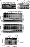
Cytosolic Sp100 remained stable during HSV-1 infection. (A) HEp-2 cells were transfected with plasmids coding for individual Sp100 isoforms (Flag tagged) for 24 h and mock infected or infected with HSV-1 at an MOI of 5. At 8 hpi, cell lysates were collected and immunoblotted with antibodies against Flag and β-actin. (B) HEp-2+A cells were nontreated, treated with 5 μg/mL actinomycin D (ActD) for 2 h, infected with HSV-1 at an MOI of 10 in the presence or absence of ActD, or inoculated with UV-inactivated HSV-1 of the same amount. Subcellular fractions of different groups of cells were collected at 6 hpi, and the protein levels of Sp100A in the nucleus (Nu-Sp100A) and the cytoplasm (Cyto-Sp100A) were analyzed by immunoblotting with polyclonal anti-Sp100 antibody. Histone and GAPDH served as loading controls. (C) HEp-2 cells were infected with HSV-1 at an MOI of 10. Subcellular fractions were collected at the indicated time points postinfection, and protein levels of Sp100, PML, and ICP0 were analyzed by immunoblotting with polyclonal anti-Sp100 antibody and anti-PML and anti-ICP0 antibodies. Histone and GAPDH served as markers for subcellular compartments. Cytosolic Sp100A was loaded at portions and exposed under conditions similar to those of nuclear Sp100. (D) HEp-2 cells were cotransfected with plasmids coding for Flag-Sp100A or GFP and plasmids coding for HA-SUMO1 or HA-SUMO2/3, as indicated. At 24 h posttransfection, Sp100A was immunoprecipitated (IP) down from the cytosolic and nuclear compartments by anti-Flag antibody and immunoblotted (IB) with antibodies against the HA tag and Flag tag. Fivefold more total cytosolic immunoprecipitated lysate was loaded so that cytosolic Sp100A was at an amount similar to that of nuclear Sp100A. The arrow points to the SUMOylated Sp100A band.
FIG 4
FIG 4
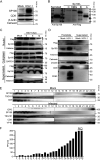
Sp100A was secreted into the extracellular space during HSV-1 infection. (A) HEp-2 cells were mock infected or infected with HSV-1 at an MOI of 0.1. At 50 hpi, culture medium was collected, and cell debris was removed as described in Materials and Methods. Proteins secreted into the extracellular space were concentrated and examined by immunoblotting with polyclonal anti-Sp100 antibody. β-Actin, CD9, and calnexin served as controls. (B) HEp-2 cells were transfected with plasmids coding for individual Flag-tagged Sp100 isoforms for 24 h. Cell lysates were collected; separated in a polyacrylamide gel side by side with the concentrated, cell debris-free extracellular proteins from panel A; and immunoblotted with anti-Flag or anti-Sp100 antibodies, as indicated. (C) HEp-2+A cells were infected with HSV-1 at an MOI of 10. Subcellular fractions and culture medium were collected at the indicated time points and analyzed by immunoblotting with anti-Flag and anti-Sp100 antibodies. Histone, GAPDH, calnexin, and TSG101 served as markers for the origin and purity of the samples. (D) HEp-2+A cells were infected with HSV-1 at an MOI of 10 for 24 h. Extracellular vesicles ranging in sizes between30 and 200 nm in diameter were isolated using a commercial exosome extraction kit according to the manufacturer’s protocol. The protein contents were analyzed by immunoblotting with the indicated antibodies. (E) HEp-2 cells were mock infected or infected with HSV-1 at an MOI of 0.1 for 50 h. Culture medium was collected and processed as described in Materials and Methods. Extracellular vesicles and HSV-1 virions were separated by iodixanol-sucrose gradient-based ultracentrifugation. Twenty-four fractions, each in 500 μL, were collected from top to bottom from the density gradient, labeled 1 to 24. The proteins present in each fraction were analyzed by immunoblotting with the indicated antibodies. (F) Infectious HSV-1 virions in the 24 fractions from panel E were titrated by a plaque assay on Vero cells. The cells exposed to the bottom 2 fractions of the gradient (fractions 23 and 24) were fully infected, so plaques could not be determined. N.D., not determined.
FIG 5
FIG 5
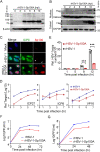
Effect of EVs from Sp100−/−, HEp-2, and HEp-2+A cells on HSV-1 replication in the recipient HEp-2 cells. (A) Sp100−/−, HEp-2, and HEp-2+A cells were mock infected or infected (In) with HSV-1(F) at an MOI of 0.1 for 50 h. Exosome-enriched fractions (fractions 1 to 6 in Fig. 4E) were used. The Sp100 protein level in an equal volume of EVs was examined by immunoblotting with anti-Sp100 antibody. (B and C) A total of 4 × 105 Sp100−/− cells were incubated with an equal volume (500 μL) of EVs from mock-infected or HSV-1-infected Sp100−/−, HEp-2, and HEp-2+A cells without infection (B) or with 4,000 PFU of HSV-1 (MOI of 0.01) (C) at 37°C for 2 h. NC represents negative control. Five hundred microliters of EVs from different infected groups was contaminated with on average 7 to 173 PFU (EVs from infected HEp-2 cells, 172 to 173 PFU/500 μL; EVs from infected SP100−/− cells, 7 to 8 PFU/500 μL; EVs from infected HEp-2+A cells, not detectable). (B) Cells were extensively washed and collected for immunoblotting with antibodies against Sp100 and β-actin. (C) At 24 hpi, cell-associated viruses were titrated by a plaque assay.
FIG 6
FIG 6
rHSV-1-Sp100A was attenuated compared to rHSV-1. (A and B) Protein levels of Sp100A in rHSV-1-Sp100A-infected HEp-2 cells (MOI of 10) at the indicated time points postinfection were examined by immunoblotting with anti-Flag antibody, in whole-cell lysates (A) or subcellular fractions (B). (C) HEp-2 cells were mock infected or infected with rHSV-1 or rHSV-1-Sp100A at an MOI of 10. At 12 hpi, cells were fixed and stained with anti-Flag (red) and anti-ICP0 (green) antibodies. White arrowheads mark the faint cytosolic staining of proteins reactive to anti-Sp100 antibody. (D to F) HEp-2 cells were infected with rHSV-1-Sp100A at an MOI of 0.01. (D) Expression levels of the representative α (ICP27), β (ICP8), and γ (VP16) genes at the indicated time points were quantified by quantitative PCR (qPCR). (E) Viral DNA levels at the indicated time points postinfection were quantified by qPCR using primers targeting the ICP27 gene region. (F) Growth kinetics of rHSV-1 and rHSV-1-Sp100A in HEp-2 cells at an MOI of 0.01 by a plaque assay. (G) Growth kinetics of rHSV-1 and rHSV-1-Sp100A in PML−/− cells at an MOI of 0.01 by a plaque assay.

Similar articles

Cited by

References

    1. Szostecki C, Guldner HH, Netter HJ, Will H. 1990. Isolation and characterization of cDNA encoding a human nuclear antigen predominantly recognized by autoantibodies from patients with primary biliary cirrhosis. J Immunol 145:4338–4347. - PubMed
    1. Guldner HH, Szostecki C, Grotzinger T, Will H. 1992. IFN enhance expression of Sp100, an autoantigen in primary biliary cirrhosis. J Immunol 149:4067–4073. - PubMed
    1. Grotzinger T, Jensen K, Will H. 1996. The interferon (IFN)-stimulated gene Sp100 promoter contains an IFN-gamma activation site and an imperfect IFN-stimulated response element which mediate type I IFN inducibility. J Biol Chem 271:25253–25260. 10.1074/jbc.271.41.25253. - DOI - PubMed
    1. Puvion-Dutilleul F, Venturini L, Guillemin MC, de The H, Puvion E. 1995. Sequestration of PML and Sp100 proteins in an intranuclear viral structure during herpes simplex virus type 1 infection. Exp Cell Res 221:448–461. 10.1006/excr.1995.1396. - DOI - PubMed
    1. Chelbi-Alix MK, de The H. 1999. Herpes virus induced proteasome-dependent degradation of the nuclear bodies-associated PML and Sp100 proteins. Oncogene 18:935–941. 10.1038/sj.onc.1202366. - DOI - PubMed

MeSH terms