. 2022 Apr 27;96(8):e0025022.
doi: 10.1128/jvi.00250-22. Epub 2022 Mar 30.

Possible Cross-Reactivity of Feline and White-Tailed Deer Antibodies against the SARS-CoV-2 Receptor Binding Domain

Affiliations

Possible Cross-Reactivity of Feline and White-Tailed Deer Antibodies against the SARS-CoV-2 Receptor Binding Domain

Trevor J Hancock et al. J Virol. .

Abstract

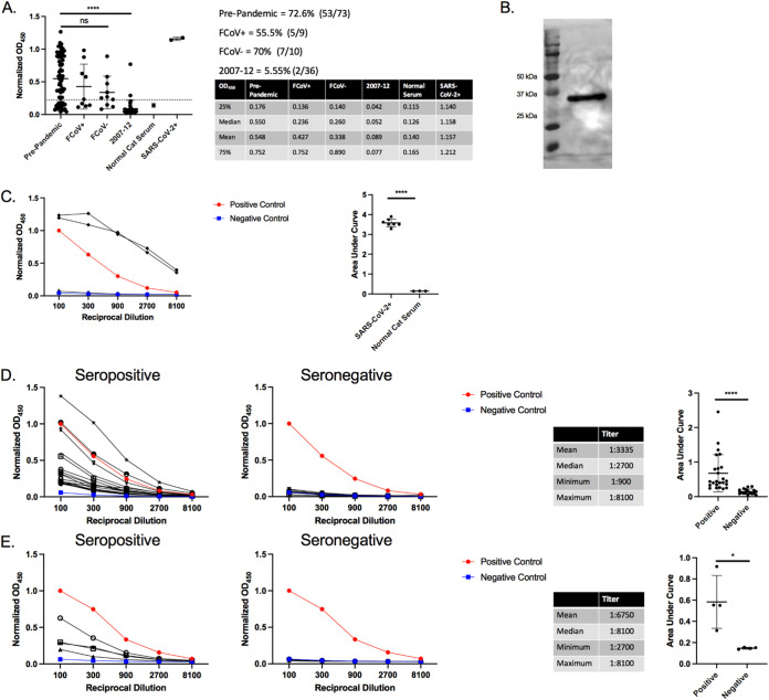
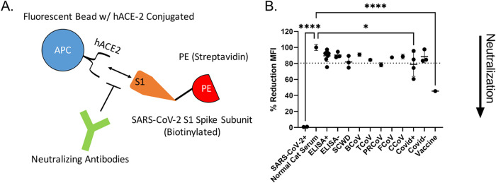
In late 2019, a novel coronavirus began circulating within humans in central China. It was designated SARS-CoV-2 because of its genetic similarities to the 2003 SARS coronavirus (SARS-CoV). Now that SARS-CoV-2 has spread worldwide, there is a risk of it establishing new animal reservoirs and recombination with native circulating coronaviruses. To screen local animal populations in the United States for exposure to SARS-like coronaviruses, we developed a serological assay using the receptor binding domain (RBD) from SARS-CoV-2. SARS-CoV-2's RBD is antigenically distinct from common human and animal coronaviruses, allowing us to identify animals previously infected with SARS-CoV or SARS-CoV-2. Using an indirect enzyme-linked immunosorbent assay (ELISA) for SARS-CoV-2's RBD, we screened serum from wild and domestic animals for the presence of antibodies against SARS-CoV-2's RBD. Surprisingly prepandemic feline serum samples submitted to the University of Tennessee Veterinary Hospital were ∼50% positive for anti-SARS RBD antibodies. Some of these samples were serologically negative for feline coronavirus (FCoV), raising the question of the etiological agent generating anti-SARS-CoV-2 RBD cross-reactivity. We also identified several white-tailed deer from South Carolina with anti-SARS-CoV-2 antibodies. These results are intriguing, as cross-reactive antibodies toward SARS-CoV-2 RBD have not been reported to date. The etiological agent responsible for seropositivity was not readily apparent, but finding seropositive cats prior to the current SARS-CoV-2 pandemic highlights our lack of information about circulating coronaviruses in other species. IMPORTANCE We report cross-reactive antibodies from prepandemic cats and postpandemic South Carolina white-tailed deer that are specific for that SARS-CoV RBD. There are several potential explanations for this cross-reactivity, each with important implications to coronavirus disease surveillance. Perhaps the most intriguing possibility is the existence and transmission of an etiological agent (such as another coronavirus) with similarity to SARS-CoV-2's RBD region. However, we lack conclusive evidence of prepandemic transmission of a SARS-like virus. Our findings provide impetus for the adoption of a One Health Initiative focusing on infectious disease surveillance of multiple animal species to predict the next zoonotic transmission to humans and future pandemics.

Keywords: ELISA; RBD; SARS-CoV-2; antibodies; bovine; canine; coronavirus; cross-reactive; feline; white-tailed deer.


Conflict of interest statement

The authors declare no conflict of interest.

Figures

FIG 1
FIG 1
Anti-SARS-CoV-2 ELISA sensitivity and specificity. (A) Cross reactivity of anti-CoV antibodies against SARS-CoV-2 RBD. Polyclonal sera from guinea pigs immunized with common animal coronaviruses (turkey coronavirus, TCoV; porcine respiratory coronavirus, PRCoV; canine coronavirus, CCoV; feline coronavirus, FCoV; bovine coronavirus, BCoV) were used in a SARS-CoV-2 RBD indirect ELISA. Positive samples consisted of polyclonal serum from a SARS-CoV-2-infected patient and a monoclonal antibody to SARS-CoV (CR3022). The negative-control group comprised prepandemic human sera. Secondary antibodies were either anti-human IgG (1:10,000) (Rockland Immunochemicals, USA) or anti-guinea pig IgG (1:10,000) (Life Technologies Corp., USA). Bars represent the mean and standard deviation (n > 3 for all samples). (B) ELISA validation using 66 human COVID-positive plasma and 22 negative serum samples. Human antibodies against the SARS-CoV-2 RBD were detected with an indirect RBD-specific ELISA. Secondary antibody was the anti-human IgG (1:10,000) (Rockland Immunochemicals, USA). ROC analysis determined the positive OD450 cutoff value (dashed line). Positive plasma samples were donated by COVID-recovered patients, and prepandemic serum samples were the negative controls. Based on the experimentally determined cutoff value, 64 of the 66 positive samples were anti-RBD positive, giving a sensitivity value of 96.96%. All but one of the negative samples were below the cutoff value for a specificity of 95.45%. The adjacent tables list the first and third quartiles along with mean and median OD450 values of COVID-positive and -negative human samples. Bars represent the mean and standard deviation (n > 3). (C) Anti-6×His Western blot on HEK-293T17 purified RBD from 5% serum conditions. Samples were run on a 12% denaturing SDS-PAGE gel. Protein was transferred to nitrocellulose and probed with anti-6×His antibody at 1:10,000 (Proteintech, USA). White light and chemiluminescent images were overlaid and from left to right, are ladder (lane 1) and purified RBD (lane 2). (D) Silver stain of purified recombinant SARS-Cov-2 RBD produced in HEK-293T17. From left to right, ladder, HEK-RBD under 5% serum conditions, HEK-RBD from 2% serum conditions. Samples were denatured and run on a 12% SDS-PAGE gel and silver-stained (Thermo Fisher Scientific, USA). For panels B and C, representative data are shown.
FIG 2
FIG 2
Prepandemic feline antibodies cross-react with SARS-CoV-2 RBD. (A) ELISA results of cat serum RBD reactivity. A total of 93 prepandemic feline serum samples were tested for reactivity in our anti-RBD ELISA with anti-felid IgG secondary antibody (1:10,000) (Invitrogen, USA). Cutoff values were determined by receiver operator curve (ROC) analysis. OD450 values for samples in each group were plotted, with the dotted line representing the positive threshold. Two sets of prepandemic cat samples were collected. Prepandemic cat convenience samples (n = 73) were collected in local clinics and sent to the University of Tennessee for diagnostic testing or during feral cat studies (2007 to 2012) (n = 36). Prepandemic convenience samples were subdivided into feline coronavirus-positive (FCoV+) and -negative (FCoV–) subgroups. Normal cat serum (Jackson ImmunoResearch Laboratories, USA) serves as the negative control, and SARS-CoV-2+ serum from two cats experimentally inoculated with SARS-CoV-2 serves as positive controls. The side table lists the first and third quartiles and mean and median OD450 values for all samples. Bars represent the mean ± standard deviation (n > 3 for all samples). (B) Western blot of purified RBD using serum from a single positive cat sample. Purified RBD was run under denaturing conditions and blotted onto nitrocellulose. The RBD blot was first probed with cat serum from an ELISA positive sample (1:20 dilution) followed by anti-felid IgG-HRP conjugated antibody (1:10,000 dilution) (Invitrogen, USA). White light and chemiluminescent images were overlaid. Lane 1 is the molecular weight ladder and lane 2 is purified RBD. (C to E) Titration of seropositive and seronegative serums assessed via RBD ELISA. OD450 values were plotted against the reciprocal dilution. Samples were considered positive if they were 3 standard deviations above the negative average for each dilution. Anti-RBD titer was designated the last dilution above the negative cutoff. Positive controls were human COVID-positive serum, and negative controls were normal human and cat serum (Jackson ImmunoResearch Laboratories, USA). Statistics for the positive sample titrations are included in the table along with AUC analysis. (C) Serum from two SARS-CoV-2-infected cats (red circles) and normal cat serum (blue squares) were titrated in an anti-RBD ELISA. (D) Titration of 17 seropositive and 10 seronegative, prepandemic cat samples. (E) Titration of four seropositive and seronegative cat samples collected from 2007 to 2012. For panels A and B, representative data are shown. For panel A, Tukey’s one-way ANOVA with multiple comparisons was performed. For panels C, D, and E AUC analysis and Student’s one-tailed t test with Welch’s correction were performed. *, P < 0.05; **, P < 0.01; ***, P < 0.001.
FIG 3
FIG 3
Dog serum cross-reacts to a copurified protein. (A) Anti-SARS-CoV-2 RBD ELISA with dog serum. Serum from 36 client-owned and two purpose-bred research dogs were tested in an anti-RBD ELISA with anti-canine IgG secondary HRP (1:10,000) (Bethyl Laboratories, USA). The table to the right lists the first and third quartile, median, and mean OD450 values for all samples. Bars represent the mean ± standard deviation (n > 3). (B) Western blot of purified RBD using serum from a positive dog sample. Purified RBD was probed with dog serum from an ELISA positive sample (1:20 dilution) followed by anti-canine IgG HRP (1:10,000 dilution) (Bethyl Laboratories, USA). White light and chemiluminescent images were overlaid. Lane 1 (from left to right), ladder; lane 2, purified RBD. For all figures, representative data are shown.
FIG 4
FIG 4
Serological testing of other regional animals. (A). Anti-SARS-CoV-2 RBD ELISA with bovine, elk, tiger, and deer serum; 33 prepandemic East Tennessee cows, 12 postpandemic East Tennessee elk, 9 prepandemic East Tennessee tigers, and 22 postpandemic South Carolina deer serum samples were tested for anti-RBD antibodies. Species-specific secondary antibodies were used at the following dilutions: anti-bovine, 1:250 (Bethyl Laboratories, USA); anti-elk/deer, 1:250 (KPL, USA); anti-tiger/cat, 1:10,000 (Invitrogen, USA); and anti-deer, 1:250 (KPL, USA). Bars represent the mean ± standard deviation (n > 3 for all samples). (B) Titration of two seropositive (red circles) and four seronegative (blue squares) deer samples. OD450 values are plotted against the reciprocal dilution of each sample. Samples were considered positive if they were 3 standard deviations above the negative average for each dilution. Positive and negative controls were human COVID-positive and -negative samples, respectively. Statistics for the positive sample titrations are included in the table. The AUC analysis for titrations of deer ELISA positive and negative samples is shown to the right. For all figures, representative data are shown. For AUC analysis Student’s one-tailed t test with Welch’s correction was performed. *, P < 0.05; **, P < 0.01; ***, P < 0.001.
FIG 5
FIG 5
Neutralization assays. (A) Schematic of the neutralization assay. Neutralization is measured as the decrease in binding of phycoerythrin (PE)-labeled SARS-CoV-2 S1 subunit to human ACE-2 conjugated beads. Addition of neutralizing antibodies results in a decreased mean fluorescent intensity (MFI) as measured by flow cytometry. (B) Neutralization of SARS-CoV-2 S1 subunit interaction with hACE2. Serum from several ELISA positive and negative cats (ELISA+ and ELISA–, respectively), serum from South Carolina white-tailed deer (SCWD, 2 ELISA positive and 1 negative), mice immunized with other common coronaviruses (BCoV, bovine coronavirus; TCoV, turkey coronavirus; PRCoV, porcine respiratory coronavirus; FCoV, feline coronavirus; CCoV, canine coronavirus), and human serum samples (Covid+, convalescent plasma from Covid+ humans; Covid–, pre-SARS-CoV-2 serum samples; vaccine, serum postvaccination against SARS-CoV-2 spike protein) were used. SARS-CoV-2-infected cats and normal cat serum served as positive and negative controls, respectively. Data were normalized to normal cat serum representing 100% binding of SARS-CoV-2 S1 subunit to hACE2 beads. ROC analysis was used to generate a positive reduction threshold (dotted line). Each point is an average of 2 replicates. Tukey’s one-way ANOVA with multiple comparisons was used to analyze experimental groups. *, P < 0.05; ****, P < 0.0001.
FIG 6
FIG 6
Pan-coronavirus screen of East Tennessee felines. Fecal samples from healthy cats were collected and screened for conserved coronavirus sequences. Phylogenetic trees were generated consisting of the following common human and animal coronaviruses: CRCoV (canine respiratory coronavirus), BCoV (bovine coronavirus), OC43 (human beta-coronavirus), MHV (murine hepatitis virus), MERS-CoV (Middle East respiratory coronavirus), SARS-CoV-2 (severe acute respiratory coronavirus-2), SARS-CoV (severe acute respiratory coronavirus), AvianCoV (duck coronavirus), NL63 (human alphacoronavirus), 229E (human alphacoronavirus), TGEV (transmissible gastroenteritis virus), PRCoV (porcine respiratory coronavirus), FCoV (feline coronavirus strains UU19, Felix, RM, Black, and UU8), CCov (canine coronavirus), HKU15 (porcine delta-coronavirus), as well as a locally identified coronavirus (CP-20-26). Sequences from five coronavirus loci were independently aligned, trimmed, and concatenated. Concatenated sequences were aligned, and phylogenetic trees were generated with the maximum-likelihood method with bootstrap analysis in MEGA X. Bootstrap values for each branch are shown with lengths to scale. Coronavirus lineages are annotated on the tree.
FIG 7
FIG 7
RBD coronavirus screen of East Tennessee felines. (top) Fecal samples from healthy cats were collected and screened for S1/RBD coronavirus sequences. Phylogenetic tree consisting of common human and animal coronaviruses: CRCoV (canine respiratory coronavirus), BcoV (bovine coronavirus), OC43 (human betacoronavirus), MHV (murine hepatitis virus), MERS-CoV (Middle East respiratory coronavirus), SARS-CoV-2 (severe acute respiratory coronavirus-2), SARS-CoV (severe acute respiratory coronavirus), AvianCoV (duck coronavirus), NL63 (human alpha-coronavirus), 229E (human alphacoronavirus), TGEV (transmissible gastroenteritis virus), PRCoV (porcine respiratory coronavirus), FcoV (feline coronavirus strains UU19, Felix, RM, Black, and UU8), CCoV (canine coronavirus), HKU15 (porcine deltacoronavirus), as well as locally identified coronaviruses (CP-20-13, CP-20-19, and CP-20-23). Sequences from the S1 region were aligned and trimmed. Maximum-Likelihood phylogenetic trees were generated with bootstrap analysis in MEGA X. Bootstrap values for each branch are shown with lengths to scale. (Bottom) Percent similarity matrix of select coronaviruses. Aligned and truncated RBD regions from the shown coronaviruses were analyzed via Clustal Omega to determine the percent identity matrix.

Similar articles

Cited by

References

    1. Poon LLM, Peiris M. 2020. Emergence of a novel human coronavirus threatening human health. Nat Med 26:317–319. 10.1038/s41591-020-0796-5. - DOI - PMC - PubMed
    1. Guan W-J, Ni Z-Y, Hu Y, Liang W-H, Ou C-Q, He J-X, Liu L, Shan H, Lei C-L, Hui DSC, Du B, Li L-J, Zeng G, Yuen K-Y, Chen R-C, Tang C-L, Wang T, Chen P-Y, Xiang J, Li S-Y, Wang J-L, Liang Z-J, Peng Y-X, Wei L, Liu Y, Hu Y-H, Peng P, Wang J-M, Liu J-Y, Chen Z, Li G, Zheng Z-J, Qiu S-Q, Luo J, Ye C-J, Zhu S-Y, Zhong N-S, China Medical Treatment Expert Group for Covid-19. 2020. Clinical characteristics of coronavirus disease 2019 in China. N Engl J Med 382:1708–1720. 10.1056/NEJMoa2002032. - DOI - PMC - PubMed
    1. Latinne A, Hu B, Olival KJ, Zhu G, Zhang L, Li H, Chmura AA, Field HE, Zambrana-Torrelio C, Epstein JH, Li B, Zhang W, Wang L-F, Shi Z-L, Daszak P. 2020. Origin and cross-species transmission of bat coronaviruses in China. Nat Commun 11:4235. 10.1038/s41467-020-17687-3. - DOI - PMC - PubMed
    1. Li X, Giorgi EE, Marichannegowda MH, Foley B, Xiao C, Kong X-P, Chen Y, Gnanakaran S, Korber B, Gao F. 2020. Emergence of SARS-CoV-2 through recombination and strong purifying selection. Sci Adv 6:eabb9153. 10.1126/sciadv.abb9153. - DOI - PMC - PubMed
    1. Andersen KG, Rambaut A, Lipkin WI, Holmes EC, Garry RF. 2020. The proximal origin of SARS-CoV-2. Nat Med 26:450–452. 10.1038/s41591-020-0820-9. - DOI - PMC - PubMed

MeSH terms

Substances

Supplementary concepts

LinkOut - more resources