. 2022 Mar 3;14(3):514.
doi: 10.3390/v14030514.

Pseudorabies Virus Inhibits Expression of Liver X Receptors to Assist Viral Infection

Affiliations

Pseudorabies Virus Inhibits Expression of Liver X Receptors to Assist Viral Infection

Yi Wang et al. Viruses. .

Abstract

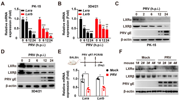
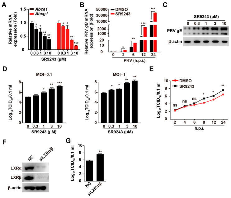
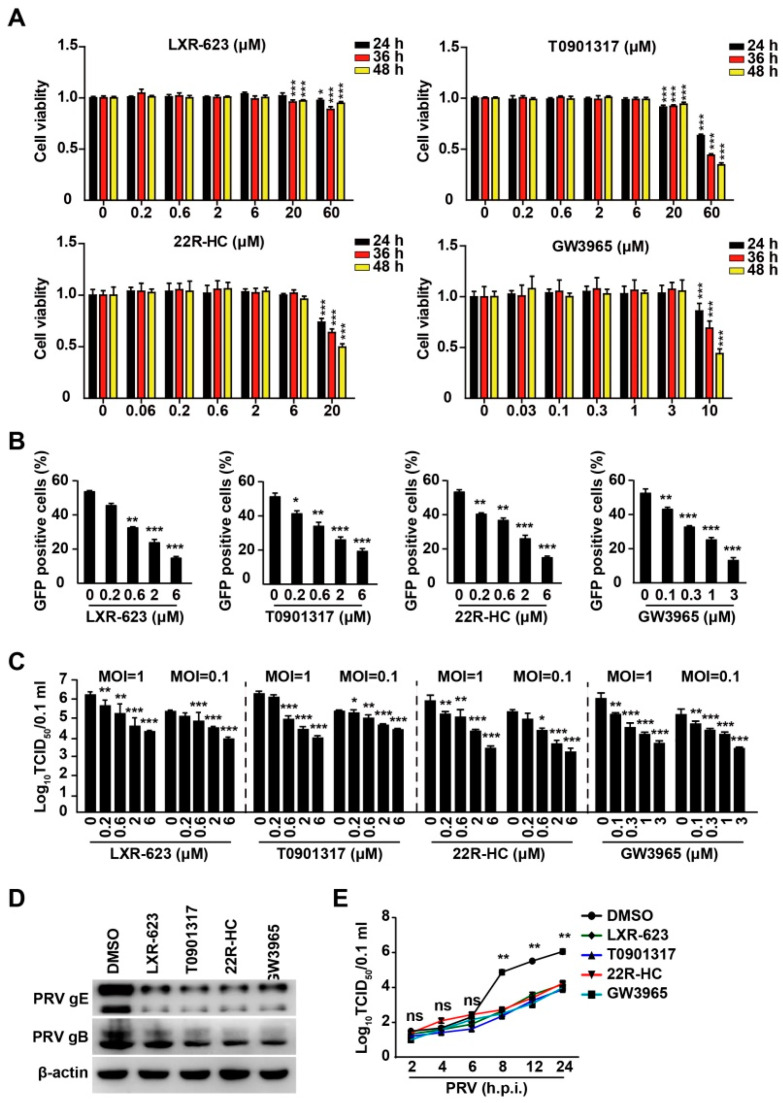
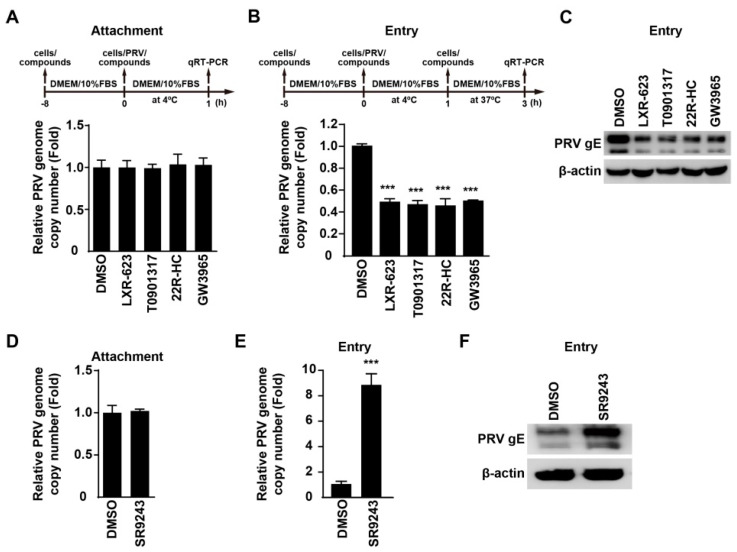
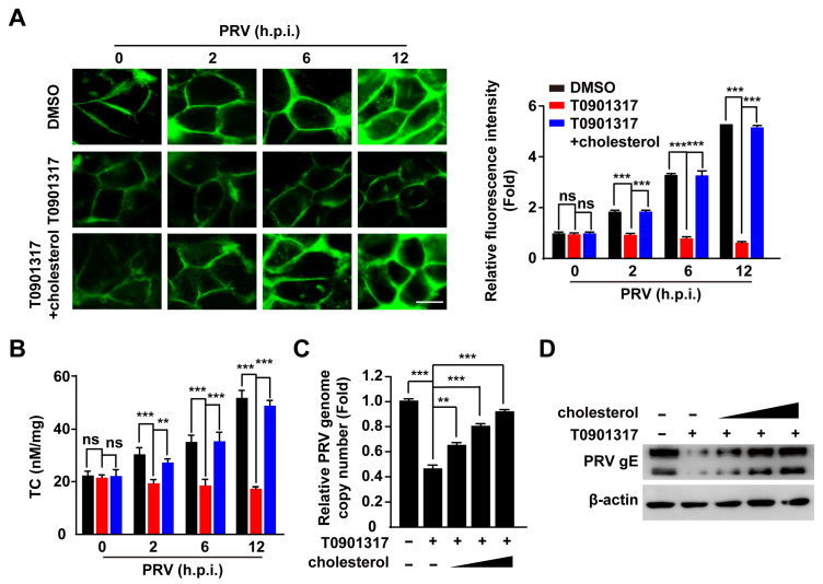
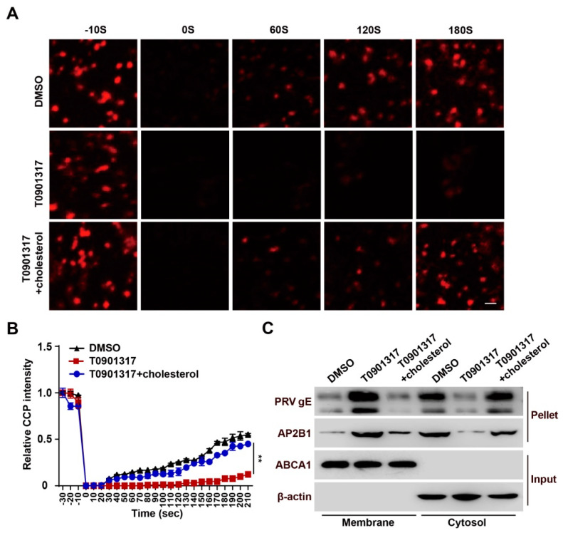
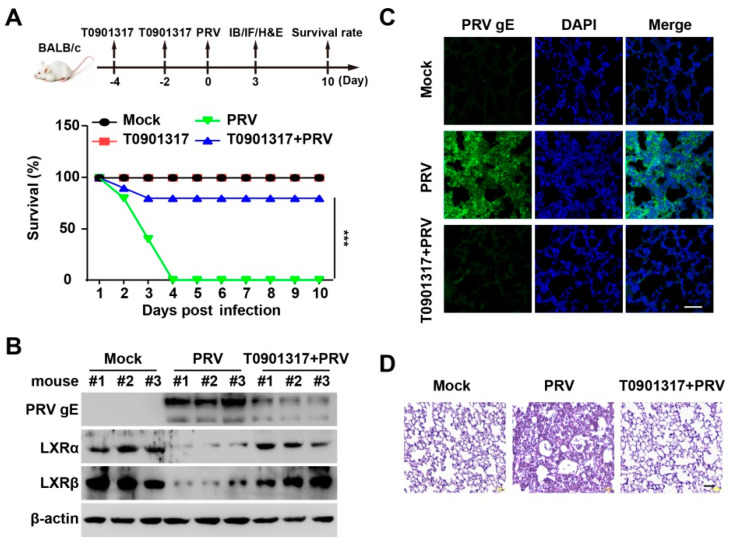
Pseudorabies virus (PRV) is a contagious herpesvirus that causes Aujeszky's disease and economic losses worldwide. Liver X receptors (LXRs) belong to the nuclear receptor superfamily and are critical for the control of lipid homeostasis. However, the role of LXR in PRV infection has not been fully established. In this study, we found that PRV infection downregulated the mRNA and protein levels of LXRα and LXRβ in vitro and in vivo. Furthermore, we discovered that LXR activation suppressed PRV proliferation, while LXR inhibition promoted PRV proliferation. We demonstrated that LXR activation-mediated reduction of cellular cholesterol was critical for the dynamics of PRV entry-dependent clathrin-coated pits. Replenishment of cholesterol restored the dynamics of clathrin-coated pits and PRV entry under LXR activation conditions. Interestingly, T0901317, an LXR agonist, prevented PRV infection in mice. Our results support a model that PRV modulates LXR-regulated cholesterol metabolism to facilitate viral proliferation.

Keywords: Liver X receptors; clathrin-coated pits; pseudorabies virus; viral entry.


Conflict of interest statement

The authors declare that they have no conflict of interest.

Figures

Figure 1
Figure 1
PRV infection downregulates LXR expression. (A,B) PK-15 (A) and 3D4/21 (B) cells were infected with PRV-QXX (MOI = 0.1) for 0–24 h. The mRNA levels of Lxra and Lxrb were assessed by qRT-PCR analysis. * p < 0.05, ** p < 0.01, *** p < 0.001. (C,D) PK-15 (C) and 3D4/21 (D) cells were infected with PRV-QXX (MOI = 0.1) for 0–24 h. LXRα, LXRβ, and PRV gE were assessed by immunoblotting analysis. (E) Mice were mock infected or intranasally infected with PRV-QXX (5 × 10³ TCID50/50 μL per mouse) for 3 days. The mRNA levels of Lxra and Lxrb in the lung were assessed by qRT-PCR analysis (n = 4 per group). * p < 0.05. (F) Mice were treated as in G. LXRα, LARβ, and PRV gE in the lung were assessed by immunoblotting analysis (n = 4 per group).
Figure 2
Figure 2
LXR inverse agonist SR9243 promotes PRV proliferation. (A) PK-15 cells were treated with SR9243 (0–10 µM) for 24 h. Abca1 and Abcg1 mRNA was assessed by qRT-PCR analysis. * p < 0.05, ** p < 0.01, *** p < 0.001. (B) PK-15 cells were infected with PRV-QXX (MOI = 0.1) and simultaneously treated with DMSO or SR9243 (10 µM) for 0–24 h. PRV gB mRNA was assessed by qRT-PCR analysis. * p < 0.05, ** p < 0.01, *** p < 0.001. (C) PK-15 cells were infected with PRV-QXX (MOI = 0.1) and simultaneously treated with SR9243 (0–10 µM) for 24 h. PRV gE was assessed by immunoblotting analysis. (D) PK-15 cells were infected with PRV-QXX (MOI = 0.1 and 1) and simultaneously treated with SR9243 (0–10 µM) for 24 h. Viral titers were assessed by a TCID50 assay. * p < 0.05, ** p < 0.01, *** p < 0.001. (E) PK-15 cells were infected with PRV-QXX (MOI = 0.1) and simultaneously treated with SR9243 (10 µM) for 2–24 h. One-step growth curves of PRV-QXX were assessed using a TCID50 assay of viral titers. * p < 0.05, ** p < 0.01. ns, no significance. (F) PK-15 cells were transfected with NC and siLXRα/β for 48 h. LXRα and LXRβ were assessed by immunoblotting analysis. (G) PK-15 cells were transfected with NC and siLXRα/β. At 24 h post-transfection, cells were infected with PRV-QXX (MOI = 1) for another 24 h. Viral titers were assessed by a TCID50 assay. ** p < 0.01.
Figure 3
Figure 3
LXR agonists inhibit PRV infection. (A) PK-15 cells were treated with LXR-623, T0901317, 22R-HC, and GW3965 at indicated concentrations for 24–48 h. Cell viability was assessed with CCK-8 cell counting assays. * p < 0.05, ** p < 0.01, *** p < 0.001. (B) PK-15 cells were infected with PRV-GFP (MOI = 0.01) and simultaneously treated with LXR-623 (0–6 μM), T0901317 (0–6 μM), 22R-HC (0–6 μM), and GW3965 (0–3 μM) for 36 h. GFP-positive cells were analyzed by flow cytometry. * p < 0.05, ** p < 0.01, *** p < 0.001. (C) PK-15 cells were infected with PRV-QXX (MOI = 0.1 and 1) and simultaneously treated with LXR-623 (0–6 μM), T0901317 (0–6 μM), 22R-HC (0–6 μM) and GW3965 (0–3 μM) for 24 h. Viral titers were assessed by a TCID50 assay. * p < 0.05, ** p < 0.01, *** p < 0.001. (D) PK-15 cells were infected with PRV-QXX (MOI = 0.1) and simultaneously treated with DMSO, LXR-623 (6 μM), T0901317 (6 μM), 22R-HC (6 μM), and GW3965 (3 μM) for 24 h. PRV gB and gE were assessed by immunoblotting analysis. (E) PK-15 cells were infected with PRV-QXX (MOI = 0.1) and simultaneously treated with LXR-623 (6 μM), T0901317 (6 μM), 22R-HC (6 μM) and GW3965 (3 μM) for 2–24 h. One-step growth curves of PRV-QXX were assessed using a TCID50 assay of viral titers. ** p < 0.01. ns, no significance.
Figure 4
Figure 4
Activation of LXR inhibits PRV entry. (A) PK-15 cells were pretreated with DMSO, LXR-623 (6 μM), T0901317 (6 μM), 22R-HC (6 μM) and GW3965 (3 μM) for 8 h at 37 °C. Cells were incubated with PRV-QXX (MOI = 0.1) combined with DMSO, LXR-623 (6 μM), T0901317 (6 μM), 22R-HC (6 μM), and GW3965 (3 μM) for 1 h at 4 °C. After three washes with ice-cold PBS, the viral genome was isolated. PRV genome copy numbers on cells were assessed by qRT-PCR analysis. (B) PK-15 cells were pretreated with DMSO, LXR-623 (6 μM), T0901317 (6 μM), 22R-HC (6 μM), and GW3965 (3 μM) for 8 h at 37 °C. Cells were incubated with PRV-QXX (MOI = 0.1) combined with DMSO, LXR-623 (6 μM), T0901317 (6 μM), 22R-HC (6 μM), and GW3965 (3 μM) for 1 h at 4 °C. After three washes with ice-cold PBS, cells were cultured in prewarmed medium containing DMSO, LXR-623 (6 μM), T0901317 (6 μM), 22R-HC (6 μM), and GW3965 (3 μM) for 2 h at 37 °C. PRV genome copy numbers in cells were assessed by qRT-PCR analysis. *** p < 0.001. (C) PK-15 cells were treated as in (B). PRV gE in cells was assessed by immunoblotting analysis. (D) PK-15 cells were pretreated with DMSO and SR9243 (10 µM) for 8 h at 37 °C. Cells were then incubated with PRV-QXX (MOI = 0.1) combined with DMSO and SR9243 (10 µM) for 1 h at 4 °C. After three washes with ice-cold PBS, the viral genome was isolated. PRV genome copy numbers on cells were assessed by qRT-PCR analysis. (E) PK-15 cells were pretreated with DMSO and SR9243 (10 µM) for 8 h at 37 °C. Cells were incubated with PRV-QXX (MOI = 0.1) combined with DMSO and SR9243 (10 µM) for 1 h at 4 °C. After three washes with ice-cold PBS, cells were cultured in prewarmed medium containing DMSO and SR9243 (10 µM) for 2 h at 37 °C. PRV genome copy numbers in cells were assessed by qRT-PCR analysis. *** p < 0.001. (F) PK-15 cells were infected and treated as in (E). PRV gE in cells was assessed by immunoblotting analysis.
Figure 5
Figure 5
Activation of LXR decreases cellular cholesterol to inhibit PRV entry. (A) PK-15 cells were infected with PRV-QXX (MOI = 0.1) and simultaneously treated with DMSO, T0901317 (6 μM), and T0901317 (6 μM) + cholesterol (0.003 μg/mL) for 0–12 h. Cholesterol was detected by filipin staining (left). Quantification of the relative fluorescence intensity of filipin is shown on the right. *** p < 0.001. ns, no significance. Scale bar: 10 μm. (B) PK-15 cells were treated as in (A). Quantification of cellular cholesterol was performed by biochemical determination. ** p < 0.01, *** p < 0.001. ns, no significance. (C) PK-15 cells were incubated with PRV-QXX (MOI = 0.1) for 1 h at 4 °C and then in medium containing T0901317 (6 μM) and cholesterol (0, 0.0003, 0.001, and 0.003 μg/mL) as indicated for 2 h at 37 °C. PRV genome copy numbers in cells were assessed by qRT-PCR analysis. ** p < 0.01, *** p < 0.001. (D) PK-15 cells were infected and treated as in (C). PRV gE in cells was assessed by immunoblotting analysis.
Figure 6
Figure 6
T0901317 blocks CCP dynamics-dependent viral entry. (A) HeLa cells were transfected with AP2B1-mCherry plasmid for 24 h followed by treatment with DMSO, T0901317 (6 μM) and T0901317 (6 μM) + cholesterol (0.003 μg/mL) for a further 8 h. The CCP dynamics were assessed by FRAP analysis. Scale bar: 1 μm. (B) Quantification of the relative fluorescent intensity of AP2B1 puncta in the FRAP region over time from (A) (n = 10). ** p < 0.01. (C) PK-15 cells were incubated with PRV-QXX (MOI = 0.1) for 1 h at 4 °C and then in medium containing DMSO T0901317 (6 μM) and T0901317 (6 μM) + cholesterol (0.003 μg/mL) at 37 °C. The internalization of PRV gE and AP2B1 was assessed by cell surface biotinylation assay after viral entry for 15 min.
Figure 7
Figure 7
T0901317 inhibits PRV infection in vivo. (A) Mice were intraperitoneally injected with vehicle or T0901317 (30 mg/kg) on days 4 and 2. On day 0, the mice were mock infected or intranasally infected with PRV-QXX (5 × 10³ TCID50/50 μL per mouse). The survival rate was monitored daily for 10 days (n = 12 per group). *** p < 0.001. (B) PRV gE, LXRα, and LXRβ in the lungs were assessed by immunoblotting analysis at 3 days post-infection (n = 3 per group). (C) PRV gE in the lungs was assessed by immunofluorescence at 3 days post-infection (n = 3 per group). Scale bar: 100 μm. (D) The lung injury was assessed by H&E staining at 3 days post-infection (n = 3 per group). Scale bar: 100 μm.

Similar articles

Cited by

References

    1. Pomeranz L.E., Reynolds A.E., Hengartner C.J. Molecular biology of pseudorabies virus: Impact on neurovirology and veterinary medicine. Microbiol. Mol. Biol. Rev. 2005;69:462–500. doi: 10.1128/MMBR.69.3.462-500.2005. - DOI - PMC - PubMed
    1. Wozniakowski G., Samorek-Salamonowicz E. Animal herpesviruses and their zoonotic potential for cross-species infection. Ann. Agric. Environ. Med. 2015;22:191–194. doi: 10.5604/12321966.1152063. - DOI - PubMed
    1. Ai J.W., Weng S.S., Cheng Q., Cui P., Li Y.J., Wu H.L., Zhu Y.M., Xu B., Zhang W.H. Human Endophthalmitis Caused By Pseudorabies Virus Infection, China, 2017. Emerg. Infect. Dis. 2018;24:1087–1090. doi: 10.3201/eid2406.171612. - DOI - PMC - PubMed
    1. Yang X., Guan H., Li C., Li Y., Wang S., Zhao X., Zhao Y., Liu Y. Characteristics of human encephalitis caused by pseudorabies virus: A case series study. Int. J. Infect. Dis. 2019;87:92–99. doi: 10.1016/j.ijid.2019.08.007. - DOI - PubMed
    1. Wong G., Lu J., Zhang W., Gao G.F. Pseudorabies virus: A neglected zoonotic pathogen in humans? Emerg. Microbes Infect. 2019;8:150–154. doi: 10.1080/22221751.2018.1563459. - DOI - PMC - PubMed

Publication types

LinkOut - more resources