

SARS-CoV-2 Spike Protein and Mouse Coronavirus Inhibit Biofilm Formation by Streptococcus pneumoniae and Staphylococcus aureus
- PMID: 35328711
- PMCID: PMC8950232
- DOI: 10.3390/ijms23063291
SARS-CoV-2 Spike Protein and Mouse Coronavirus Inhibit Biofilm Formation by Streptococcus pneumoniae and Staphylococcus aureus
Abstract
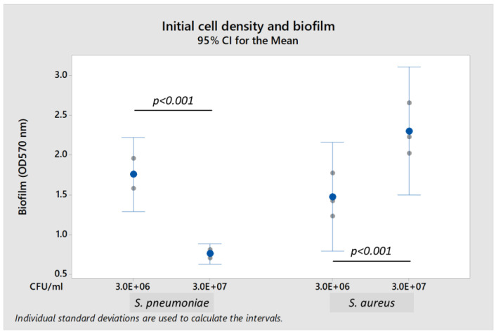
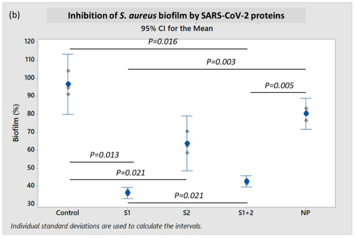
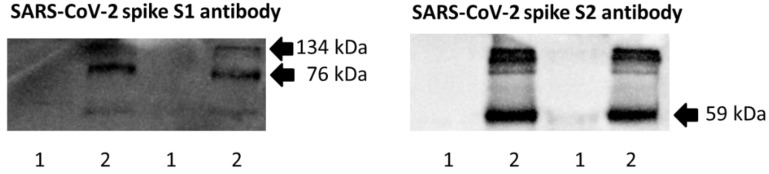
The presence of co-infections or superinfections with bacterial pathogens in COVID-19 patients is associated with poor outcomes, including increased morbidity and mortality. We hypothesized that SARS-CoV-2 and its components interact with the biofilms generated by commensal bacteria, which may contribute to co-infections. This study employed crystal violet staining and particle-tracking microrheology to characterize the formation of biofilms by Streptococcus pneumoniae and Staphylococcus aureus that commonly cause secondary bacterial pneumonia. Microrheology analyses suggested that these biofilms were inhomogeneous soft solids, consistent with their dynamic characteristics. Biofilm formation by both bacteria was significantly inhibited by co-incubation with recombinant SARS-CoV-2 spike S1 subunit and both S1 + S2 subunits, but not with S2 extracellular domain nor nucleocapsid protein. Addition of spike S1 and S2 antibodies to spike protein could partially restore bacterial biofilm production. Furthermore, biofilm formation in vitro was also compromised by live murine hepatitis virus, a related beta-coronavirus. Supporting data from LC-MS-based proteomics of spike-biofilm interactions revealed differential expression of proteins involved in quorum sensing and biofilm maturation, such as the AI-2E family transporter and LuxS, a key enzyme for AI-2 biosynthesis. Our findings suggest that these opportunistic pathogens may egress from biofilms to resume a more virulent planktonic lifestyle during coronavirus infections. The dispersion of pathogens from biofilms may culminate in potentially severe secondary infections with poor prognosis. Further detailed investigations are warranted to establish bacterial biofilms as risk factors for secondary pneumonia in COVID-19 patients.
Keywords: COVID-19; MHV; S1 and S2 subunits; SARS-CoV-2; Staphylococcus aureus; Streptococcus pneumoniae; biofilm; coronavirus; nucleocapsid; spike protein.
Conflict of interest statement
The authors declare no conflict of interest.
Figures


Similar articles
-
Streptococcus pneumoniae Modulates Staphylococcus aureus Biofilm Dispersion and the Transition from Colonization to Invasive Disease.mBio. 2018 Jan 9;9(1):e02089-17. doi: 10.1128/mBio.02089-17. mBio. 2018. PMID: 29317512 Free PMC article.
-
The LuxS/AI-2 Quorum-Sensing System of Streptococcus pneumoniae Is Required to Cause Disease, and to Regulate Virulence- and Metabolism-Related Genes in a Rat Model of Middle Ear Infection.Front Cell Infect Microbiol. 2018 May 4;8:138. doi: 10.3389/fcimb.2018.00138. eCollection 2018. Front Cell Infect Microbiol. 2018. PMID: 29780750 Free PMC article.
-
Streptococcus pneumoniae Eradicates Preformed Staphylococcus aureus Biofilms through a Mechanism Requiring Physical Contact.Front Cell Infect Microbiol. 2016 Sep 27;6:104. doi: 10.3389/fcimb.2016.00104. eCollection 2016. Front Cell Infect Microbiol. 2016. PMID: 27730096 Free PMC article.
-
The expression of hACE2 receptor protein and its involvement in SARS-CoV-2 entry, pathogenesis, and its application as potential therapeutic target.Tumour Biol. 2021;43(1):177-196. doi: 10.3233/TUB-200084. Tumour Biol. 2021. PMID: 34420993 Review.
-
Microbial co-infections in COVID-19: Associated microbiota and underlying mechanisms of pathogenesis.Microb Pathog. 2021 Jul;156:104941. doi: 10.1016/j.micpath.2021.104941. Epub 2021 May 4. Microb Pathog. 2021. PMID: 33962007 Free PMC article. Review.
Cited by
-
Calcium Rescues Streptococcus pneumoniae D39 ΔmntE Manganese-Sensitive Growth Phenotype.Microorganisms. 2024 Sep 1;12(9):1810. doi: 10.3390/microorganisms12091810. Microorganisms. 2024. PMID: 39338484 Free PMC article.
-
Genomics: Infectious Disease and Host-Pathogen Interaction.Int J Mol Sci. 2023 Jan 16;24(2):1748. doi: 10.3390/ijms24021748. Int J Mol Sci. 2023. PMID: 36675263 Free PMC article.
-
An Update on the Mutual Impact between SARS-CoV-2 Infection and Gut Microbiota.Viruses. 2022 Aug 15;14(8):1774. doi: 10.3390/v14081774. Viruses. 2022. PMID: 36016396 Free PMC article. Review.
-
From Co-Infections to Autoimmune Disease via Hyperactivated Innate Immunity: COVID-19 Autoimmune Coagulopathies, Autoimmune Myocarditis and Multisystem Inflammatory Syndrome in Children.Int J Mol Sci. 2023 Feb 3;24(3):3001. doi: 10.3390/ijms24033001. Int J Mol Sci. 2023. PMID: 36769320 Free PMC article. Review.
References
-
- Zhou F., Yu T., Du R., Fan G., Liu Y., Liu Z., Xiang J., Wang Y., Song B., Gu X., et al. Clinical course and risk factors for mortality of adult inpatients with COVID-19 in Wuhan, China: A retrospective cohort study. Lancet. 2020;395:1054–1062. doi: 10.1016/S0140-6736(20)30566-3. - DOI - PMC - PubMed
-
- Russell C.D., Fairfield C.J., Drake T.M., Turtle L., Seaton R.A., Wootton D.G., Sigfrid L., Harrison E.M., Docherty A.B., de Silva T.I., et al. Co-infections, secondary infections, and antimicrobial use in patients hospitalised with COVID-19 during the first pandemic wave from the ISARIC WHO CCP-UK study: A multicentre, prospective cohort study. Lancet Microbe. 2021;2:e354–e365. doi: 10.1016/S2666-5247(21)00090-2. - DOI - PMC - PubMed
MeSH terms
Substances
Grants and funding
LinkOut - more resources
Full Text Sources
Molecular Biology Databases
Miscellaneous
