Review
. 2022 May 5;221(1):iyac014.
doi: 10.1093/genetics/iyac014.

Nematode chromosomes

Affiliations
Review

Nematode chromosomes

Peter M Carlton et al. Genetics. .

Abstract

The nematode Caenorhabditis elegans has shed light on many aspects of eukaryotic biology, including genetics, development, cell biology, and genomics. A major factor in the success of C. elegans as a model organism has been the availability, since the late 1990s, of an essentially gap-free and well-annotated nuclear genome sequence, divided among 6 chromosomes. In this review, we discuss the structure, function, and biology of C. elegans chromosomes and then provide a general perspective on chromosome biology in other diverse nematode species. We highlight malleable chromosome features including centromeres, telomeres, and repetitive elements, as well as the remarkable process of programmed DNA elimination (historically described as chromatin diminution) that induces loss of portions of the genome in somatic cells of a handful of nematode species. An exciting future prospect is that nematode species may enable experimental approaches to study chromosome features and to test models of chromosome evolution. In the long term, fundamental insights regarding how speciation is integrated with chromosome biology may be revealed.

Keywords: WormBook; centromere; holocentric; meiosis; programmed DNA elimination; repetitive DNA; synteny; telomere.


Figures

Fig. 1.
Fig. 1.
The 6 chromosomes of C. elegans at the pachytene stage of meiosis, after pairing and synapsis, visualized with 3D-SIM microscopy. Chromosomes have been pseudocolored after manual tracing and segmentation.
Fig. 2.
Fig. 2.
A schematic of the 6 chromosomes of C. elegans, based on data collected from Wormbase (Harris et al. 2020) version WS280. The chromosomes are shown with their genetically defined left ends at left. The X-axis is the physical (genomic) size in megabases. Various landmarks discussed in the text are shown, including large noncoding RNA loci (21U RNAs and ribosomal RNAs) and PC motifs associated with particular ZIM and HIM-8 zinc-finger proteins, shown as density within 100-kb regions (bars). The genetic map position (including both measured and estimated data) of all protein-coding genes in Wormbase is also shown as a yellow line; each line is scaled to fill the height of the horizontal bar and covers ∼50 cM.
Fig. 3.
Fig. 3.
Balancer chromosomes are translocations that suppress crossing over on chromosomes. Light blue: Chromosome II, purple: Chromosome III. Dotted chromosome arms: crossover suppression. Arc/angle symbols at the left end of each chromosome indicate the PC ends which are required in cis to initiate synapsis; therefore, the configurations marked with a red X are not observed.
Fig. 4.
Fig. 4.
Comparisons of monocentromeres vs holocentromeres in mitotic disjunction. a) Chromosomes with monocentromeres (left) are pulled poleward by the centromere region only, while the rest of the chromosome lags behind; chromosomes with holocentromeres (right) are pulled along their entire lengths and move lengthwise toward the poles. b) Double-strand break damage is especially dangerous to organisms with monocentromeres, since it can result in loss of the entire acentric chromosome fragment distal to the break (left), whereas chromosome fragments with holocentromeres segregate as whole chromosomes (right). c) Fusion of 2 chromosomes with monocentromeres results in dicentrics (left) that can be pulled to opposite poles of the mitotic spindle and break in anaphase, whereas holocentric chromosome fusions segregate as usual (right).
Fig. 5.
Fig. 5.
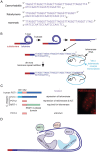
Nematode telomeres. a) Predicted telomere structures from different systems. b) 3′ overhangs of C. elegans telomeres may be protected as T-loops that unfold during S-phase, when the 3′ overhang can be extended by telomerase reverse transcriptase. Telomerase RNA template is predicted. c) Caenorhabditis elegans Pot1 homologs either limit or are required for telomerase activity (left) and can form foci at telomeres (right). d) Double-stranded DNA telomere-binding proteins TEBP-1/DTN-1 and TEBP-2/DTN-2 interact with one another and with the POT-1 single-stranded telomere-binding protein. POT-2 may interact with POT-1, and MRT-1 may interact with the chromosome terminus in the context of telomere repeat addition.
Fig. 6.
Fig. 6.
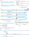
Chromosomal dynamics mediated by telomeric and guanine-rich repetitive DNA. a) ITS tracts at the right ends of C. elegans chromosomes I and X. Light blue text highlights 3 or more nucleotides of perfect telomere sequence. b) ITS tracts on the left arms of C. elegans chromosomes III, IV, and V. Length of tracts is depicted. ITS tracts that are oriented toward the chromosome terminus are positive lines above the chromosome axis, whereas those that face away from the chromosome terminus are negative lines. c) Functions of ITS tracts. In the absence of telomerase, a telomere (dark blue) becomes critically short and initiates DNA synthesis at an ITS tract (yellow). Right pink arrow: induction of the TALT telomere maintenance pathway if 2 ITS tracts as well as subtelomeric DNA between these tracts (turquoise arrows) are amplified and added to all telomeres. Left pink arrows: blue and purple chromosomes fuse in response to telomere erosion. DNA synthesis initiates at an ITS tract to create a subtelomeric duplication that bridges the fused chromosomes. d) A homopolymeric guanine tract may create a G quadruplex structure that induces deletions of (most of) the guanine tract as well as 50–300 bp of adjacent DNA.
Fig. 7.
Fig. 7.
A composite cladogram with local details taken from different studies depicts the phylogenetic structure of some nematode species with sequenced genomes. The overall structure of the clades is based on nuclear small subunit ribosomal RNA analyses and interpretation of taxon relationships derived from morphology. Taxon systematic names are given for the major nodes in the phylogeny. Clades I, II, III, IV, and V were first defined in Blaxter et al. (1998). This cladogram shows predicted relationships between species based on comparisons of several contemporary phylogenies (Blaxter and Koutsovoulos 2015; Tian et al. 2015; Haag et al. 2018; McLean et al. 2018; International Helminth Genomes Consortium 2019). The ecosystem and trophic habits are indicated by small icons, as defined at the bottom of the diagram. Some parasites that reproduce in vertebrates spend stages of their lifecycle in invertebrate hosts, as indicated by invertebrate parasite icons [from Blaxter and Koutsovoulos (2015)]. For Ascaris and Parascaris, germline chromosome number and DNA content are shown (see Table 1). ND: not determined or conflicting data.
Fig. 8.
Fig. 8.
Comparison of chromosome segregation for different types of centromeres and microtubule attachment modes. In all cases, sister chromatid cohesion must be removed at the bivalent interface, and protected on at least some part of the remaining linked chromatid pairs, for correct 2-step segregation. The diagram for telokinetic segregation is speculative, based on Figure 3a of Goday and Pimpinelli (1989).
Fig. 9.
Fig. 9.
Programmed DNA elimination in early development of Parascaris univalens. (1) Two-cell stage (St, somatic cell; P, germline cell). (2a) Two-cell stage with the somatic cell in metaphase of a mitotic division. (2b) Higher-resolution illustration of diploid chromosomes illustrating the thick heterochromatic arms that undergo DNA elimination and the bead-like nature of the central region of the chromosomes. (3) Anaphase of somatic cell division illustrating smaller and multiple chromosomes being segregated with large chromosome fragments remaining at the metaphase plate. The germline cell has entered into mitosis. (4) Telophase and cytokinesis of the somatic cell and anaphase of the germ cell. (5) Four-cell stage illustrating the 2 somatic cells (A and B). On formation of the nuclear membrane, there will be 29 haploid somatic chromosomes and 6 or 12 X chromosomes, male or female, respectively. The large fragments of DNA that will be lost are relegated to the cytoplasm and eventually degraded. The germline cell has undergone division into a somatic cell (EmSt) and a germline cell (P2). See text and Figs. 10 and 11 for additional description of DNA elimination [from Boveri (1899) as modified by Satzinger (2008)]
Fig. 10.
Fig. 10.
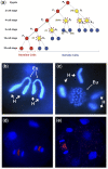
Ascaris early embryo development, cell lineage, and DNA elimination. a) Primordial germ cells (P) are in red, cells undergoing DNA elimination are represented by yellow-filled circles surrounded by red dots (representing the eliminated DNA), and blue cells (S) are precursor somatic cells and lineages. The primordial germ cell numbers correspond to their division state. P0 is the zygote, whereas P1 through P4 represent the primordial germ cell derived from each subsequent cleavage of the germ cells as illustrated. S1–S4 cells are successive precursor somatic cells derived from each division of a germ cell. Adapted from original Boveri presentation [see Streit et al. (2016)]. b and c) Parascaris univalens. b) Two-cell embryo showing one cell with the single pair of germline chromosomes. The big arrows indicate the heterochromatic arms of the chromosomes (H) and the small arrows point to the euchromatic regions of the genome (Eu). c) Somatic cell undergoing programmed DNA elimination from a 2-cell embryo. The retained portions of the germline chromosomes (Eu) are fragmented into ∼2n = 70 chromosomes. The heterochromatic arms that will be eliminated (H, big arrows) remain visible. d, e) Ascaris suum. d) Four-cell embryo with 2 cells undergoing DNA elimination (65 h) and e) 6-cell embryo with one cell undergoing DNA elimination (∼80 h). Note that DNA to be eliminated is present as fragments (artificially colored in red) between segregating chromosomes in early anaphase. DNA fragments derived from a previous elimination event can still be seen in the cytoplasm of cells in (e). Modified from Streit et al. (2016).
Fig. 11.
Fig. 11.
Models for Ascaris DNA elimination and mechanism for the loss of chromosomal regions from holocentric chromosomes. a) Model of internal DNA elimination (Wang et al. 2017). Following alignment, chromosomes in somatic cells undergo chromosome breaks producing fragments of chromosomes. Chromosome fragments that are retained (blue) have centromeric sites for microtubule attachment to facilitate chromosome segregation, whereas chromosomal fragments that will be eliminated (red) remain at the metaphase plate, are not segregated, and are lost. b) Monocentric chromosomes have a single centromere (blue box) where spindle microtubule attachment occurs. Fragmentation of a monocentric chromosome would likely lead to a loss of acentric chromosomal regions during chromosome segregation. c) Holocentric chromosomes have multiple centromeric regions distributed along the chromosome length that serve as sites for microtubule attachment. This distribution of microtubule attachment sites would predict no loss of chromosomal fragments following chromosome breakage during DNA elimination. d) CENP-A is reduced in genomic regions that are not segregated to daughter nuclei in a DNA elimination mitosis. These genomic regions remain at the metaphase plate during anaphase and will be lost during DNA elimination. Immunostaining of CENP-A in a 4-cell Ascaris embryo with 2 cells undergoing DNA elimination mitoses (anaphase) indicates the DNA to be eliminated (red arrows) has much less CENP-A than the DNA that will be segregated and retained. e) CENP-A and centromeres/kinetochores in germline Ascaris chromosomes are distributed along the length of the chromosomes. During development, CENP-A deposition is reduced on regions of chromosomes that will be lost during DNA elimination. Thus, dynamic CENP-A deposition defines and regulates which portions of chromosomes will be retained and which will be lost during DNA elimination. f) Model of chromosome end remodeling. All chromosome ends undergo subtelomeric DNA breaks. The broken ends of the chromosomes are healed by telomere addition. g) Integrated model illustrating internal chromosome breaks leading to 2 new chromosomes and chromosome end remodeling. Modified from Streit et al. (2016).
Fig. 12.
Fig. 12.
DNA elimination in Strongyloides spp. Chromosomal configuration in females (left) and males (right) in S. ratti (top) and S. papillosus (bottom). Chromosomes and chromosomal regions present in 2 copies in both sexes are in blue; chromosomes and regions present in 2 copies in females but only 1 copy in males are in red. Note in S. ratti, 1 whole chromosome is lost in males, while in S. papillosus, only part of a chromosome is lost during programmed DNA elimination [from Streit et al. (2016)]

Similar articles

Cited by

References

    1. Ahmed S, Hodgkin J.. MRT-2 checkpoint protein is required for germline immortality and telomere replication in C. elegans. Nature. 2000;403(6766):159–164. - PubMed
    1. Ahringer J, Gasser SM.. Repressive chromatin in Caenorhabditis elegans: establishment, composition, and function. Genetics. 2018;208(2):491–511. - PMC - PubMed
    1. Albertson DG, Nwaorgu OC, Sulston JE.. Chromatin diminution and a chromosomal mechanism of sexual differentiation in Strongyloides papillosus. Chromosoma. 1979;75(1):75–87. - PubMed
    1. Albertson DG, Rose AM, Villeneuve AM.. Chromosome organization, mitosis, and meiosis. In: Riddle DL, Blumenthal T, Meyer BJ, Priess JR, editors. C. elegans II. Cold Spring Harbor (NY: ): Cold Spring Harbor Laboratory Press; 2011. - PubMed
    1. Albertson DG, Thomson JN.. The kinetochores of Caenorhabditis elegans. Chromosoma. 1982;86(3):409–428. - PubMed

Publication types