. 2022 May 1;322(5):C833-C848.
doi: 10.1152/ajpcell.00414.2021. Epub 2022 Mar 23.

Free fatty acid treatment of mouse preimplantation embryos demonstrates contrasting effects of palmitic acid and oleic acid on autophagy

Affiliations

Free fatty acid treatment of mouse preimplantation embryos demonstrates contrasting effects of palmitic acid and oleic acid on autophagy

Zuleika C L Leung et al. Am J Physiol Cell Physiol. .

Abstract

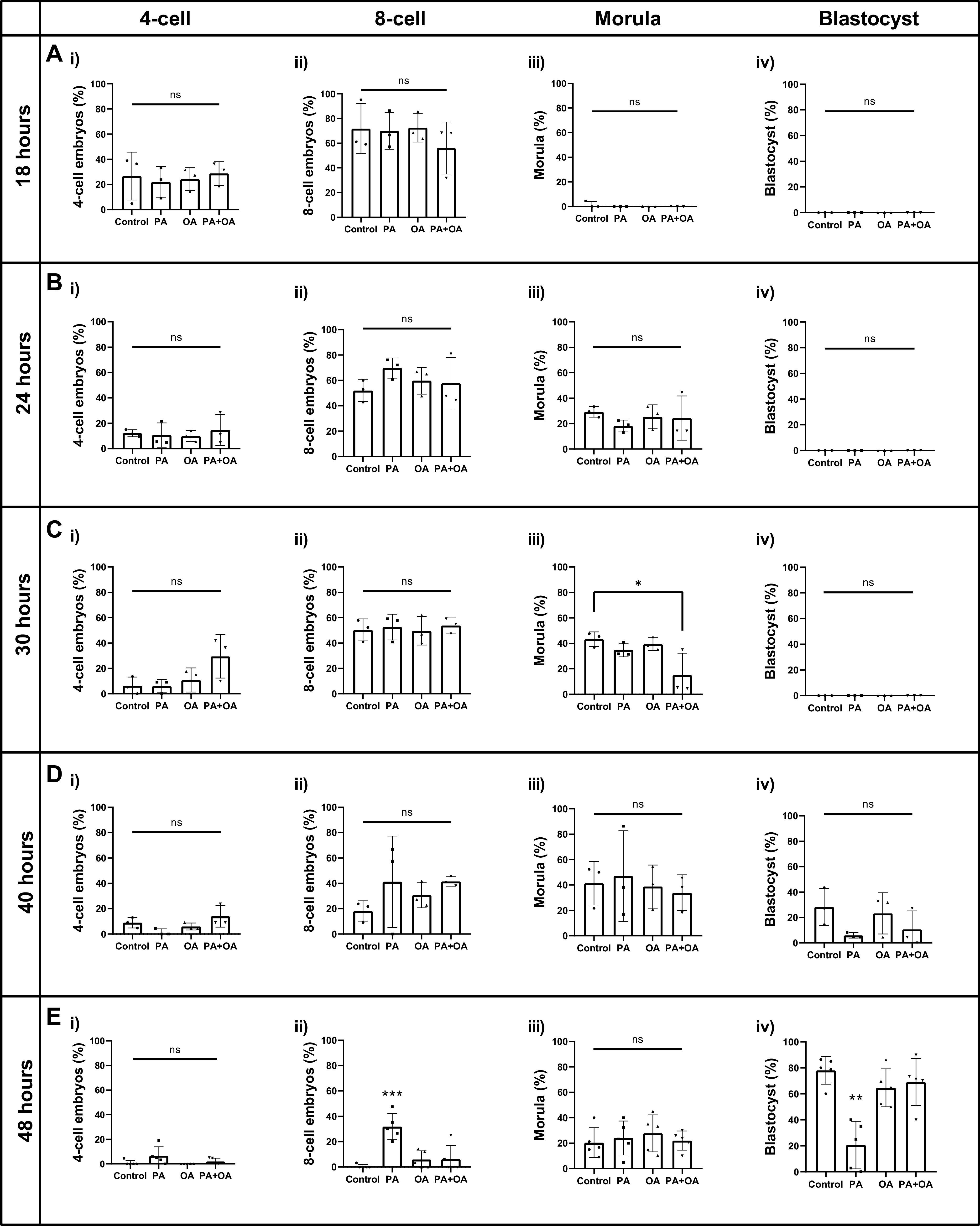
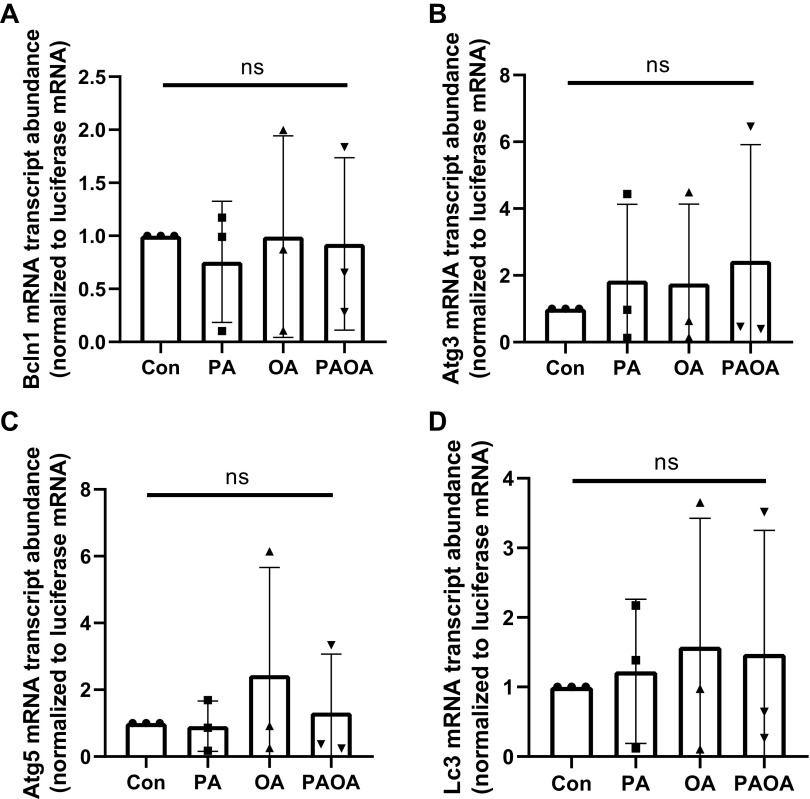
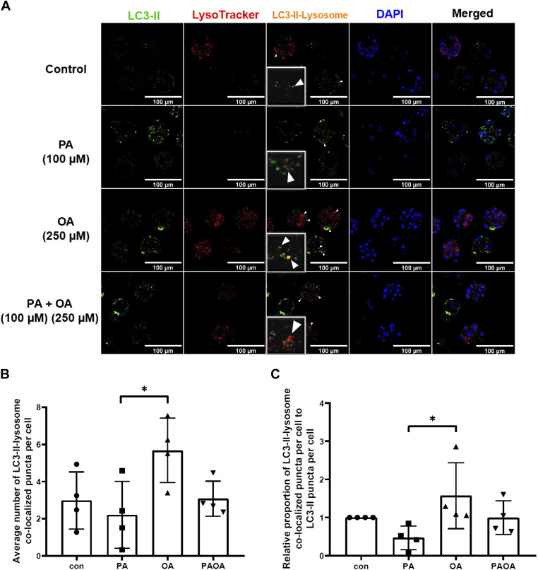
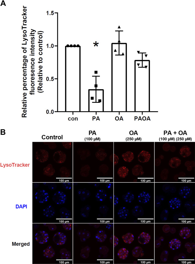
Treatment of mouse preimplantation embryos with elevated palmitic acid (PA) reduces blastocyst development, whereas cotreatment with PA and oleic acid (OA) together rescues blastocyst development to control frequencies. To understand the mechanistic effects of PA and OA treatment on early mouse embryos, we investigated the effects of PA and OA, alone and in combination, on autophagy during preimplantation development in vitro. We hypothesized that PA would alter autophagic processes and that OA cotreatment would restore control levels of autophagy. Two-cell stage mouse embryos were placed into culture medium supplemented with 100 μM PA, 250 μM OA, 100 μM PA and 250 μM OA, or potassium simplex optimization media with amino acid (KSOMaa) medium alone (control) for 18-48 h. The results demonstrated that OA cotreatment slowed developmental progression after 30 h of cotreatment but restored control blastocyst frequencies by 48 h. PA treatment elevated light chain 3 (LC3)-II puncta and p62 levels per cell whereas OA cotreatment returned to control levels of autophagy by 48 h. Autophagic mechanisms are altered by nonesterified fatty acid (NEFA) treatments during mouse preimplantation development in vitro, where PA elevates autophagosome formation and reduces autophagosome degradation levels, whereas cotreatment with OA reversed these PA effects. Autophagosome-lysosome colocalization only differed between PA and OA alone treatment groups. These findings advance our understanding of the effects of free fatty acid exposure on preimplantation development, and they uncover principles that may underlie the associations between elevated fatty acid levels and overall declines in reproductive fertility.

Keywords: autophagy; nonesterified fatty acids; obesity; preimplantation development; preimplantation embryos.


Conflict of interest statement

No conflicts of interest, financial or otherwise, are declared by the authors.

Figures

Figure 1.
Figure 1.
NEFA treatment effects on mouse preimplantation development. Relative percentage (±SD) of 4-cell, 8-cell, morula, and blastocyst stage embryos following treatment of 2-cell mouse embryos in 100 µM PA, 250 µM OA, 100 µM PA and 250 µM OA, or KSOMaa medium alone (control) for 18 (A), 24 (B), 30 (C), 40 (D), 48 (E) h. n > 3, one-way ANOVA with Tukey’s HSD post hoc test. Significant differences are indicated by *P < 0.05, **P < 0.01, ***P < 0.001. By 30 h of treatment (Ciii), the percentage of morula stage embryos was significantly lower in the PA + OA group than control (P = 0.0317). By 48 h, PA treatment displayed a significant higher percentage of 8-cell embryos [Eii; P = 0.0001 (vs. control), P = 0.0007 (vs. OA), P = 0.0008 (vs. PA + OA)] and a significantly lower percentage of blastocysts development [Eiv; P = 0.0002 (vs. control), P = 0.0021 (vs. OA), P = 0.0009 (vs. PA+OA)]. The percentage of morula and blastocysts development in the PA + OA group no longer varied significantly from control at 48 h. HSD, honestly significant difference; KSOMaa, potassium simplex optimization media with amino acid; NEFA, nonesterified fatty acid; ns, not significant; OA, oleic acid; PA, palmitic acid.
Figure 2.
Figure 2.
Autophagic marker transcript abundance across mouse preimplantation development. Relative autophagic marker transcript levels (±SD) of preimplantation mouse embryos at 1-cell, 2-cell, 4-cell, 8-cell, morula, and blastocyst stage. n > 3, one-way ANOVA with Tukey’s HSD post hoc test. Significant differences are indicated by *P < 0.05. A: Bcln1 relative transcript abundance was significantly decreased at the first cleavage (2-cell) stage and maintained at low levels before returning to the blastocyst stage (P < 0.05). B: relative Atg3 transcript levels did not significantly differ across any preimplantation development stages. C: relative Atg5 transcript abundance levels significantly dropped from the zygote stage and stayed at a low relative level throughout preimplantation development (P < 0.05). D: relative Lc3 transcript levels significantly increased from the 4-cell stage to the blastocyst stage (P < 0.05). Atg, autophagy related protein; Bcln1, Beclin-1; HSD, honestly significant difference; Lc3, light chain 3; ns, not significant.
Figure 3.
Figure 3.
Autophagic marker transcript levels of mouse preimplantation embryos following 48 h of NEFA treatments. Relative transcript levels (±SD) for autophagic marker transcripts in preimplantation mouse embryos treated with 100 µM PA, 250 µM OA, 100 µM PA and 250 µM OA, or KSOMaa medium alone (control) for 48 h. n = 3, one-way ANOVA. Relative transcript levels for Bcln1 (A), Atg3 (B), Atg5 (C), and Lc3 (D) did not vary significantly across treatment groups compared with controls. Atg, autophagy related protein; Bcln1, Beclin-1; Con, control; KSOMaa, potassium simplex optimization media with amino acid; Lc3, light chain 3; NEFA, nonesterified fatty acid; ns, not significant; OA, oleic acid; PA, palmitic acid.
Figure 4.
Figure 4.
Autophagy marker of LC3-II across mouse preimplantation embryo development. A: relative percentage of LC3-II aggregate puncta formation per cell (±SD) at each stage of preimplantation mouse embryos. n > 3, one-way ANOVA and Tukey’s HSD post hoc test. Significant differences are indicated by ***P < 0.01 and ****P < 0.0001. Relative percent aggregate puncta formation of LC3-II per cell significantly declined from 2-cell to 4-cell stages and maintained at a low level at all later stages (P < 0.01). B: representative images of LC3-II puncta at each stage of embryo development. Scale bars = 100 µm. HSD, honestly significant difference; LC3, light chain 3.
Figure 5.
Figure 5.
LC3-II puncta per cell in NEFA-treated embryos after 30–48 h of NEFA treatment. Relative percent aggregate of LC3-II per cell (±SD) of 2-cell mouse embryos after exposure to 100 µM PA, 250 µM OA, 100 µM PA and 250 µM OA, or KSOMaa medium alone (control) for 30, 40, and 48 h. Stage-specific embryos from each time point were assessed. n > 3, two-way ANOVA with Tukey’s HSD post hoc test. Significant differences are indicated by *P < 0.05, **P < 0.01, ***P < 0.001, and ****P < 0.0001. A: at the 30-h time point, PA and PA + OA groups resulted in significantly more LC3-II puncta accumulated per cell than OA-alone group [P = 0.0370 (vs. PA); P = 0.0098 (vs. PA + OA)]. B: at the 40-h time point, both PA and PA + OA groups sustained a significantly higher LC3-II puncta aggregation per cell than control [P < 0.0001 (vs. PA); P = 0.0002 (vs. PA + OA)] and OA-alone groups [P < 0.0001 (vs. PA); P = 0.0002 (vs. PA + OA)]. C: after 48 h of NEFA exposure, the PA-alone group resulted in a significantly higher LC3-II puncta count per cell compared with both the OA-alone group (P = 0.0131) and the PA + OA combination group (P = 0.0251). Representative images of LC3-II puncta aggregate after NEFA treatments for 30 (Aii), 40 (Bii), and 48 h (Cii) of exposure. Scale bars = 100 µm. HSD, honestly significant difference; KSOMaa, potassium simplex optimization media with amino acid; LC3, light chain 3; NEFA, nonesterified fatty acid; OA, oleic acid; PA, palmitic acid.
Figure 6.
Figure 6.
Immunofluorescence intensity of p62 in preimplantation embryos after 48 h of NEFA treatment. Relative p62 immunofluorescence intensity per cell (±SD) of 2-cell mouse embryos after exposure to 100 µM PA, 250 µM OA, 100 µM PA and 250 µM OA, or KSOMaa medium alone (control) for 48 h. Only blastocyst embryos were assessed. n = 3, one-way ANOVA with Tukey’s HSD post hoc test. Significant differences are indicated by **P < 0.01. A: after 48 h of NEFA exposure, the PA-alone group resulted in a significantly higher LC3-II puncta count per cell compared with the control group (P = 0.0062), the OA-alone group (P = 0.0014), and the PA + OA combination group (P = 0.0017). B: representative images of p62 immunofluorescence after 48 h of exposure to NEFA treatments. Scale bars = 100 µm. Con, control; HSD, honestly significant difference; KSOMaa, potassium simplex optimization media with amino acid; LC3, light chain 3; NEFA, nonesterified fatty acid; OA, oleic acid; PA, palmitic acid.
Figure 7.
Figure 7.
Autophagosome formation of embryos after NEFA ± CQ treatment for 40 h. A: LC3-II puncta aggregate fluorescence per cell (±SD) after 40 h of treatment with 100 µM PA, 250 µM OA, 100 µM PA and 250 µM OA, or KSOMaa medium alone (control), with 75 µM chloroquine (CQ) treatment for the last 2 h. Only embryos of 8-cell stage and later were included. n = 3, one-way ANOVA and Tukey’s HSD post hoc test. Significant differences are indicated by *P < 0.05 and **P < 0.01. After CQ treatment, fluorescence of LC3-II puncta aggregate per cell of PA group was significantly higher than other treatment groups [P = 0.0047 (vs. control + CQ); P = 0.0032 (vs. OA + CQ); P = 0.0106 (vs. PA + OA + CQ)], suggesting a higher level of autophagosome formation in the PA group. B: representative images of LC3-II aggregate puncta formation after NEFA ± CQ treatment for 40 h. Scale bars = 100 µm. Con, control; HSD, honestly significant difference; KSOMaa, potassium simplex optimization media with amino acid; LC3, light chain 3; NEFA, nonesterified fatty acid; OA, oleic acid; PA, palmitic acid.
Figure 8.
Figure 8.
Autophagosome-lysosome colocalization in mouse preimplantation embryos after 40 h of NEFA treatments. LC3-II puncta that are colocalized with lysosomes were quantified after treatment of mouse embryos with 100 µM PA, 250 µM OA, 100 µM PA and 250 µM OA, or KSOMaa medium alone (control) for 40 h. n = 4, one-way ANOVA and Tukey’s HSD post hoc test. Significant differences are indicated by *P < 0.05. Only embryos of 8-cell stage and later were included. A: representative images of LC3-II-lysosome colocalization in mouse embryos after NEFA exposure. Arrowheads indicate for LC3-II-lysosome colocalized puncta. Scale bars = 100 µm. B: average number of LC3-II-lysosome colocalized puncta per cell (±SD) after NEFA treatments for 40 h. PA-treated group resulted in a significantly lowered number of LC3-II-lysosome colocalized puncta per cell than OA-alone group (P = 0.0346). C: relative proportion of LC3-II-lysosome colocalized puncta per cell to total LC3-II puncta per cell (±SD) of preimplantation mouse embryos after exposure to 40 h of NEFA treatments. Exposure to 40 h of PA treatment results in a significantly lower proportion of autolysosome compared with OA-only group (P = 0.0414). Con, control; HSD, honestly significant difference; KSOMaa, potassium simplex optimization media with amino acid; LC3, light chain 3; NEFA, nonesterified fatty acid; OA, oleic acid; PA, palmitic acid.
Figure 9.
Figure 9.
Relative level of LysoTracker fluorescence in mouse preimplantation embryos after NEFA treatments for 40 h. Fluorescence of LysoTracker were quantified in mouse preimplantation embryos after treatment with 100 µM PA, 250 µM OA, 100 µM PA and 250 µM OA, or KSOMaa medium alone (control) for 40 h. n = 4, one-way ANOVA and Tukey’s HSD post hoc test. Significant differences are indicated by *P < 0.05. Only embryos of 8-cell stage and later were included. A: relative level of LysoTracker fluorescence signal (±SD) after 40 h of NEFA treatments. Exposure to 40 h of PA treatment results in a significantly lower LysoTracker signal compared with all other treatment groups, suggesting a lowered lysosomal activity in mouse preimplantation embryos after PA exposure [P = 0.0002 (vs. control), P < 0.0001 (vs. OA), P = 0.0048 (vs. PA + OA)]. B: representative images of LysoTracker fluorescence staining in mouse embryos following 40 h of NEFA treatment. Scale bars = 100 µm. con, control; HSD, honestly significant difference; KSOMaa, potassium simplex optimization media with amino acid; NEFA, nonesterified fatty acid; OA, oleic acid; PA, palmitic acid.
Figure 10.
Figure 10.
Model of PA and OA treatment effects on autophagy during mouse preimplantation development. Autophagy begins when a phagosome is initiated to form an autophagosome. Phagosomes recruit autophagy-related proteins to direct contents into the phagosome as it elongates and seals off to become an autophagosome. After autophagosome formation is completed, autophagosomes are transported to a lysosome for autophagosome maturation. Fusion of autophagosomes and lysosomes creates an autolysosome. Acidic contents of the lysosome degrade the autophagosome, then releasing the degraded contents into the cytosol for recycling. PA treatment of mouse preimplantation embryos increased autophagosome accumulation by elevating autophagosome formation, reducing autophagosome maturation, and reducing autophagosome degradation by disruption of lysosomal activity. In contrast, OA treatment alone had no obvious effect on these events. However, PA + OA cotreatment restricted PA-induced effects on preimplantation embryo autophagy, but only after 40 h of cotreatment time, resulting in the successful reversal of PA-induced effects by 48 h of exposure. Created with BioRender.com. LC3, light chain 3; OA, oleic acid; PA, palmitic acid.

Similar articles

Cited by

References

    1. Wilson PWF, D’Agostino RB, Sullivan L, Parise H, Kannel WB. Overweight and obesity as determinants of cardiovascular risk: the Framingham experience. Arch Intern Med 162: 1867–1872, 2002. doi:10.1001/archinte.162.16.1867. - DOI - PubMed
    1. Ubags ND, Stapleton RD, Vernooy JH, Burg E, Bement J, Hayes CM, Ventrone S, Zabeau L, Tavernier J, Poynter ME, Parsons PE, Dixon AE, Wargo MJ, Littenberg B, Wouters EF, Suratt BT. Hyperleptinemia is associated with impaired pulmonary host defense. JCI Insight 1: e82101, 2016. doi:10.1172/jci.insight.82101. - DOI - PMC - PubMed
    1. Conway B, Rene A. Obesity as a disease: no lightweight matter. Obes Rev 5: 145–151, 2004. doi:10.1111/j.1467-789X.2004.00144.x. - DOI - PubMed
    1. Lauterbach MA, Wunderlich FT. Macrophage function in obesity-induced inflammation and insulin resistance. Pflugers Arch 469: 385–396, 2017. doi:10.1007/s00424-017-1955-5. - DOI - PMC - PubMed
    1. Seif MW, Diamond K, Nickkho-Amiry M. Obesity and menstrual disorders. Best Pract Res Clin Obstet Gynaecol 29: 516–527, 2015. doi:10.1016/j.bpobgyn.2014.10.010. - DOI - PubMed

Publication types

Grants and funding

LinkOut - more resources