Review
. 2022 May;29(5):921-937.
doi: 10.1038/s41418-022-00975-4. Epub 2022 Mar 21.

Structural diversity of p63 and p73 isoforms

Affiliations
Review

Structural diversity of p63 and p73 isoforms

Christian Osterburg et al. Cell Death Differ. 2022 May.

Abstract

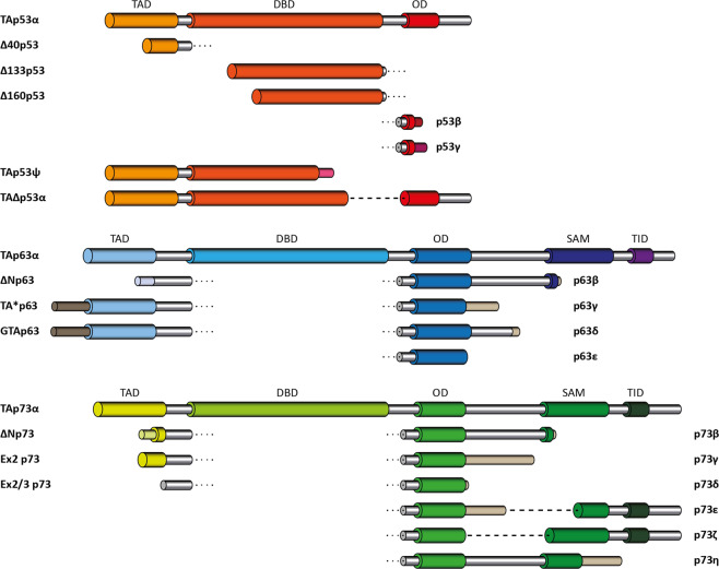
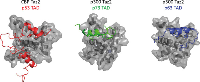
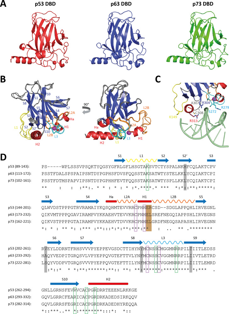
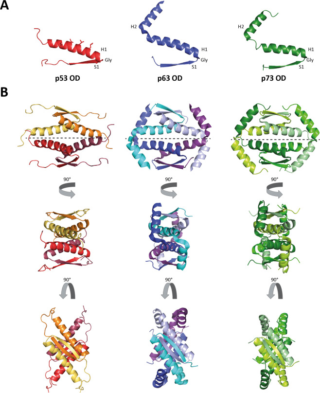
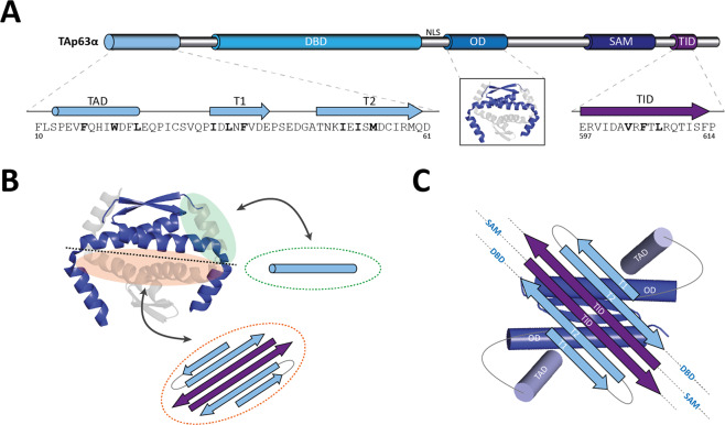
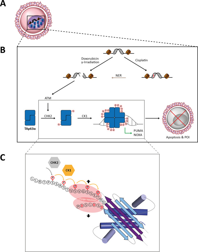
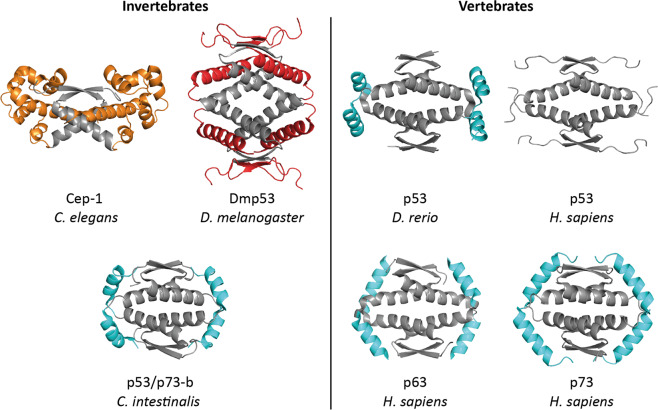
The p53 protein family is the most studied protein family of all. Sequence analysis and structure determination have revealed a high similarity of crucial domains between p53, p63 and p73. Functional studies, however, have shown a wide variety of different tasks in tumor suppression, quality control and development. Here we review the structure and organization of the individual domains of p63 and p73, the interaction of these domains in the context of full-length proteins and discuss the evolutionary origin of this protein family. FACTS: Distinct physiological roles/functions are performed by specific isoforms. The non-divided transactivation domain of p63 has a constitutively high activity while the transactivation domains of p53/p73 are divided into two subdomains that are regulated by phosphorylation. Mdm2 binds to all three family members but ubiquitinates only p53. TAp63α forms an autoinhibited dimeric state while all other vertebrate p53 family isoforms are constitutively tetrameric. The oligomerization domain of p63 and p73 contain an additional helix that is necessary for stabilizing the tetrameric states. During evolution this helix got lost independently in different phylogenetic branches, while the DNA binding domain became destabilized and the transactivation domain split into two subdomains. OPEN QUESTIONS: Is the autoinhibitory mechanism of mammalian TAp63α conserved in p53 proteins of invertebrates that have the same function of genomic quality control in germ cells? What is the physiological function of the p63/p73 SAM domains? Do the short isoforms of p63 and p73 have physiological functions? What are the roles of the N-terminal elongated TAp63 isoforms, TA* and GTA?


Conflict of interest statement

The authors declare no competing interests.

Figures

Fig. 1
Fig. 1. Domain organizations and isoforms in the p53 protein family.
For p53, p63 and p73 all so far known isoforms are shown. TAD transactivation domain, DBD DNA binding domain, OD oligomerization domain, SAM Sterile-alpha motif domain, TID transactivation inhibitory domain. Except for TAp53α and Δ40p53α, all p53 isoforms contain incomplete segments of either the DBD or the OD which leads to unfolding of the corresponding domains. For p73 isoforms with a truncated SAM domain have been found as well.
Fig. 2
Fig. 2. Structures of the TA domains of all family members bound to the Taz2 domains of either CBP (p53) or p300 (p73 and p63).
The Taz2 domains are shown as gray surfaces with the underlying α-helices in dark gray. The TA domains are presented in red (p53), green (p73) and blue (p63). In case of p53, both the TAD1 and TAD2 bind simultaneously (PDB code: 5HPD). The p73 TAD1 binds in a different location; the position of the TAD2 could so far not unambiguously be identified (PDB code: 6FGS). p63 contains a single TA domain that is longer than the individual TA subdomains of p53 and p73 and binds to the same site as the p53 TAD2, albeit with a slight reorientation (PDB code: 6FGN).
Fig. 3
Fig. 3. Structure, organization, and sequence similarity of the p53 family DBDs.
A p53 (red, PDB code: 2AC0), p63 (blue, PDB code: 3QYM) and p73 (green, PDB code: 3DV0) DBDs consist of an immunoglobulin-like β-sandwich of two β-sheets as the domain scaffold and exhibit high structural homology. B Two different orientations of the superimposed p53 family DBDs with explicit labeling of the loop-sheet-helix DNA recognition element created by the L1 loop (yellow), the S2-S2’-S10 sheet (blue) and the H2 helix (red). The loop L3 that provides additional contacts is labeled in cyan. C Close-up of the interaction of the p63 DBD with the DNA. Critical amino acids are indicated. D The DBDs show high sequence identity and conservation of secondary structural elements and residues involved in scaffold assembly and DNA binding. They are composed of ten β-strands (S1–S10), three helices (Ha, H1 and H2) and four relevant loops (L1, L2A, L2B and L3). Residues directly contacting DNA and coordinating the structural important zinc ion are framed in green and purple, respectively. Amino acids responsible for thermodynamic stability differences are highlighted in gray. The two charged residues, which are forming a salt bridge in the intra-dimer interface of p53 and that are crucial for its DNA binding cooperativity, are marked in brown.
Fig. 4
Fig. 4. Structures of the oligomerization domains of all p53 family members.
A The p53 oligomerization domain (OD) contains a β-strand S1 followed by an α-helix H1 (red). The p63 and p73 ODs are elongated by an addition helix H2 (blue and green). For p53 the residues constituting the nuclear export signal (NES) are depicted as sticks. In all three structures S1 is separated from H1 by a structurally important Gly residue. B Four monomers assemble into tetramers as a dimer of dimers with a D2 symmetry. One dimer is built by the formation of an antiparallel intermolecular β-sheet and an antiparallel two helix bundle. The hydrophobic surface presented by the helices engages in tetramerization leading to a four-helix bundle, thereby burying the NES. The second helix H2 of p63 and p73 stabilizes the tetramer further by reaching across the tetramerization interface and clutching the respective opposite dimer. The tetramerization interfaces are depicted by dashed lines (PDB code 1SAF, 4A9Z and 2KBY).
Fig. 5
Fig. 5. Structures of the p63 and p73 SAM domains.
A The p63 SAM domain (blue; PDB code: 2Y9T) and the p73 SAM domain (green; PDB code: 1COK) are shown in different orientations. The domains each consist of four α-helices and one 310-helix. B Superposition of the p63 and p73 SAM domains showing the high structural similarity. C Comparison of the p63 and p73 sequences of the SAM domains with indicated secondary structure elements. The two stretches in the p63 SAM domain with a high aggregation propensity are marked with gray. These sequences cause aggregation initiated by mutations in the SAM domain in patients suffering from ankyloblepharon-ectodermal defects-cleft lip/palate (AEC) syndrome.
Fig. 6
Fig. 6. Model of the autoinhibitory complex of the inactive TAp63α dimer.
A TAp63α is kept in an inactive dimeric conformation unable to bind DNA and transactivate target genes. Several (sub-)domains are involved in formation of the dimer: the transactivation (TA) domain, the β-strand T1, the β-strand T2, the oligomerization domain OD and the transcriptional inhibitory (TI) domain. The TA domain forms an α-helix, T1, T2 and the TI domain β-strands. The hydrophobic core motifs mediating the inter- and intramolecular interactions between these subdomains are highlighted in bold. B The current model of the inactive TAp63α dimer proposes the blockage of the tetramerization interface of the OD. The β-strands of T1, T2 and TI domain of two p63 molecules form a six-stranded antiparallel β-sheet that utilizes its hydrophobic side to interact with the also hydrophobic tetramerization interface of an OD dimer (orange). The helical TA domain simultaneously binds the interface of the OD which is normally bound by the second helix of the OD of the opposing dimer (green). Thereby both interfaces used for efficient dimerization of two OD dimers (blue and gray) to form a stable tetramer are blocked. C In the detailed model of the autoinhibitory complex, the six-stranded β-sheet sits on top of the tetramerization interface of the dimeric OD and both TA domains reach around the OD blocking the binding site of the second helix of the OD.
Fig. 7
Fig. 7. Model of the phosphorylation dependent activation of TAp63α.
A The dimeric and inactive TAp63α is highly expressed in oocytes during dictyate arrest. B Anti-cancer therapy with chemotherapeutic agents or γ-irradiation causes DNA damage. Doxorubicin or γ-irradiation directly induces DSBs. Cisplatin, however, creates covalent inter- and intra-strand DNA adducts, which are then turned into DSBs by the nucleotide excision repair (NER) pathway. The kinase ATM is recruited and activated by DSBs and activates its downstream kinase Chk2 by phosphorylation. Chk2 in turn phosphorylates TAp63α at a single residue (S582) creating a consensus sequence for the constitutively active kinase CK1. CK1 then consecutively phosphorylates S585, S588, S591 and T594 as with each step it creates a new consensus sequence to phosphorylate the next residue at position i + 3. The accumulated negative charge of the phosphate groups triggers the formation of an active tetramer inducing the transcription of the pro-apoptotic Bcl-2 family members PUMA and NOXA [181]. This leads to apoptosis of the primordial follicles and ultimately to premature ovarian insufficiency (POI) upon cancer therapy [, , –232]. C The priming kinase Chk2 and the executioner kinase CK1 phosphorylate serine and threonine residues in a consecutive manner. In the autoinhibitory complex the phosphorylated sequence is adjacent to the TI domain and thereby in proximity to the negatively charged residues following the β-strand T2 (D61, D63 and D66). The charge repulsion with these residues breaks open the autoinhibitory complex leading to irreversible tetramerization.
Fig. 8
Fig. 8. Structural comparison of the ODs of different invertebrate and vertebrate species.
The structures of the p53 ODs of different invertebrate species are compared with the ODs of human p53 (PDB code: 1SAF), p63 (PDB code: 4A9Z) and p73 (PDB code: 2KBY) as well as with p53 from Danio rerio (zebrafish; PDB code: 4D1M). In the structure of Cep-1 from Caenorhabditis elegans (PDB code: 2RP5) the OD forms only a dimer that is tightly coupled to a SAM domain (yellow). In the structure of the OD from Drosophila melanogaster (PDB code: 2RP4) each secondary structure element known from human p53 is doubled. The extra β-sheets and α-helices per monomer are shown in red. The OD of p53/p73-b from the tunicate Ciona intestinalis (PDB code: 2MW4) is structurally similar to human p63 and p73 showing a second helix, despite the replacement of amino acids crucial for forming this second helix in vertebrate species. The structure of the p53 OD from Danio rerio contains a second helix that, however, is differently orientated compared to the second helices in human p63 and p73.

Similar articles

Cited by

References

    1. Kaghad M, Bonnet H, Yang A, Creancier L, Biscan JC, Valent A, et al. Monoallelically expressed gene related to p53 at 1p36, a region frequently deleted in neuroblastoma and other human cancers. Cell. 1997;90:809–19.. doi: 10.1016/S0092-8674(00)80540-1. - DOI - PubMed
    1. Schmale H, Bamberger C. A novel protein with strong homology to the tumor suppressor p53. Oncogene. 1997;15:1363–7. doi: 10.1038/sj.onc.1201500. - DOI - PubMed
    1. Trink B, Okami K, Wu L, Sriuranpong V, Jen J, Sidransky D. A new human p53 homologue. Nat Med. 1998;4:747–8. doi: 10.1038/nm0798-747. - DOI - PubMed
    1. Osada M, Ohba M, Kawahara C, Ishioka C, Kanamaru R, Katoh I, et al. Cloning and functional analysis of human p51, which structurally and functionally resembles p53. Nat Med. 1998;4:839–43. doi: 10.1038/nm0798-839. - DOI - PubMed
    1. Yang A, Kaghad M, Wang Y, Gillett E, Fleming MD, Dotsch V, et al. p63, a p53 homolog at 3q27-29, encodes multiple products with transactivating, death-inducing, and dominant-negative activities. Mol Cell. 1998;2:305–16. doi: 10.1016/S1097-2765(00)80275-0. - DOI - PubMed

Publication types

MeSH terms