. 2022 Jan-Mar;13(1):19476035221081466.
doi: 10.1177/19476035221081466.

Stiffness- and Bioactive Factor-Mediated Protection of Self-Assembled Cartilage against Macrophage Challenge in a Novel Co-Culture System

Affiliations

Stiffness- and Bioactive Factor-Mediated Protection of Self-Assembled Cartilage against Macrophage Challenge in a Novel Co-Culture System

Ryan P Donahue et al. Cartilage. 2022 Jan-Mar.

Abstract

Objective: Tissue-engineered cartilage implants must withstand the potential inflammatory and joint loading environment for successful long-term repair of defects. The work's objectives were to develop a novel, direct cartilage-macrophage co-culture system and to characterize interactions between self-assembled neocartilage and differentially stimulated macrophages.

Design: In study 1, it was hypothesized that the proinflammatory response of macrophages would intensify with increasing construct stiffness; it was expected that the neocartilage would display a decrease in mechanical properties after co-culture. In study 2, it was hypothesized that bioactive factors would protect neocartilage properties during macrophage co-culture. Also, it was hypothesized that interleukin 10 (IL-10)-stimulated macrophages would improve neocartilage mechanical properties compared to lipopolysaccharide (LPS)-stimulated macrophages.

Results: As hypothesized, stiffer neocartilage elicited a heightened proinflammatory macrophage response, increasing tumor necrosis factor alpha (TNF-α) secretion by 5.47 times when LPS-stimulated compared to construct-only controls. Interestingly, this response did not adversely affect construct properties for the stiffest neocartilage but did correspond to a significant decrease in aggregate modulus for soft and medium stiffness constructs. In addition, bioactive factor-treated constructs were protected from macrophage challenge compared to chondrogenic medium-treated constructs, but IL-10 did not improve neocartilage properties, although stiff constructs appeared to bolster the anti-inflammatory nature of IL-10-stimulated macrophages. However, co-culture of bioactive factor-treated constructs with LPS-treated macrophages reduced TNF-α secretion by over 4 times compared to macrophage-only controls.

Conclusions: In conclusion, neocartilage stiffness can mediate macrophage behavior, but stiffness and bioactive factors prevent macrophage-induced degradation. Ultimately, this co-culture system could be utilized for additional studies to develop the burgeoning field of cartilage mechano-immunology.

Keywords: cartilage; immunology; macrophage; tissue engineering.


Conflict of interest statement

Declaration of Conflicting Interests: The author(s) declared the following potential conflicts of interest with respect to the research, authorship, and/or publication of this article: Kyriacos A. Athanasiou and Jerry C. Hu are scientific consultants for Cartilage Inc.

Figures

Figure 1.
Figure 1.
Stiffness is modulated via C-ABCcomp., and macrophages are adhered and stimulated in a direct co-culture system. Constructs were cultured in either CHG only or with TCL treatment. After 27 days, compressive stiffness was modulated via C-ABCcomp. application. Constructs were assayed for baseline properties (t = 0). The next day, macrophages adhered and were cultured in a 1:1 mix of CHG and MΦM medium, then stimulated with LPS in study 1 and LPS or IL-10 in study 2. Unstimulated macrophage controls, construct-only controls, and macrophage-only controls were also included. After 2 weeks of co-culture, constructs were assayed again (t = 2W). C-ABCcomp. = chondroitinase ABC to modulate compressive stiffness; CHG = chondrogenic medium; TCL = transforming growth factor beta 1/chondroitinase ABCeng./lysyl oxidase-like 2; MΦM = macrophage medium; LPS = lipopolysaccharide; IL-10 = interleukin 10.
Figure 2.
Figure 2.
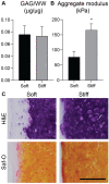
C-ABCcomp. modulates the compressive stiffness of CHG-treated constructs. (A) GAG/WW increased across soft to stiff groups and decreased as higher C-ABCcomp. concentrations were used to modulate compressive stiffness. (B) Similarly, aggregate modulus also trended higher from soft to stiff, as expected. Statistics: One-way ANOVA with post hoc Tukey’s HSD, α = 0.05, n = 3 per group. C-ABCcomp. = chondroitinase ABC to modulate compressive stiffness; CHG = chondrogenic medium; GAG = glycosaminoglycan; WW = wet weight; ANOVA = analysis of variance; HSD = honestly significant difference.
Figure 3.
Figure 3.
CHG-treated constructs of soft and medium stiffness differ in staining intensity. Following 2 weeks of co-culture, stiff constructs maintain cell morphology and tissue staining intensity in co-culture groups more than the soft and medium groups. Scale bar = 200 µm. CHG = chondrogenic medium; LPS = lipopolysaccharide.
Figure 4.
Figure 4.
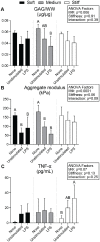
Soft and medium stiffness CHG-treated constructs suffer losses in aggregate modulus values despite no increases in TNF-α production. (A) GAG/WW was significantly affected by the ΜΦ factor, with significant decreases within the medium stiffness constructs between the construct-only control and LPS-stimulated co-culture group. (B) Trending with GAG/WW, aggregate modulus significantly decreased with the addition of macrophages (unstimulated or LPS-stimulated) in soft and medium stiffness constructs. (C) Conversely, after 48 hours of co-culture, only the stiff construct group had significant increases in TNF-α secretion between the construct-only control and the LPS-stimulated group. Statistics: Two-way ANOVA with post hoc Tukey’s HSD among groups within a stiffness (dotted lines), α = 0.05, n = 3-6 per group. CHG = chondrogenic medium; TNF-α = tumor necrosis factor alpha; GAG = glycosaminoglycan; WW = wet weight; ΜΦ = macrophage; LPS = lipopolysaccharide; ANOVA = analysis of variance; HSD = honestly significant difference.
Figure 5.
Figure 5.
C-ABCcomp. modulates the compressive stiffness of TCL-treated constructs. (A) GAG/WW does not differ significantly for TCL-treated constructs at t = 0. (B) Aggregate modulus significantly decreases with application of C-ABCcomp. (C) Soft constructs show less intense and H&E and Saf-O staining due to C-ABCcomp. application and only peripheral loss of GAG. Also, some cells are visible near the soft construct edge. Conversely, stiff constructs show intense Saf-O staining at the periphery indicating high GAG content, and cells are not present at the periphery of the construct. Scale bar = 100 µm. Statistics: Student t test, α = 0.05. C-ABCcomp. = chondroitinase ABC to modulate compressive stiffness; TCL = transforming growth factor beta 1/chondroitinase ABCeng./lysyl oxidase-like 2; GAG = glycosaminoglycan; WW = wet weight; H&E = hematoxylin and eosin; Saf-O = Safranin O.
Figure 6.
Figure 6.
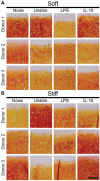
Saf-O staining after 2 weeks of macrophage co-culture shows donor-related variation, with diminished staining among LPS-stimulated co-cultures. Both construct-only controls and co-culture groups show variability between donors in both (A) soft and (B) stiff TCL-treated constructs. However, on average, staining is slightly diminished in some LPS-stimulated groups compared to construct-only controls. Scale bar = 100 µm. Saf-O = Safranin O; LPS = lipopolysaccharide; Unstim. = unstimulated; IL-10 = interleukin 10; TCL = transforming growth factor beta 1/chondroitinase ABCeng./lysyl oxidase-like 2.
Figure 7.
Figure 7.
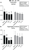
Aggregate modulus values of TCL-treated constructs only decrease in soft construct, LPS-stimulated co-cultures. (A) The construct-only control and macrophage co-cultures in the soft constructs are significantly different in GAG/WW, while the stiff group does not exhibit significant differences between groups. (B) Aggregate modulus also trends downward for the soft group when co-cultured with macrophages, but only the LPS-stimulated co-culture group is significantly different from the construct-only control. Stiff group aggregate moduli were largely unaffected by macrophage treatment. Statistics: Two-way ANOVA with post hoc Tukey’s HSD among groups within a stiffness (dotted lines), α = 0.05, n = 5-6 per group. TCL = transforming growth factor beta 1/chondroitinase ABCeng./lysyl oxidase-like 2; LPS = lipopolysaccharide; GAG = glycosaminoglycan; WW = wet weight; MΦ = macrophage; IL-10 = interleukin 10; ANOVA = analysis of variance; HSD = honestly significant difference.
Figure 8.
Figure 8.
TNF-α secretion of TCL-treated construct co-cultures increases with stiffness, decreases with construct co-culture, and diminishes when macrophages are stimulated toward an anti-inflammatory phenotype. In unstimulated co-culture conditions, TNF-α production increases with stiffness. Similarly, TNF-α secretion also trends higher for stiff construct co-culture compared to the soft construct condition in the LPS-stimulated groups. Interestingly, construct addition significantly decreases the TNF-α production compared to macrophage-only controls in LPS-stimulated groups. For IL-10 stimulation, a decrease in TNF-α levels is seen with increasing construct stiffness. Statistics: One-way ANOVA with post hoc Tukey’s HSD (for each stimulation condition), α = 0.05, n = 5-6 per group. TNF-α = tumor necrosis factor alpha; TCL = transforming growth factor beta 1/chondroitinase ABCeng./lysyl oxidase-like 2; MΦ = macrophage, LPS = lipopolysaccharide; IL-10 = interleukin 10; ANOVA = analysis of variance; HSD = honestly significant difference.

Similar articles

Cited by

References

    1. Koh TJ, DiPietro LA. Inflammation and wound healing: the role of the macrophage. Expert Rev Mol Med. 2011;13:e23. - PMC - PubMed
    1. Kennedy A, Fearon U, Veale DJ, Godson C. Macrophages in synovial inflammation. Front Immunol. 2011;2:52. - PMC - PubMed
    1. Chen Y, Jiang W, Yong H, He M, Yang Y, Deng Z, et al.. Macrophages in osteoarthritis: pathophysiology and therapeutics. Am J Transl Res. 2020;12(1):261-8. - PMC - PubMed
    1. Wang KH, Wan R, Chiu LH, Tsai YH, Fang CL, Bowley JF, et al.. Effects of collagen matrix and bioreactor cultivation on cartilage regeneration of a full-thickness critical-size knee joint cartilage defects with subchondral bone damage in a rabbit model. PLoS One. 2018;13(5):e0196779. - PMC - PubMed
    1. Kondo M, Kameishi S, Kim K, Metzler NF, Maak TG, Hutchinson DT, et al.. Safety and efficacy of human juvenile chondrocyte-derived cell sheets for osteochondral defect treatment. NPJ Regen Med. 2021;6(1):65. - PMC - PubMed

Publication types

MeSH terms