. 2022 Jun 15:168:105694.
doi: 10.1016/j.nbd.2022.105694. Epub 2022 Mar 18.

The innate immune system stimulating cytokine GM-CSF improves learning/memory and interneuron and astrocyte brain pathology in Dp16 Down syndrome mice and improves learning/memory in wild-type mice

Affiliations

The innate immune system stimulating cytokine GM-CSF improves learning/memory and interneuron and astrocyte brain pathology in Dp16 Down syndrome mice and improves learning/memory in wild-type mice

Md Mahiuddin Ahmed et al. Neurobiol Dis. .

Abstract

Down syndrome (DS) is characterized by chronic neuroinflammation, peripheral inflammation, astrogliosis, imbalanced excitatory/inhibitory neuronal function, and cognitive deficits in both humans and mouse models. Suppression of inflammation has been proposed as a therapeutic approach to treating DS co-morbidities, including intellectual disability (DS/ID). Conversely, we discovered previously that treatment with the innate immune system stimulating cytokine granulocyte-macrophage colony-stimulating factor (GM-CSF), which has both pro- and anti-inflammatory activities, improved cognition and reduced brain pathology in a mouse model of Alzheimer's disease (AD), another inflammatory disorder, and improved cognition and reduced biomarkers of brain pathology in a phase II trial of humans with mild-to-moderate AD. To investigate the effects of GM-CSF treatment on DS/ID in the absence of AD, we assessed behavior and brain pathology in 12-14 month-old DS mice (Dp[16]1Yey) and their wild-type (WT) littermates, neither of which develop amyloid, and found that subcutaneous GM-CSF treatment (5 μg/day, five days/week, for five weeks) improved performance in the radial arm water maze in both Dp16 and WT mice compared to placebo. Dp16 mice also showed abnormal astrocyte morphology, increased percent area of GFAP staining in the hippocampus, clustering of astrocytes in the hippocampus, and reduced numbers of calretinin-positive interneurons in the entorhinal cortex and subiculum, and all of these brain pathologies were improved by GM-CSF treatment. These findings suggest that stimulating and/or modulating inflammation and the innate immune system with GM-CSF treatment may enhance cognition in both people with DS/ID and in the typical aging population.

Keywords: Alzheimer's disease (AD); Astrocyte; Calretinin; Cognition; Down syndrome (DS); Dp(16)1Yey (Dp16); Glial-fibrillary acidic protein (GFAP); Granulocyte-macrophage colony-stimulating factor (GM-CSF); Intellectual disability (ID); Interneuron.


Conflict of interest statement

Declaration of Interests

The authors declare no competing interests. Dr. Boyd was recently hired by Partner Therapeutics, the manufacturer of human recombinant GM-CSF.

Figures

Figure 1.
Figure 1.. Schematic of the Study Timeline
Injections began on day 1 (D1) and ended on day 24 (D24). After the baseline pre-treatment assessments, the cohorts of Dp16 and WT control mice were injected five days/week with either GM-CSF (5 μg/day) or an equal volume of saline (200 μl/day), Monday-Friday, for five weeks for a total of 24 injections (ending on D24) over a period of 32 days including non-treatment days. There were four treatment groups for both female and male mice: (1) WT mice injected with saline (9 males; 6 females), (2) WT mice injected with GM-CSF (8 males; 7 females), (3) Dp16 mice injected with saline (5 males; 6 females), and (4) Dp16 mice injected with GM-CSF (6 males; 6 females). Both male and female mice were tested in the Open Field task at baseline and in the Y-maze task at baseline and post-treatment. Only male mice were used for the RAWM task at baseline and post-treatment because the female mice have poor swimming abilities at this age.
Figure 2.
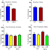
Figure 2.. Male and Female Dp16 Mice Show Elevated Locomotor Activity in the Open Field Task at Baseline
Locomotor and exploratory activity were measured in the Open Field task at baseline. Both male and female Dp16 mice showed a significant increase in total distance traveled (A and D, respectively) and speed of movement (C and F, respectively) compared to male and female WT mice, respectively. No significant differences were observed in the time spent in the center for male and female Dp16 mice (B and E, respectively) compared to male and female WT littermates, respectively. Data are represented as mean ± SEM for separate groups of mice (male WT: n=19; female WT: n=13; male Dp16: n=11; and female Dp16: n=13). Statistical significance was determined by the unpaired Student’s t-test for comparison between Dp16 and WT littermates.
Figure 3.
Figure 3.. Spontaneous Alternation Rates in the Y-maze Task at Baseline and After GM-CSF Treatment: GM-CSF Treatment Does Not Rescue Spatial Working Memory Deficits in Female Dp16 Mice
Female Dp16 mice showed a significantly lower alternation rate in the Y-maze compared to female WT littermates at baseline (A). No significant differences in alternation rates in the Y-maze were observed in male Dp16 mice compared to male WT littermates at baseline (B). No significant differences in spontaneous alternation rates in the Y-maze were observed in GM-CSF-treated (GM) female WT or Dp16 mice compared to saline-injected (Sal) female WT or Dp16 mice (F (3,18)=0.49; p=0.69) (C). No significant differences in spontaneous alternation rates in the Y-maze were observed in GM-CSF-treated (GM) male WT or Dp16 mice compared to saline-injected (Sal) male WT or Dp16 mice (p=0.18) (F (3,25)=1.60; p=0.21) (D). Data are represented as mean ± SEM for the separate groups of mice, including WT-Sal (9 males; 6 females), WT-GM (8 males; 7 females), Dp16-Sal (5 males; 6 females), and Dp16-GM (6 males; 6 females). Statistical significance was determined by the unpaired Student’s t-test for baseline comparisons between Dp16 and WT mice (A, B) and by ANOVA followed by Turkey’s multiple comparisons test between groups (C, D). For evaluating the interactions between treatment and genotype, Two-Way ANOVA followed by Bonferroni’s multiple comparisons test were performed. No significant differences in female mice in spontaneous alternation rates of Y-maze were observed between genotype (F (1,18)=0.28; p=0.60), and the interactions between treatment and genotype (F (1.18)=1.23; p=0.28) (C). Similarly, no significant differences in male mice in spontaneous alternation rates of Y-maze were observed between genotype (F (1,25)=0.27; p=0.61), and the interactions between treatment and genotype (F (1,25)=1.75; p=0.20) (D).
Figure 4.
Figure 4.. Male Dp16 Mice Show Deficits in the RAWM Task at Baseline
Hippocampal-based spatial learning and memory performance were evaluated in male Dp16 mice for two days using the radial arm water maze (RAWM) task. Data were analyzed considering each group of three out of 15 trials as a block for Day 1, the training day (A), and for Day 2, the testing day (B). On day 2, in blocks 1 and 2, there was a significant difference in memory performance between the Dp16 and WT littermates. After averaging the data from 15 trials using the time values for each individual mouse, the mean time to reach the platform for each day was compared between Dp16 and WT mice for Day 1 (C) and Day 2 (D). Dp16 mice required a significantly longer time to reach the platform than WT mice on both days, although they ultimately learned as well as WT mice, as indicated by non-significant differences in latencies in Block 5. Data are represented as mean ± SEM for separate groups of mice (WT, n=17; Dp16, n=11). Statistical significance was determined by the unpaired Student’s t-test for comparison between Dp16 and WT mice (C, D) and by ANOVA followed by Tukey’s multiple comparisons test (A, B). *p<0.05; ***p<0.001.
Figure 5.
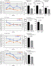
Figure 5.. GM-CSF Treatment Improves RAWM Performance in Male Dp16 and Wild-type Mice
To evaluate the effect of GM-CSF treatment on spatial memory, latencies on post-treatment Day 1 for Dp16 mice and their WT littermates injected with saline or GM-CSF were normalized to their mean latency in block 5 of pre-treatment Day 2, and the data were then compared between saline and GM-CSF injection for each genotype. For quantitative analyses, latencies on post-treatment Day 1 were calculated relative to 100% for block 5 of pre-treatment Day 2 data. Normalized latencies were analyzed in blocks of three trials (with a total of five blocks from 15 trials) and the GM-CSF-treated group was compared with the saline-injected group of Dp16 mice (A; n=5–6) and WT mice (E; n=8–9). After averaging the normalized latencies of post-treatment Day 1 from all blocks, the mean values were compared in Dp16 mice treated with saline or with GM-CSF (B; n=5–6). Combined data from blocks 4–5 of Dp16 mice were compared for saline and GM-CSF injection (C; n=5–6). Combined data from blocks 1–2 were compared with the combined data from blocks 4–5 for Dp16 mice treated with GM-CSF (D; n=5–6). Similarly, combined data from blocks 1–2 of WT mice were compared for saline injection and GM-CSF treatment (F; n=8–9). Combined data from blocks 1–2 were compared with the combined data from blocks 4–5 for WT mice injected with saline (G; n=8–9). The effect of GM-CSF treatment on learning flexibility was evaluated by normalizing the latency of each saline-injected or GM-CSF-treated mouse at post-treatment day 2 with its latency at block 5 of post-treatment day 1 and calculated data relative to 100% of block 5 post-treatment day 1 (H-K). Normalized latencies were calculated in blocks of three trials, and the GM-CSF-injected group was compared with the saline-injected group of Dp16 mice (H; n=5–6) and WT mice (J; n=8–9). Combined data from blocks 1–5 were compared for saline-injected and GM-CSF-treated Dp16 mice (I; n=5–6) and WT mice (K; n=8–9). Data are represented as mean ± SEM for the different groups of mice. Statistical significance was determined by the unpaired Student’s t-test for comparison between groups. *p <0.05; **p <0.01.
Figure 6.
Figure 6.. GM-CSF Treatment is Associated with Higher Plasma Levels of Cytokines in Both Wild-Type and Dp16 Mice
The Meso-Scale Discovery (MSD) platform was used to determine plasma levels of six key inflammation-associated cytokines (i.e., IL-6, IL-2, TNFα, IP-10, MCP-1, and MDC) following GM-CSF treatment or saline injection. No significant differences were observed in the plasma levels of these six cytokines in saline-injected WT mice compared to saline-injected Dp16 mice (A-F). Treatment with GM-CSF led to significantly higher levels of interleukin-2 (IL-2) and macrophage-derived chemokine (MDC) in the plasma of both GM-CSF-treated WT and Dp16 mice compared to saline-injected WT and Dp16 mice (B & F), respectively, whereas the plasma levels of interferon γ-induced protein 10 (IP-10) were significantly higher only in GM-CSF-treated Dp16 mice compared to saline-injected Dp16 mice (D). In addition, there was a strong trend toward higher plasma levels of interleukin-6 (IL-6) and tumor necrosis factor α (TNFα) in the plasma of both GM-CSF-treated WT and Dp16 mice compared to saline-injected WT and Dp16 mice (A & C), respectively. No significant differences were detected in the plasma levels of MCP-1 in GM-CSF-treated or saline-injected WT or Dp16 mice (E).
Figure 7.
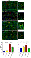
Figure 7.. GM-CSF Treatment Partially Restores the Distribution of Hippocampal Astrocytes and Reverses Astrogliosis in Male Dp16 Mice
The patterns and distributions of glial fibrillary acidic protein (GFAP)-positive astrocytes (green) were examined in the hippocampi of male WT and Dp16 mice injected with saline or with GM-CSF. Data are shown for WT mice injected with saline (A), WT mice treated with GM-CSF (B), Dp16 mice injected with saline (C), and Dp16 mice treated with GM-CSF (D). Dp16 mice injected with saline (C) showed abnormal clusters of GFAP-positive astrocytes compared to WT mice injected with saline (A). GM-CSF treatment of WT mice had no effect on hippocampal astrocyte distribution (B) compared to WT mice injected with saline. Dp16 mice treated with GM-CSF showed significantly reduced levels of the abnormal clusters of GFAP-positive astrocytes (D) compared to Dp16 mice injected with saline (C). Arrows show abnormal clusters of GFAP-positive astrocytes. Quantitative analyses showed significantly higher numbers of abnormal clusters of GFAP-positive astrocytes in Dp16 mice injected with saline compared to WT mice injected with saline (p=0.001), which were reduced in Dp16 mice treated with GM-CSF (p=0.03) (E). Quantitative analyses of the percent area of GFAP-positive staining showed a significant increase in Dp16 mice injected with saline compared to WT mice injected with saline (p=0.041), which were reduced by GM-CSF treatment (p=0.011) (F). For each bar, the data are represented as mean ± SEM for the separate groups of mice. Statistical significance was determined by the unpaired Student’s t-test for comparison between groups. Scale bar: 200 μm (20X magnification). All experiments were repeated 2–4 times with similar results (n=3 male mice in each of the four groups).
Figure 8.
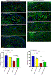
Figure 8.. GM-CSF Treatment Partially Restores the Number of Calretinin-Positive Interneurons in Male Dp16 Mice
The patterns and distributions of calretinin-positive interneurons (green) were examined in the entorhinal cortex (EC) of male WT mice injected with saline (A), male WT mice treated with GM-CSF (B), male Dp16 mice injected with saline (C), and male Dp16 mice treated with GM-CSF (D). Quantitative analyses showed a trend towards a lower number of calretinin-positive interneurons in the entorhinal cortex of Dp16 mice injected with saline compared to WT mice injected with saline (p=0.06) (E). The number of calretinin-positive interneurons in the entorhinal cortex of the Dp16 mice was significantly increased following GM-CSF treatment compared to Dp16 mice injected with saline (p=0.0406) and was statistically no different than in WT mice injected with saline or treated with GM-CSF (E). The expression patterns and distributions of calretinin-positive interneurons (green) were also examined in the subiculum of male WT mice injected with saline (F), male WT mice treated with GM-CSF (G), male Dp16 mice injected with saline (H), and male Dp16 mice treated with GM-CSF (I). There was a significantly reduced number of calretinin-positive interneurons in the subiculum of Dp16 mice injected with saline compared to WT mice (p=0.01) (J). The number of calretinin-positive interneurons in the subiculum of Dp16 mice was significantly increased following GM-CSF treatment (p=0.04), but it was still significantly lower than in WT mice injected with saline or with GM-CSF (J). For each bar, data are represented as mean ± SEM for separate groups of mice. Statistical significance was determined by the unpaired Student’s t-test for comparison between groups. Scale bar: A-D=100 μm (20X magnification) and F-I=50 μm (20X magnification). All experiments were repeated 2–4 times with similar results (n=3 male mice in each of the four groups).

Similar articles

Cited by

References

    1. Antonarakis SE, Lyle R, Dermitzakis ET, Reymond A, Deutsch S. Chromosome 21 and down syndrome: from genomics to pathophysiology. Nat Rev Genet 2004;5(10):725–38. Epub 2004/10/29. doi: 10.1038/nrg1448. - DOI - PubMed
    1. Opitz JM, Gilbert-Barness EF. Reflections on the pathogenesis of Down syndrome. Am J Med Genet Suppl 1990;7:38–51. - PubMed
    1. Epstein CJ. The consequences of chromosome imbalance. Am J Med Genet Suppl 1990;7:31–7. Epub 1990/01/01. doi: 10.1002/ajmg.1320370706. - DOI - PubMed
    1. Chapman RS, Hesketh LJ. Behavioral phenotype of individuals with Down syndrome. Ment Retard Dev Disabil Res Rev 2000;6(2):84–95. Epub 2000/07/19. doi: 10.1002/1098-2779(2000)6:2<84::Aid-mrdd2>3.0.Co;2-p. - DOI - PubMed
    1. Silverman W Down syndrome: cognitive phenotype. Ment Retard Dev Disabil Res Rev 2007;13(3):228–36. Epub 2007/10/03. doi: 10.1002/mrdd.20156. - DOI - PubMed

Publication types

MeSH terms

Substances