Histone deacetylase 7: a signalling hub controlling development, inflammation, metabolism and disease
- PMID: 35303381
- PMCID: PMC10952174
- DOI: 10.1111/febs.16437
Histone deacetylase 7: a signalling hub controlling development, inflammation, metabolism and disease
Abstract
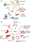
Histone deacetylases (HDACs) catalyse removal of acetyl groups from lysine residues on both histone and non-histone proteins to control numerous cellular processes. Of the 11 zinc-dependent classical HDACs, HDAC4, 5, 7 and 9 are class IIa HDAC enzymes that regulate cellular and developmental processes through both enzymatic and non-enzymatic mechanisms. Over the last two decades, HDAC7 has been associated with key roles in numerous physiological and pathological processes. Molecular, cellular, in vivo and disease association studies have revealed that HDAC7 acts through multiple mechanisms to control biological processes in immune cells, osteoclasts, muscle, the endothelium and epithelium. This HDAC protein regulates gene expression, cell proliferation, cell differentiation and cell survival and consequently controls development, angiogenesis, immune functions, inflammation and metabolism. This review focuses on the cell biology of HDAC7, including the regulation of its cellular localisation and molecular mechanisms of action, as well as its associative and causal links with cancer and inflammatory, metabolic and fibrotic diseases. We also review the development status of small molecule inhibitors targeting HDAC7 and their potential for intervention in different disease contexts.
Keywords: HDAC7; class IIa HDAC; gene regulation; immunometabolism; macrophage.
© 2022 The Authors. The FEBS Journal published by John Wiley & Sons Ltd on behalf of Federation of European Biochemical Societies.
Conflict of interest statement
The authors declare no conflict of interest.
Figures


Similar articles
-
Effects of novel HDAC inhibitors on urothelial carcinoma cells.Clin Epigenetics. 2018 Jul 31;10(1):100. doi: 10.1186/s13148-018-0531-y. Clin Epigenetics. 2018. PMID: 30064501 Free PMC article.
-
Protein kinase D1 mediates class IIa histone deacetylase phosphorylation and nuclear extrusion in intestinal epithelial cells: role in mitogenic signaling.Am J Physiol Cell Physiol. 2014 May 15;306(10):C961-71. doi: 10.1152/ajpcell.00048.2014. Epub 2014 Mar 19. Am J Physiol Cell Physiol. 2014. PMID: 24647541 Free PMC article.
-
The histone deacetylase Hdac7 supports LPS-inducible glycolysis and Il-1β production in murine macrophages via distinct mechanisms.J Leukoc Biol. 2022 Feb;111(2):327-336. doi: 10.1002/JLB.2MR1021-260R. Epub 2021 Nov 23. J Leukoc Biol. 2022. PMID: 34811804
-
Histone deacetylases as regulators of inflammation and immunity.Trends Immunol. 2011 Jul;32(7):335-43. doi: 10.1016/j.it.2011.04.001. Epub 2011 May 12. Trends Immunol. 2011. PMID: 21570914 Review.
-
Zinc-dependent histone deacetylases: Potential therapeutic targets for arterial hypertension.Biochem Pharmacol. 2022 Aug;202:115111. doi: 10.1016/j.bcp.2022.115111. Epub 2022 May 28. Biochem Pharmacol. 2022. PMID: 35640713 Review.
Cited by
-
HDAC7 is a potential therapeutic target in acute erythroid leukemia.Leukemia. 2024 Dec;38(12):2614-2627. doi: 10.1038/s41375-024-02394-5. Epub 2024 Sep 15. Leukemia. 2024. PMID: 39277669 Free PMC article.
-
Targeting histone deacetylases in head and neck squamous cell carcinoma: molecular mechanisms and therapeutic targets.J Transl Med. 2024 May 3;22(1):418. doi: 10.1186/s12967-024-05169-9. J Transl Med. 2024. PMID: 38702756 Free PMC article. Review.
-
Zinc-Dependent Histone Deacetylases in Lung Endothelial Pathobiology.Biomolecules. 2024 Jan 23;14(2):140. doi: 10.3390/biom14020140. Biomolecules. 2024. PMID: 38397377 Free PMC article. Review.
-
PROTAC-Mediated HDAC7 Protein Degradation Unveils Its Deacetylase-Independent Proinflammatory Function in Macrophages.Adv Sci (Weinh). 2024 Sep;11(36):e2309459. doi: 10.1002/advs.202309459. Epub 2024 Jul 25. Adv Sci (Weinh). 2024. PMID: 39049738 Free PMC article.
-
Targeting histone deacetylases for cancer therapy: Trends and challenges.Acta Pharm Sin B. 2023 Jun;13(6):2425-2463. doi: 10.1016/j.apsb.2023.02.007. Epub 2023 Feb 18. Acta Pharm Sin B. 2023. PMID: 37425042 Free PMC article. Review.
References
-
- Altorok N, Almeshal N, Wang Y, Kahaleh B. Epigenetics, the holy grail in the pathogenesis of systemic sclerosis. Rheumatology (Oxford). 2015;54:1759–70. - PubMed
-
- Shakespear MR, Halili MA, Irvine KM, Fairlie DP, Sweet MJ. Histone deacetylases as regulators of inflammation and immunity. Trends Immunol. 2011;32:335–43. - PubMed
Publication types
MeSH terms
Substances
Associated data
- Actions
- Actions
- Actions
LinkOut - more resources
Full Text Sources

