. 2022 Mar 16;12(1):4464.
doi: 10.1038/s41598-022-08445-0.

Increased O-GlcNAcylation promotes IGF-1 receptor/PhosphatidyI Inositol-3 kinase/Akt pathway in cervical cancer cells

Affiliations

Increased O-GlcNAcylation promotes IGF-1 receptor/PhosphatidyI Inositol-3 kinase/Akt pathway in cervical cancer cells

Victoria Jiménez-Castillo et al. Sci Rep. .

Abstract

O-linked β-N-acetylglucosaminylation (O-GlcNAcylation) is a reversible post-translational modification on serine and threonine residues of cytosolic, nuclear and mitochondrial proteins. O-GlcNAcylation level is regulated by OGT (O-GlcNAc transferase), which adds GlcNAc on proteins, and OGA (O-GlcNAcase), which removes it. Abnormal level of protein O-GlcNAcylation has been observed in numerous cancer cell types, including cervical cancer cells. In the present study, we have evaluated the effect of increasing protein O-GlcNAcylation on cervical cancer-derived CaSki cells. We observed that pharmacological enhancement of protein O-GlcNAcylation by Thiamet G (an inhibitor of OGA) and glucosamine (which provides UDP-GlcNAc substrate to OGT) increases CaSki cells proliferation, migration and survival. Moreover, we showed that increased O-GlcNAcylation promotes IGF-1 receptor (IGF1R) autophosphorylation, possibly through inhibition of protein tyrosine-phosphatase 1B activity. This was associated with increased IGF-1-induced phosphatidyl-Inositol 3-phosphate production at the plasma membrane and increased Akt activation in CaSki cells. Finally, we showed that protein O-GlcNAcylation and Akt phosphorylation levels were higher in human cervical cancer samples compared to healthy cervix tissues, and a highly positive correlation was observed between O-GlcNAcylation level and Akt phosphorylation in theses tissues. Together, our results indicate that increased O-GlcNAcylation, by activating IGF1R/ Phosphatidyl inositol 3-Kinase (PI-3K)/Akt signaling, may participate in cervical cancer cell growth and proliferation.


Conflict of interest statement

The authors declare no competing interests.

Figures

Figure 1
Figure 1
Effect of Thiamet-G and glucosamine on O-GlcNAcylation levels in CaSki cells. (a) Glucose and glutamine metabolism in the hexosamine biosynthesis pathway (HBP) leads to the production of UDP-GlcNAc (Uridine 5-diphospho N-acetylglucosamine), the substrate used by OGT for O-GlcNAcylation of cytosolic, nuclear and mitochondrial proteins. The rate-limiting step of the HBP is catalysed by GFAT (glutamine fructose-6-phosphate amidotransferase) which uses glutamine to convert fructose-6 phosphate into glucosamine-6 phosphate. Experimentally, the level of O-GlcNAcylation of proteins can be increased by incubating cells with glucosamine (which bypasses the GFAT rate-limiting enzyme), or with Thiamet G, which inhibits deglycosylation of proteins by OGA. (b) CaSki cells were grown in 1% FBS and cultured in the absence or presence of TG (10 µM for 24 h) and GlcN (5 mM for 6 h) and then stimulated with IGF-1 (5 nM for 10 min). Cells were fixed and immunofluorescence imaging was performed using anti-O-GlcNAc antibody (green). DAPI (blue) was used to visualize the nucleus. Representative images are shown. (c) Cells were cultured in 1% FBS and cultured in the absence or presence of TG (10 µM) for 24 h, GlcN (5 mM) for 6 h and IGF-1 (5 nM) for 10 min. Cell lysates were collected and analysed by western blot with anti-O-GlcNAc antibody (the uncropped western-blots are shown on the Supplementary Information file S1). The histogram represents the means ± SEM of 8 independent experiments. (d) The BRET O-GlcNAc biosensor is composed of Rluc8 luciferase fused to a lectin domain (GafD), followed by a known OGT substrate peptide derived from casein kinase II (CKII), and then by the Venus variant of the yellow fluorescent protein. Upon its O-GlcNAcylation, the casein kinase peptide binds to the lectin, resulting into a conformational change detected as an increased BRET signal. Cells were transfected with cDNAs coding for cytosol-, nucleus- and plasma membrane-targeted BRET O-GlcNAc-biosensors. Results are expressed in milliBRET units (mBU) as increased BRET above basal induced by the different agents, and are the means ± SEM of 6 independent experiments. Statistical analysis was performed using ANOVA followed by Tukey’s post-test. *, **, ***p < 0.05, p < 0.01 and p < 0.001, respectively. NS non-significant.
Figure 2
Figure 2
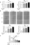
O-GlcNAcylation-inducing treatments increase cell growth and migration and inhibit apoptosis in CaSki cells. (a) Cells were cultured in 1% FBS and cultured in the absence and presence of O-GlcNAcylation-inducing agents (TG 10 µM and GlcN 5 mM) and IGF-1 (5 nM). MTT assay was used to determine the cell growth at 24 h and 48 h. Results are the mean ± SEM of 5 independent experiments. (b) Wound healing assay was performed as described in “Methods”. Representative images of the wound at 0, 24 and 48 h are shown. Migration is expressed as the wound closure percentage at 24 h and 48 h. The results are presented as mean ± SEM of 4 independent experiments. (c) CaSki cells were treated for 24 h with O-GlcNAcylation-inducing agents, IGF-1 and Cisplatin, and then stained with Annexin V and propidium iodide for FACS analysis. The results are presented as mean ± SEM of 4 independent experiments. Statistical analysis was performed using ANOVA followed by Tukey’s post-test. *, **, ***p < 0.05, p < 0.01 and p < 0.001, respectively.
Figure 3
Figure 3
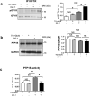
Effect of O-GlcNAcylation-inducing treatments on IGF-1R phosphorylation and PTP1B expression and activity in CaSki cells. CaSki cells were cultured in the presence of 1% FBS in the absence or presence of TG (10 µM for 24 h) and GlcN (5 mM for 6 h). Cells were then stimulated with IGF-1 (5 nM) during 10 min. (a) CaSki cells were lysed and IGF1R were immunoprecipitated with an anti-IGF1R antibody. IGF1R phosphorylation was analysed by western blotting using an anti-phospho-IGF1R antibody. The blots were reprobed with an anti-IGF1R antibody (the uncropped western-blots are shown on the Supplementary Information file S1). The histogram represents the ratio of P-IGF1R/IGF1R signal obtained by densitometric analysis of the blots. Results are the means ± SEM of 3 independent experiments. (b) CaSki cells were cultured in presence of 1% FBS in the absence or presence of TG (10 µM for 24 h) and GlcN (5 mM for 6 h). Cells were then stimulated with IGF-1 (5 nM) during 10 min. Cells were lysed and PTP1B expression was evaluated by western-blotting (the uncropped western-blots are shown on the Supplementary Information file S1). The histogram represents the ratio of PTP1B/GAPDH signal obtained by densitometric analysis of the blots. Results are the means ± SEM of 4 independent experiments. (c) Cell lysates were immunoprecipitated using an anti-PTP1B antibody and PTP1B activity was measured using p-nitrophenyl-phosphate (pNPP) as a substrate. Histograms represent the means ± SEM of PTP1B enzymatic activity (optical density at 405 nm) in 4 independent experiments. Statistical analysis was performed using ANOVA followed by Tukey’s post-test. *; **; ***p < 0.05, p < 0.01, and p < 0.001, respectively. NS non-significant.
Figure 4
Figure 4
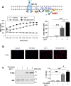
O-GlcNAcylation-inducing treatments increase IGF-1 effects on PI-3 kinase/Akt pathway in CaSki cells. (a) Activation of PI-3 kinase induces the phosphorylation of phosphatidyl-inositol 2 phosphate (PIP2) into phosphatidyl-inositol 3 phosphate (PIP3) and subsequent recruitment of Akt to the plasma membrane through its pleckstrin homology (PH) domain. CaSki cells were co-transfected with cDNAs coding for the PH domain of Akt fused to a luciferase (Luc-Akt-PH) and a plasma membrane-targeted YFP (YFP-mem). 24 h after transfection, cells were cultured in the presence of 1% FBS in the absence or presence of TG (10 µM for 24 h) and GlcN (5 mM for 6 h). Cells were incubated with coelenterazine for 10 min, and then stimulated with IGF-1 (5 nM). Light emission acquisition at 480 nm and 532 nm was started immediately after IGF-1 addition. Results were expressed in miliBRET units (mBU). Left panel: a typical real-time experiment showing the effect of O-GlcNAcylation-inducing treatment on IGF-1-induced PIP3 production in CaSki cells. Right panel: Results are expressed as the delta BRET (increased BRET above basal) and are the means ± SEM of 6 independent experiments. (b) CaSki cells were cultured in the presence of TG (10 µM) for 24 h and GlcN (5 mM) for 6 h, stimulated with IGF-1 for 10 min, and then fixed and incubated with P-Akt antibody. Representative images of immunofluorescence with P-Akt (red) and dapi staining of cell nuclei (blue) are shown. (c) CaSki cells were cultured in the presence of TG (10 µM for 24 h) and GlcN (5 mM for 6 h), stimulated with IGF-1 for 10 min, and then lysed. Proteins were submitted to western-blotting and Akt phosphorylation was detected with anti-phospho-Akt antibody (the uncropped western-blots are shown on the Supplementary Information file S1). The histogram represents the ratio of P-Akt/Akt signals obtained by densitometric analysis of the blots. Results are the means ± SEM of 8 independent experiments. Statistical analysis was performed using ANOVA followed by Tukey’s post-test. *, ***p < 0.05, p < 0.001, respectively. NS non-significant.
Figure 5
Figure 5
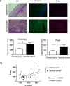
O-GlcNAcylation and Akt phosphorylation levels in cervical cancer tissues. (a) Tissues were stained with Hematoxylin/Eosin (HE). Indirect inmunofluorescence was performed using indicated primary antibodies followed by secondary antibodies (FITC-conjugated anti-mouse and biotinylated anti-rabbit/Alexa Fluor 594). Representative merged images of immunofluorescence staining for O-GlcNAc (green), P-Akt (red) and cell nuclei (blue) of normal and cervical cancer-tissues are shown. The histograms display the relative quantification of the mean fluorescence integrated density by Image J software. Data are means ± SEM of n = 5 tissues, analysing each tissue in at least 3 different optical fields. Statistical analysis was performed using a Student’s t-test. *p < 0.05. (b) Positive correlation between O-GlcNAc and phopho-Akt signals in samples from normal and cervical cancer-tissues evaluated by Pearson analysis.

Similar articles

Cited by

References

    1. Issad T, Kuo M. O-GlcNAc modification of transcription factors, glucose sensing and glucotoxicity. Trends Endocrinol. Metab. 2008;19:380–389. - PubMed
    1. Fardini Y, Dehennaut V, Lefebvre T, Issad T. O-GlcNAcylation: A new cancer hallmark? Front. Endocrinol. (Lausanne). 2013;4:99. - PMC - PubMed
    1. Nin D, Huang W, Ali M, Yew C, Kutateladze T, Deng L. O-GlcNAcylation of MLL5β is essential for MLL5β-AP-1 transcription complex assembly at the HPV16/18-long control region. J. Mol. Cell. Biol. 2015;7:180–183. - PMC - PubMed
    1. Kim M, Kim Y, Kim H, Kang M, Park J, Lee D, Roh G, Kim H, Kang S, Cho G, Park J, Cho J, Shin J, Choi W. O-linked N-acetylglucosamine transferase promotes cervical cancer tumorigenesis through human papillomaviruses E6 and E7 oncogenes. Oncotarget. 2016;7:44596–44607. - PMC - PubMed
    1. Zeng Q, Zhao R, Chen J, Li Y, Li X, Liu X, Zhang W, Quan C, Wang Y, Zhai Y, Wang J, Youssef M, Cui R, Liang J, Genovese N, Chow L, Li Y, Xu Z. O-linked GlcNAcylation elevated by HPV E6 mediates viral oncogenesis. Proc. Natl. Acad. Sci. USA. 2016;113:9333–9338. - PMC - PubMed

Publication types

MeSH terms