Structure-based design of prefusion-stabilized human metapneumovirus fusion proteins
- PMID: 35288548
- PMCID: PMC8921277
- DOI: 10.1038/s41467-022-28931-3
Structure-based design of prefusion-stabilized human metapneumovirus fusion proteins
Abstract
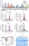
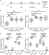
The human metapneumovirus (hMPV) fusion (F) protein is essential for viral entry and is a key target of neutralizing antibodies and vaccine development. The prefusion conformation is thought to be the optimal vaccine antigen, but previously described prefusion F proteins expressed poorly and were not well stabilized. Here, we use structures of hMPV F to guide the design of 42 variants containing stabilizing substitutions. Through combinatorial addition of disulfide bonds, cavity-filling substitutions, and improved electrostatic interactions, we describe a prefusion-stabilized F protein (DS-CavEs2) that expresses at 15 mg/L and has a melting temperature of 71.9 °C. Crystal structures of two prefusion-stabilized hMPV F variants reveal that antigenic surfaces are largely unperturbed. Importantly, immunization of mice with DS-CavEs2 elicits significantly higher neutralizing antibody titers against hMPV A1 and B1 viruses than postfusion F. The improved properties of DS-CavEs2 will advance the development of hMPV vaccines and the isolation of therapeutic antibodies.
© 2022. The Author(s).
Conflict of interest statement
C.-L.H., S.A.R., and J.S.M. are inventors on U.S. patent application no. 63/089,978 (Prefusion-stabilized hMPV F Proteins). The remaining authors declare no competing interests.
Figures


Similar articles
-
Structure-based design of a single-chain triple-disulfide-stabilized fusion-glycoprotein trimer that elicits high-titer neutralizing responses against human metapneumovirus.PLoS Pathog. 2023 Sep 22;19(9):e1011584. doi: 10.1371/journal.ppat.1011584. eCollection 2023 Sep. PLoS Pathog. 2023. PMID: 37738240 Free PMC article.
-
Interprotomer disulfide-stabilized variants of the human metapneumovirus fusion glycoprotein induce high titer-neutralizing responses.Proc Natl Acad Sci U S A. 2021 Sep 28;118(39):e2106196118. doi: 10.1073/pnas.2106196118. Proc Natl Acad Sci U S A. 2021. PMID: 34551978 Free PMC article.
-
Structure, Immunogenicity, and Conformation-Dependent Receptor Binding of the Postfusion Human Metapneumovirus F Protein.J Virol. 2021 Aug 25;95(18):e0059321. doi: 10.1128/JVI.00593-21. Epub 2021 Aug 25. J Virol. 2021. PMID: 34160259 Free PMC article.
-
Clinical Potential of Prefusion RSV F-specific Antibodies.Trends Microbiol. 2018 Mar;26(3):209-219. doi: 10.1016/j.tim.2017.09.009. Epub 2017 Oct 17. Trends Microbiol. 2018. PMID: 29054341 Review.
-
Human Metapneumovirus.Microbiol Spectr. 2014 Oct;2(5). doi: 10.1128/microbiolspec.AID-0020-2014. Microbiol Spectr. 2014. PMID: 26104361 Review.
Cited by
-
Structural basis for respiratory syncytial virus and human metapneumovirus neutralization.Curr Opin Virol. 2023 Aug;61:101337. doi: 10.1016/j.coviro.2023.101337. Curr Opin Virol. 2023. PMID: 37544710 Free PMC article. Review.
-
Mechanistic insights into structure-based design of a Lyme disease subunit vaccine.bioRxiv [Preprint]. 2024 Oct 28:2024.10.23.619738. doi: 10.1101/2024.10.23.619738. bioRxiv. 2024. PMID: 39554036 Free PMC article. Preprint.
-
Potent cross-neutralization of respiratory syncytial virus and human metapneumovirus through a structurally conserved antibody recognition mode.Cell Host Microbe. 2023 Aug 9;31(8):1288-1300.e6. doi: 10.1016/j.chom.2023.07.002. Epub 2023 Jul 28. Cell Host Microbe. 2023. PMID: 37516111 Free PMC article.
-
Structure-based design of a single-chain triple-disulfide-stabilized fusion-glycoprotein trimer that elicits high-titer neutralizing responses against human metapneumovirus.PLoS Pathog. 2023 Sep 22;19(9):e1011584. doi: 10.1371/journal.ppat.1011584. eCollection 2023 Sep. PLoS Pathog. 2023. PMID: 37738240 Free PMC article.
-
Systematic computer-aided disulfide design as a general strategy to stabilize prefusion class I fusion proteins.Front Immunol. 2024 Jul 24;15:1406929. doi: 10.3389/fimmu.2024.1406929. eCollection 2024. Front Immunol. 2024. PMID: 39114655 Free PMC article.
References
-
- Deffrasnes C, Hamelin MÈ, Boivin G. Human metapneumovirus. Semin. Respiratory Crit. Care Med. 2007;28:213–221. - PubMed
-
- Skiadopoulos MH, et al. Individual contributions of the human metapneumovirus F, G, and SH surface glycoproteins to the induction of neutralizing antibodies and protective immunity. Virology. 2006;345:492–501. - PubMed
-
- Schickli JH, Kaur J, Ulbrandt N, Spaete RR, Tang RS. An S101P substitution in the putative cleavage motif of the human metapneumovirus fusion protein is a major determinant for trypsin-independent growth in vero cells and does not alter tissue tropism in hamsters. J. Virol. 2005;79:10678–10689. - PMC - PubMed
Publication types
MeSH terms
Substances
LinkOut - more resources
Full Text Sources
Other Literature Sources

