. 2022 Feb 25:12:801230.
doi: 10.3389/fonc.2022.801230. eCollection 2022.

Ionizing Radiation-Induced GDF15 Promotes Angiogenesis in Human Glioblastoma Models by Promoting VEGFA Expression Through p-MAPK1/SP1 Signaling

Affiliations

Ionizing Radiation-Induced GDF15 Promotes Angiogenesis in Human Glioblastoma Models by Promoting VEGFA Expression Through p-MAPK1/SP1 Signaling

Hyejin Park et al. Front Oncol. .

Abstract

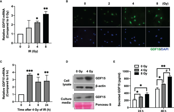
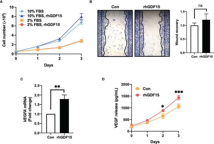
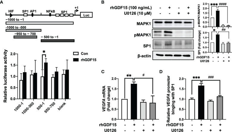
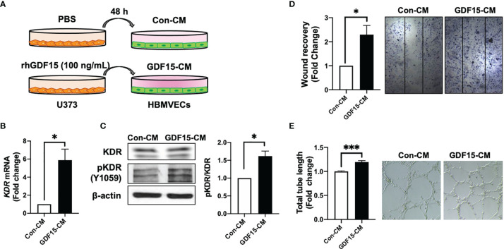
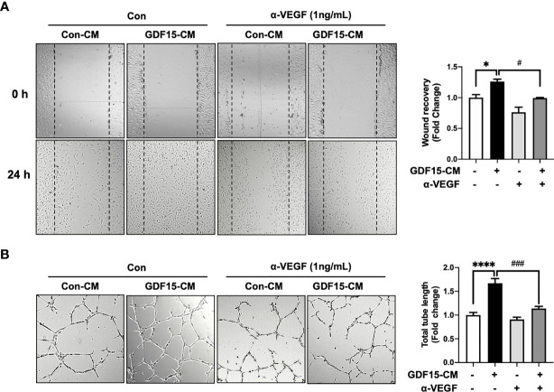
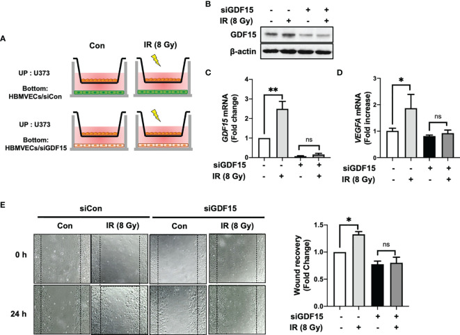
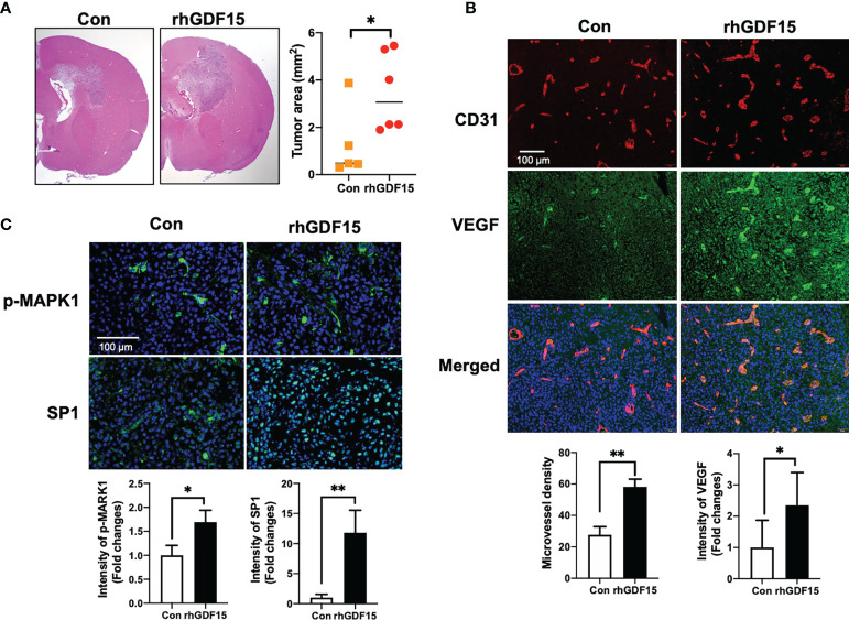
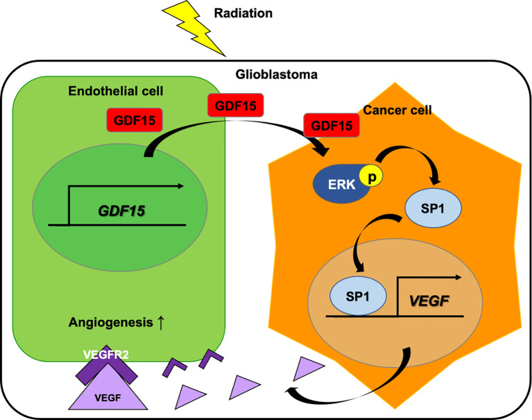
Glioblastoma multiforme (GBM), the most aggressive cancer type that has a poor prognosis, is characterized by enhanced and aberrant angiogenesis. In addition to surgical resection and chemotherapy, radiotherapy is commonly used to treat GBM. However, radiation-induced angiogenesis in GBM remains unexplored. This study examined the role of radiation-induced growth/differentiation factor-15 (GDF15) in regulating tumor angiogenesis by promoting intercellular cross-talk between brain endothelial cells (ECs) and glioblastoma cells. Radiation promoted GDF15 secretion from human brain microvascular endothelial cells (HBMVECs). Subsequently, GDF15 activated the transcriptional promoter VEGFA in the human glioblastoma cell line U373 through p-MAPK1/SP1 signaling. Upregulation of vascular endothelial growth factor (VEGF) expression in U373 cells resulted in the activation of angiogenic activity in HBMVECs via KDR phosphorylation. Wound healing, tube formation, and invasion assay results revealed that the conditioned medium of recombinant human GDF15 (rhGDF15)-stimulated U373 cell cultures promoted the angiogenic activity of HBMVECs. In the HBMVEC-U373 cell co-culture, GDF15 knockdown mitigated radiation-induced VEGFA upregulation in U373 cells and enhanced angiogenic activity of HBMVECs. Moreover, injecting rhGDF15-stimulated U373 cells into orthotopic brain tumors in mice promoted angiogenesis in the tumors. Thus, radiation-induced GDF15 is essential for the cross-talk between ECs and GBM cells and promotes angiogenesis. These findings indicate that GDF15 is a putative therapeutic target for patients with GBM undergoing radio-chemotherapy.

Keywords: GDF15; angiogenesis; endothelial cells; glioblastoma; radiotherapy.


Conflict of interest statement

The authors declare that the research was conducted in the absence of any commercial or financial relationships that could be construed as a potential conflict of interest.

Figures

Figure 1
Figure 1
Effect of ionizing radiation (IR) on GDF15 expression in human brain microvascular endothelial cells (HBMVECs). (A) Effect of different doses of IR on GDF15 mRNA expression in HBMVECs. Cultured cells were harvested 24 h post-IR exposure, and GDF15 mRNA levels were analyzed using quantitative reverse transcription polymerase chain reaction (qRT-PCR). (B) Effect of different doses of IR (0 – 8 Gy) on GDF15 protein levels in HBMVECs. GDF15 protein expression was detected by immunofluorescence analysis using anti-GDF15 antibody (green). Nuclei were stained with 4′,6-diamidino-2-phenylindole (DAPI) (blue) (Magnification: 400×); (C) Time course of GDF15 mRNA expression after irradiation with 4 Gy of IR. GDF15 mRNA levels measured using qRT-PCR at the indicated time points. (D) Immunoblotting analysis of GDF15 in the cell lysate and culture medium reveals that IR upregulated GDF15 protein levels. Cells and culture media were harvested at 24 h post-IR exposure. (E) IR promotes the secretion of GDF15. The secretion of GDF15 into the culture medium was measured using a human GDF15 enzyme-linked immunosorbent assay kit at 24 and 48 h post-irradiation with 4 or 8 Gy of IR. Data are presented as the mean ± standard deviation of three experiments. *p < 0.05, **p < 0.01, and ***p < 0.001 compared with the control. ## p < 0.05 compared with 4 Gy.
Figure 2
Figure 2
GDF15 promotes VEGFA expression in U373 glioblastoma cells but not proliferation and migration of U373 cells. (A) U373 cells were treated with 100 ng/mL rhGDF15 protein for 3 days in a medium with different serum concentrations (2% or 10% fetal bovine serum) and the cells were counted daily. (B) The cells were seeded on 12-well plates (3 × 10⁵ cells/well) and cultured for 1 day. The monolayer was scratched and incubated for 24 h in a medium containing 2% serum. Subsequently, the cells were stained with a fixation solution containing trypan blue. (C) The VEGFA mRNA levels in rhGDF15-stimulated U373 cells were measured using quantitative reverse transcription polymerase chain reaction. (D) Soluble VEGF was quantified in the enriched culture medium containing 2% serum using enzyme-linked immunosorbent assay from days 1–3. Data are presented as the mean ± standard deviation of three experiments. *p < 0.05, **p < 0.01, and ***p < 0.001 compared with control group. ns, no significance.
Figure 3
Figure 3
GDF15 upregulates VEGFA transcription by promoting the binding of SP1 to its promoter. (A) 293T cells were transfected with a luciferase construct containing different VEGFA promotor regions or control vectors and treated with recombinant human GDF15 (rhGDF15). Data are presented as the mean ± standard deviation of three experiments (*p < 0.05). (B) For immunoblotting analysis of MAPK1, p-MAPK1, and SP1 in U373 cells, the cells were incubated for 1 h in the presence or absence of a MAPK1 inhibitor (U0126) and treated with rhGDF15 for 6 h. (C) Quantitative reverse transcription polymerase chain reaction (qRT-PCR) analysis of VEGFA mRNA expression in U373 cells treated with rhGDF15 and/or U0126. qRT-PCR analysis was performed using samples extracted 12 h post-rhGDF15 treatment. For inhibition studies, U373 cells were incubated with 10 µM U0126 for 1 h before treatment with 100 ng/mL rhGDF15. (D) Cells were incubated with or without rhGDF15 and U0126 and subjected to chromatin immunoprecipitation (ChIP) assay using anti-SP1 or IgG isotype control antibodies. Bar graphs represent the qRT-PCR results for immunoprecipitated VEGFA promoter. Data are presented as the mean ± standard error (**p < 0.01 and ***p < 0.001 compared with control group; # p < 0.05, ## p < 0.01, ### p < 0.001 and #### p < 0.0001 compared with rhGDF15-treated group.
Figure 4
Figure 4
GDF15-induced VEGFA activates angiogenesis. (A) Schematic representing human brain microvascular endothelial cells (HBMVECs) cultured in control conditioned medium (Con-CM; conditioned medium from U373 cells) or GDF15-CM (conditioned medium from rhGDF15-stimulated U373 cells); (B) KDR mRNA expression in HBMVECs was measured using quantitative reverse transcription polymerase chain reaction (qRT-PCR). (C) Phosphorylation of KDR was examined using western blotting (β-actin as used as loading control); (D) Wound healing assay results. The day before the experiment, HBMVECs were seeded in 12-well plates (1.5 × 10⁵/well). The monolayer was scratched to introduce a wound and treated with Con-CM or GDF15-CM. The migration of cells into the wound area was analyzed after 24 h (significance compared with the cells cultured in Con-CM). (E) Tube formation assay results. HBMVECs (1.5 × 10⁵ cells) were incubated on Matrigel matrix for 3 days with Con-CM or GDF15-CM. The tube length was measured using ImageJ angiogenesis analyzer (significance was compared with the cells cultured in Con-CM). (*p < 0.05 and ***p < 0.001 compared with the cells cultured in Con-CM).
Figure 5
Figure 5
Anti-VEGF antibody blocks GDF15 mediated angiogenesis. (A) Wound healing assay was performed using human brain microvascular endothelial cells (HBMVECs) in the absence/presence of anti-VEGF antibody. The day before the experiment, HBMVECs were seeded in 12-well plates (1.5 × 10⁵/well). The monolayer was scratched to introduce a wound and cells were cultured with control conditioned medium (Con-CM; conditioned medium from HBMVECs) or GDF15-CM HBMVECs (conditioned medium from rhGDF15-stimulated HBMVECs) with added control antibody or α-VEGF (1 ng/mL each). The wounded areas were photographed at 0 h and 24 h and the cell migration as percent mean distance migration was assessed. (B) Representative photographs of tube formation assay. HBMVECs (1.5 × 10⁵ cells) were incubated on Matrigel matrix for 12 h with Con-CM or GDF15-CM in the presence or absence of α-VEGF (1 ng/mL). The tube length was measured using ImageJ angiogenesis analyzer (significance was compared with the group Con-CM in the absence of α-VEGF). Data are presented as the mean ± standard error (*p < 0.01 and ****p < 0.001 compared with the non-treated CM control; # p < 0.05 and ### p < 0.001 compared with rhGDF15- CM group).
Figure 6
Figure 6
GDF15 knockdown suppresses ionizing radiation (IR)-induced wound recovery. (A) Graphic experimental scheme; for the co-culture of U373 cells and human brain microvascular endothelial cells (HBMVECs), U373 cells were seeded on the upper well and the siGDF15-transfected or siCon-transfected HBMVECs were seeded on the bottom well. After irradiation with 8 Gy IR, U373 cells and HBMVECs were co-cultured for 24 h. (B) In HBMVECs, the protein levels of GDF15 were examined by immunoblotting analysis 24 h post-IR exposure. (C) In HBMVECs, the levels of GDF15 mRNA were determined using quantitative reverse transcription polymerase chain reaction 24 h post-IR exposure. (D) In U373 cells, the levels of VEGFA mRNA were determined using qRT-PCR 24 h post-IR exposure. (E) For the wound healing assay, the HBMVECs on the plate were scratched and incubated for 24 h after irradiation with 8 Gy IR. The wound recovery rates were assessed using ImageJ. Data are presented as the mean ± standard deviation of three experiments. *p < 0.05 and **p < 0.01 compared with siCon-transfected and nonirradiated HBMVECs. ns, no significance.
Figure 7
Figure 7
GDF15 promotes angiogenesis by stimulating VEGFA secretion in the brain tumor. (A) Representative section of the brain tumors from mice injected with control U373 cells and rhGDF15-stimulated U373 cells. The brain sections were stained with hematoxylin and eosin. The brain tumor area was measured using ImageJ. (B) VEGFA (green) and CD31-positive endothelial cells (red) in mouse brain tumor tissues were analyzed using immunofluorescence staining. Microvessel density was counted as the number of CD31-positive vessels. Scale bar = 100 µm. (C) Representative image of immunofluorescence analysis of the phosphorylation of MAPK1 (p-MAPK1; upper panel; green) and expression of SP1 (upper panel; green) counterstained with 4′,6-diamidino-2-phenylindole (DAPI) (blue). Scale bar = 100 µm. Fluorescence intensity was calculated using ImageJ as follows: Corrected total cell fluorescence = integrated density − (area of selected cell × mean fluorescence of background readings). Data are presented as the mean ± standard (control group, n = 5; rhGDF15-treated group; n = 6), * p < 0.05 and ** p < 0.01 compared with control group.
Figure 8
Figure 8
Role of GDF15 in ionizing radiation (IR)-induced angiogenesis in glioblastoma. Radiation upregulates GDF15 expression in the endothelial cells (ECs). EC-derived GDF15 activates the p-MAPK1/SP1 pathway in glioblastoma cells. SP1 stimulates the promoter activity of VEGF. The upregulated VEGF levels induce angiogenesis in glioblastoma through p-KDR on ECs.

Similar articles

Cited by

References

    1. Bulnes S, Bengoetxea H, Ortuzar N, Argandona EG, Garcia-Blanco A, Rico-Barrio I, et al. . Angiogenic Signalling Pathways Altered in Gliomas: Selection Mechanisms for More Aggressive Neoplastic Subpopulations With Invasive Phenotype. J Signal Transduct (2012) 2012:597915. doi: 10.1155/2012/597915 - DOI - PMC - PubMed
    1. Plate KH, Scholz A, Dumont DJ. Tumor Angiogenesis and Anti-Angiogenic Therapy in Malignant Gliomas Revisited. Acta Neuropathol (2012) 124(6):763–75. doi: 10.1007/s00401-012-1066-5 - DOI - PMC - PubMed
    1. Ahir BK, Engelhard HH, Lakka SS. Tumor Development and Angiogenesis in Adult Brain Tumor: Glioblastoma. Mol Neurobiol (2020) 57(5):2461–78. doi: 10.1007/s12035-020-01892-8 - DOI - PMC - PubMed
    1. Wong ML, Prawira A, Kaye AH, Hovens CM. Tumour Angiogenesis: Its Mechanism and Therapeutic Implications in Malignant Gliomas. J Clin Neurosci (2009) 16(9):1119–30. doi: 10.1016/j.jocn.2009.02.009 - DOI - PubMed
    1. Hida K, Maishi N, Annan DA, Hida Y. Contribution of Tumor Endothelial Cells in Cancer Progression. Int J Mol Sci (2018) 19(5):1272. doi: 10.3390/ijms19051272 - DOI - PMC - PubMed