. 2022 Feb;10(4):204.
doi: 10.21037/atm-22-6.

High glucose inhibits the osteogenic differentiation of periodontal ligament stem cells in periodontitis by activating endoplasmic reticulum stress

Affiliations

High glucose inhibits the osteogenic differentiation of periodontal ligament stem cells in periodontitis by activating endoplasmic reticulum stress

Jun Tan et al. Ann Transl Med. 2022 Feb.

Abstract

Background: Periodontitis is a highly prevalent dental disease which is associated with diabetes and is challenging to cure in diabetic patients. However, the mechanism of comorbid diabetes and periodontitis is still unclear. This study aimed to uncover the role of endoplasmic reticulum (ER) stress in high glucose-associated periodontitis.

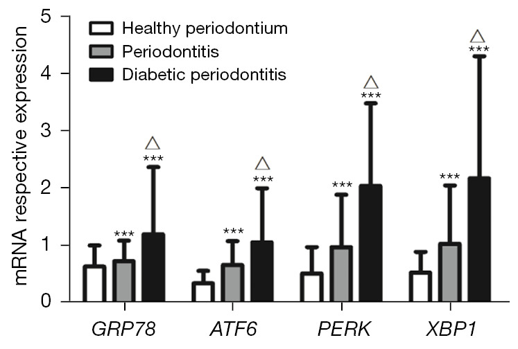
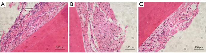
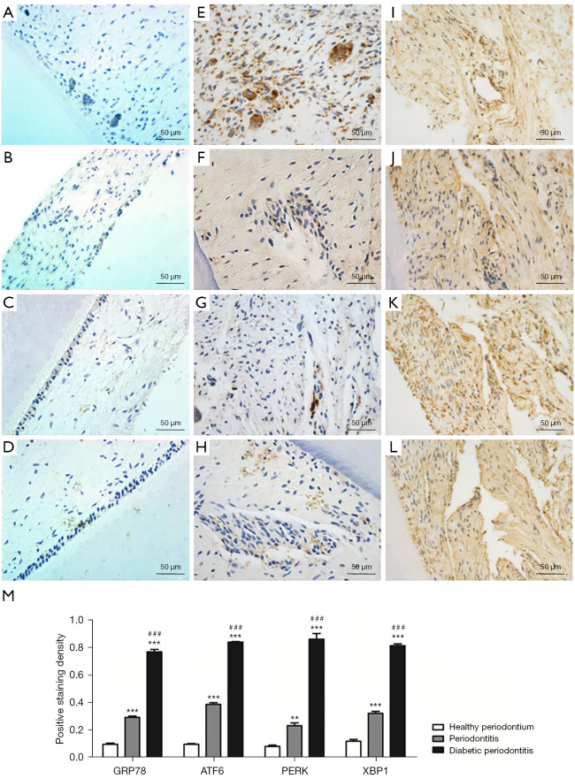
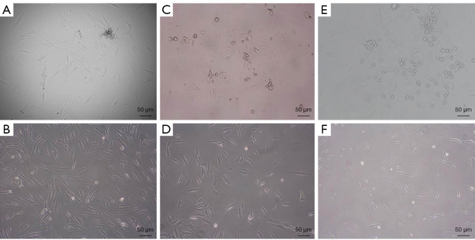
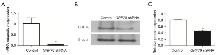
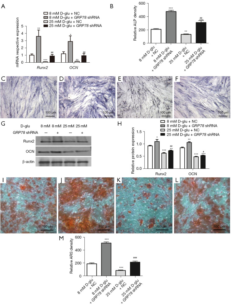
Methods: Periodontal tissues were obtained from diabetic patients with periodontitis, periodontitis patients without systemic disease, and healthy teeth. The expressions of ER stress-related factors GRP78, ATF6, PERK and XBP1 were detected by quantitative real-time polymerase chain reaction (qRT-PCR) and immumohistochemical staining. Periodontal ligament stem cells (PDLSCs) from three states of periodontal tissues were isolated and cultured as diabetic PDLSCs (dPDLSCs), inflamed PDLSCs (iPDLSCs) and healthy PDLSCs (hPDLSCs), and the cell stemness was assayed. Different concentrations (8, 11, and 25 mmol/L) of D-glucose were used on hPDLSCs to simulate high glucose microenvironment. The changes of osteogenic ability of PDLSCs were observed, and the expressions of ER stress-related factors in different time point (6, 12, 24, and 72 h) were detected. Finally, GRP78 shRNA lentivirus was used to block ER stress on PDLSCs in the 25 mmol/L D-glucose microenvironment, and the osteogenic ability of PDLSCs was observed.

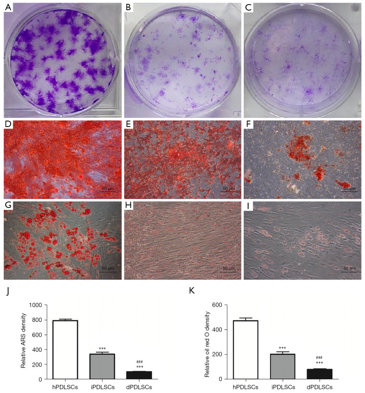
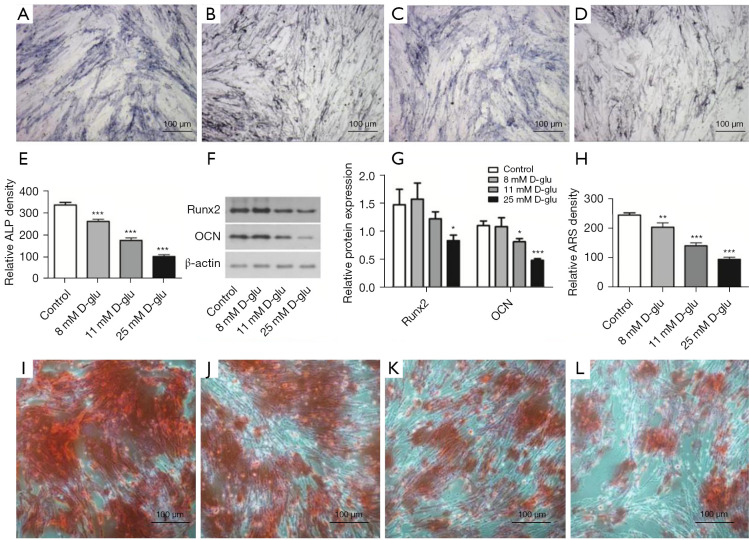
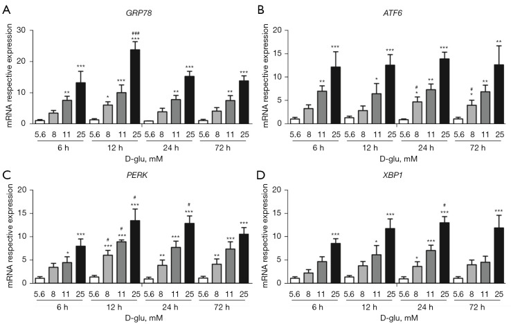
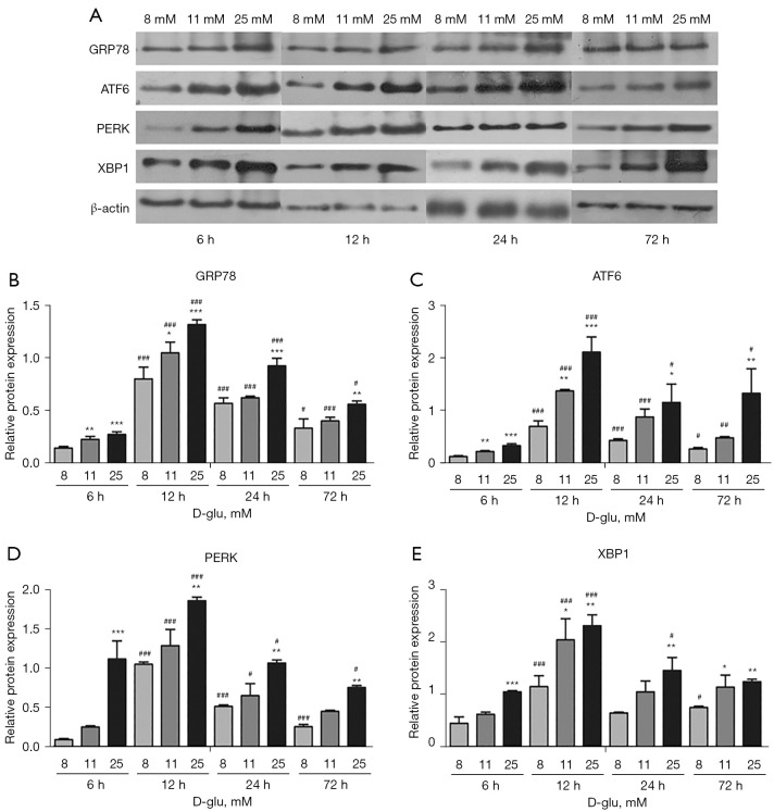
Results: The results showed that the expressions of GRP78, ATF6, PERK, and XBP1 were highest in the diabetic periodontitis group and lowest in the healthy periodontal tissue group (P<0.05). The clone formation, osteogenic and lipogenic differentiation abilities were lowest in dPDLSCs and highest in hPDLSCs. With the increase of glucose concentration, the osteogensis ability of PDLSCs decreased. After 6 hours of stimulation with D-glucose 25 mmol/L, the ER stress pathways in PDLSCs were effectively activated, and the peak value was reached at 12 hours. The decrease in the osteogensis ability of PDLSCs in a high glucose microenvironment reversed when ER stress was blocked.

Conclusions: The osteogenic differentiation ability of PDLSCs cells is inhibited in a high glucose microenvironment, and this effect is realized by ER stress activation. Blocking ER stress can partially restore the reduced osteogenic ability of PDLSCs. These results suggest that high glucose inhibits the osteogenic differentiation ability of PDLSCs by activating ER stress, which ultimately exacerbates periodontitis.

Keywords: Periodontitis; diabetes; endoplasmic reticulum stress; osteogenesis; periodontal ligament stem cells (PDLSCs).


Conflict of interest statement

Conflicts of Interest: All authors have completed the ICMJE uniform disclosure form (available at https://atm.amegroups.com/article/view/10.21037/atm-22-6/coif). The authors report that this work was supported by the National Natural Science Foundation of China (No. 81700987). The authors have no other conflicts of interest to declare.

Figures

Figure 1
Figure 1
Messenger RNA expression levels of ER stress pathway-related factors in periodontal tissues from individuals with healthy teeth, periodontitis patients without systemic diseases, and diabetic patients with periodontitis. ***, P<0.001 compared with healthy group. ∆, P<0.05 compared with inflamed group. RNA, ribonucleic acid; ER, endoplasmic reticulum.
Figure 2
Figure 2
HE staining of periodontal tissues from different sources. (A) HE staining of healthy periodontal tissue. (B) HE staining of periodontal tissue of periodontitis patients without systemic diseases. (C) HE staining of periodontal tissue of diabetic patients with periodontitis. Bar =100 µm. HE, hematoxylin and eosin.
Figure 3
Figure 3
Immunohistochemical staining of ER stress-related factors in periodontal tissues from individuals with healthy teeth, patients with periodontitis, and patients with diabetic periodontitis. GRP78 (A), ATF6 (B), PERK (C), and XBP1 (D) staining of periodontal tissue from healthy group, GRP78 (E), ATF6 (F), PERK (G), and XBP1 (H) staining of periodontal tissue from inflamed group, and GRP78 (I), ATF6 (J), PERK (K), and XBP1 (L) staining of periodontal tissue from diabetic group. (M) Analysis of immunohistochemical staining positivity in each group. Bar =50 µm. ***, P<0.001, **, P<0.01 compared with healthy group. ###, P<0.001 compared with inflamed group. ER, endoplasmic reticulum; GRP78, glucose regulated protein 78; ATF6, activating transcription factor 6; PERK, double-stranded RNA-activated protein kinase (PKR) like endoplasmic reticulum kinase; XBP1, X box-binding protein 1.
Figure 4
Figure 4
PDLSCs observed with an inverted microscope. (A) Primary PDLSCs from healthy periodontal tissue. (B) Second-generation hPDLSCs after limited dilution and purification. (C) Primary PDLSCs from periodontal tissue of inflamed group. (D) Second-generation iPDLSCs after limited dilution and purification. (E) PDLSCs from periodontal tissue of diabetic group. (F) Second -generation dPDLSCs after limited dilution and purification. Bar =50 µm. PDLSCs, periodontal ligament stem cells; hPDLSCs, healthy periodontal ligament stem cells; iPDLSCs, inflamed periodontal ligament stem cells; dPDLSCs, diabetic periodontal ligament stem cells.
Figure 5
Figure 5
Cell stemness assay of PDLSCs. Clone formation assay of hPDLSCs (A), iPDLSCs (B), and dPDLSCs (C). Alizarin red staining of osteogenic-induced hPDLSCs (D), iPDLSCs (E), and dPDLSCs (F). Bar =100 µm. Oil red O staining of adipogenic-induced hPDLSCs (G), iPDLSCs (H), and dPDLSCs (I). Bar =50 µm. (J) Comparison of the total optical density values of alizarin red positive staining in each group. (K) Comparison of the total optical density values of Oil red O positive staining in each group. ***, P<0.001 compared with hPDLSCs. ###, P<0.001 compared with iPDLSCs. PDLSCs, periodontal ligament stem cells; hPDLSCs, healthy periodontal ligament stem cells; iPDLSCs, inflamed periodontal ligament stem cells; dPDLSCs, diabetic periodontal ligament stem cells.
Figure 6
Figure 6
The osteogenic differentiation abilities of PDLSCs cultured in osteogenic inducing medium containing D-glucose at different concentrations. ALP staining of PDLSCs after osteogenic induction in 5.6 mmol/L D-glucose (control group) (A), 8 mmol/L D-glucose (B), 11 mmol/L D-glucose (C), and 25 mmol/L D-glucose (D). (E) Comparison of the total optical density values of ALP positive staining in each group. (F) The protein levels of Runx2 and OCN were detected by western blot after 2-week osteogenic induction at different D-glucose concentrations. (G) Quantification of Runx2 and OCN protein expression. The relative quantities evaluated by band grey value were normalized to β-actin and are presented as mean ± SD. (H) Comparison of the total optical density values of alizarin red staining. Alizarin red staining of PDLSCs after osteogenic induction in 5.6 mmol/L D-glucose (control group) (I), 8 mmol/L D-glucose (J), 11 mmol/L D-glucose (K), and 25 mmol/L D-glucose (L). Bar =100 µm. *, P<0.05, **, P<0.01, ***, P<0.01 compared with the control group. PDLSCs, periodontal ligament stem cells; ALP, alkaline phosphatase; OCN, osteocalcin; Runx2, runt-related transcription factor 2; SD, standard deviation.
Figure 7
Figure 7
Changes in the expression of ER stress-related genes in PDLSCs after D-glucose stimulation at different concentrations. The mRNA expression of GRP78 (A), ATF6 (B), PERK (C), and XBP1 (D) in PDLSCs after stimulation with different concentrations of D-glucose. *, P<0.05, **, P<0.01, ***, P<0.001 compared with the 5.6 mM D-glucose (control) group at the same time point. #, P<0.05, ###, P<0.001 compared with 6 hours after stimulation with the same D-glucose concentration. ER, endoplasmic reticulum; PDLSCs, periodontal ligament stem cells; mRNA, messenger ribonucleic acid; GRP78, glucose regulated protein 78; ATF6, activating transcription factor 6; PERK, double-stranded RNA-activated protein kinase (PKR) like endoplasmic reticulum kinase; XBP1, X box-binding protein 1.
Figure 8
Figure 8
Changes in the protein levels of ER stress-related factors in PDLSCs after stimulation with different concentrations of D-glucose. (A) The protein levels of ER stress-related factors after D-glucose stimulation at different concentrations, as detected by western blot. Quantification of the GRP78 (B), ATF6 (C), PERK (D), and XBP1 (E) protein levels, using β-actin for internal reference. *, P<0.05, **, P<0.01, ***, P<0.001 compared with 8 mmol/L D-glucose stimulation group at the same time point. #, P<0.05, ##, P<0.01, ###, P<0.001 compared with 6 hours after stimulation with the same D-glucose concentration. ER, endoplasmic reticulum; PDLSCs, periodontal ligament stem cells; GRP78, glucose regulated protein 78; ATF6, activating transcription factor 6; PERK, double-stranded RNA-activated protein kinase (PKR) like endoplasmic reticulum kinase; XBP1, X box-binding protein 1.
Figure 9
Figure 9
GRP78 expression levels after 48-hour lentivirus infection of PDLSCs cells. (A) The mRNA expressions of GRP78 detected by qRT-PCR. (B) The protein levels of GRP78, as detected by western blot. (C) Quantification of GRP78 protein levels, using β-actin for internal reference. **, P<0.05 compared with uninfected controls. GRP78, glucose regulated protein 78; PDLSCs, periodontal ligament stem cells; mRNA, messenger ribonucleic acid; qRT-PCR, quantitative real-time polymerase chain reaction.
Figure 10
Figure 10
The effects of GRP78 shRNA on the osteogenic differentiation ability of PDLSCs under different glucose concentrations. (A) The mRNA expression levels of osteogenic factors after 7-day osteogenic induction. (B) Comparison of the total optical density values of ALP positive staining in each group. ALP staining of PDLSCs after osteogenic induction in 8 mmol/L D-glucose + NC (control group) (C), 8 mmol/L D-glucose + GRP78 shRNA lentivirus (D), 25 mmol/L D-glucose + NC (E), and 25 mmol/L D-glucose + GRP78 shRNA lentivirus (F). (G) The protein levels of osteogenic factors, as detected by western blot. (H) Quantification of osteogenic factor protein expression, using β-actin for internal reference. Alizarin red staining of PDLSCs after osteogenic induction in 8 mmol/L D-glucose + NC (control group) (I), 8 mmol/L D-glucose + GRP78 shRNA lentivirus (J), 25 mmol/L D-glucose + NC (K), and 25 mmol/L D-glucose + GRP78 shRNA lentivirus (L). (M) Comparison of the total optical density values of alizarin red positive staining. Bar =100 µm. *, P<0.05, **, P<0.01, ***, P<0.001 compared with 8 mmol/L D-glucose + NC (control) group. #, P<0.05, ##, P<0.01, ###, P<0.001 compared with 25 mmol/L D-glucose + NC group. GRP78, glucose regulated protein 78; shRNA, short hairpin ribonucleic acid; PDLSCs, periodontal ligament stem cells; mRNA, messenger ribonucleic acid; ALP, alkaline phosphatase; NC, normal saline.

Similar articles

Cited by

References

    1. Slots J. Periodontitis: facts, fallacies and the future. Periodontol 2000 2017;75:7-23. 10.1111/prd.12221 - DOI - PubMed
    1. Pihlstrom BL, Michalowicz BS, Johnson NW. Periodontal diseases. Lancet 2005;366:1809-20. 10.1016/S0140-6736(05)67728-8 - DOI - PubMed
    1. Yoshihara A, Seida Y, Hanada N, et al. A longitudinal study of the relationship between periodontal disease and bone mineral density in community-dwelling older adults. J Clin Periodontol 2004;31:680-4. 10.1111/j.1600-051X.2004.00548.x - DOI - PubMed
    1. Mulligan R, Phelan JA, Brunelle J, et al. Baseline characteristics of participants in the oral health component of the Women's Interagency HIV Study. Community Dent Oral Epidemiol 2004;32:86-98. 10.1111/j.0301-5661.2004.00128.x - DOI - PubMed
    1. Preshaw PM, Bissett SM. Periodontitis and diabetes. Br Dent J 2019;227:577-84. 10.1038/s41415-019-0794-5 - DOI - PubMed