

Over-expression of miR-193a-3p regulates the apoptosis of colorectal cancer cells by targeting PAK3
- PMID: 35273739
- PMCID: PMC8902527
Over-expression of miR-193a-3p regulates the apoptosis of colorectal cancer cells by targeting PAK3
Abstract
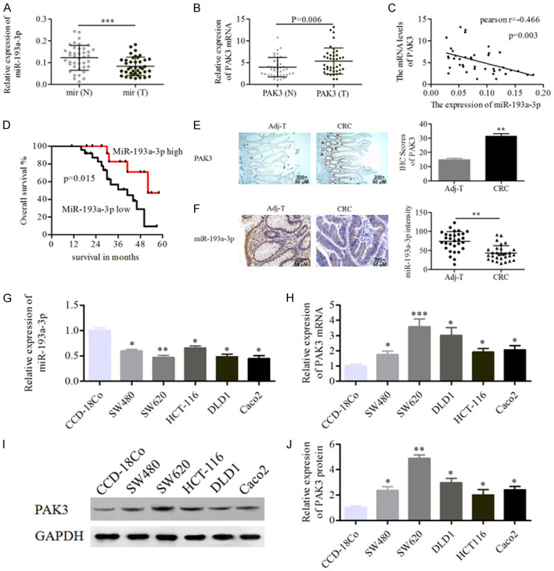
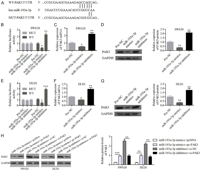
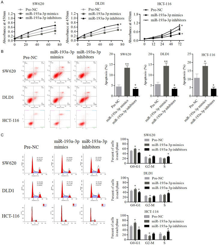
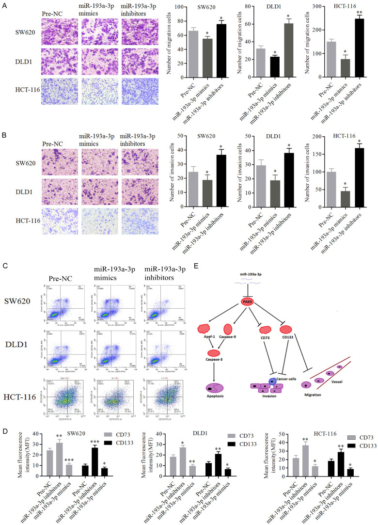
Although dysregulated expression of microRNAs (miRNA) has been investigated in colorectal cancer (CRC), MiR-193a-3p, as a tumor inhibitor, is less studied. To investigate the function and mechanism of miR-193a-3p in CRC, the potential function of miR-193a-3p in regulating PAK3 in CRC with a series of experimental assays including western blotting, qRT-PCR, bioinformatics analysis, a luciferase reporter assay, flow cytometry, Transwell assay, CCK8 assay and immunofluorescence were performed in this study. The results showed that miR-193a-3p was down-regulated in CRC tissues and cell lines, which was also correlated with tumor progression. PAK3 was predicted as a target gene of miR-193a-3p in CRC cells by TargetScan database, which was confirmed by luciferase assays. Moreover, overexpression of miR-193a-3p suppressed the viability, cell cycle progression, migration, and invasion, and induced apoptosis of CRC cells in vitro by regulating the PAK3 signaling pathway. Therefore, miR-193a-3p may serve as a tumor suppressor and potential target for CRC treatment.
Keywords: PAK3; apoptosis; colorectal cancer; miR-193a-3p; regulate.
AJTR Copyright © 2022.
Conflict of interest statement
None.
Figures

Similar articles
-
Circular RNA ACACA negatively regulated p53-modulated mevalonate pathway to promote colorectal tumorigenesis via regulating miR-193a/b-3p/HDAC3 axis.Mol Carcinog. 2023 Jun;62(6):754-770. doi: 10.1002/mc.23522. Epub 2023 Mar 15. Mol Carcinog. 2023. PMID: 36920044
-
Transcription factor SP1-induced microRNA-146b-3p facilitates the progression and metastasis of colorectal cancer via regulating FAM107A.Life Sci. 2021 Jul 15;277:119398. doi: 10.1016/j.lfs.2021.119398. Epub 2021 Apr 5. Life Sci. 2021. PMID: 33831429
-
miRNA-193a-3p Regulates the AKT2 Pathway to Inhibit the Growth and Promote the Apoptosis of Glioma Cells by Targeting ALKBH5.Front Oncol. 2021 Apr 23;11:600451. doi: 10.3389/fonc.2021.600451. eCollection 2021. Front Oncol. 2021. Retraction in: Front Oncol. 2021 Dec 29;11:840650. doi: 10.3389/fonc.2021.840650. PMID: 33968717 Free PMC article. Retracted.
-
MicroRNA-193a-3p suppresses the colorectal cancer cell proliferation and progression through downregulating the PLAU expression.Cancer Manag Res. 2019 Jun 12;11:5353-5363. doi: 10.2147/CMAR.S208233. eCollection 2019. Cancer Manag Res. 2019. PMID: 31354344 Free PMC article.
-
MiR-193a-3p functions as a tumour suppressor in human aldosterone-producing adrenocortical adenoma by down-regulating CYP11B2.Int J Exp Pathol. 2018 Apr;99(2):77-86. doi: 10.1111/iep.12267. Epub 2018 Apr 17. Int J Exp Pathol. 2018. PMID: 29665181 Free PMC article.
Cited by
-
MicroRNA-193a-3p as a Valuable Biomarker for Discriminating between Colorectal Cancer and Colorectal Adenoma Patients.Int J Mol Sci. 2024 Jul 26;25(15):8156. doi: 10.3390/ijms25158156. Int J Mol Sci. 2024. PMID: 39125725 Free PMC article.
-
Circular RNA COL1A1 promotes Warburg effect and tumor growth in nasopharyngeal carcinoma.Discov Oncol. 2024 Apr 15;15(1):120. doi: 10.1007/s12672-024-00941-1. Discov Oncol. 2024. PMID: 38619648 Free PMC article.
-
Circular RNA PGPEP1 induces colorectal cancer malignancy and immune escape.Cell Cycle. 2023 Jul-Aug;22(14-16):1743-1758. doi: 10.1080/15384101.2023.2225923. Epub 2023 Jul 9. Cell Cycle. 2023. PMID: 37424115 Free PMC article.
References
-
- Bray F, Ferlay J, Soerjomataram I, Siegel RL, Torre LA, Jemal A. Global cancer statistics 2018: GLOBOCAN estimates of incidence and mortality worldwide for 36 cancers in 185 countries. CA Cancer J Clin. 2018;68:394–424. - PubMed
-
- Ota M, Takamura N, Irimura T. Involvement of cell surface glycans in adhesion of human colon carcinoma cells to liver tissue in a frozen section assay: role of endo-β-galactosidase-sensitive structures. Cancer Res. 2000;60:5261–5268. - PubMed
-
- Kozovska Z, Gabrisova V, Kucerova L. Colon cancer: cancer stem cells markers, drug resistance and treatment. Biomed Pharmacother. 2014;68:911–916. - PubMed
-
- Chen W, Zheng R, Baade PD, Zhang S, Zeng H, Bray F, Jemal A, Yu XQ, He J. Cancer statistics in China, 2015. CA Cancer J Clin. 2016;66:115–132. - PubMed
-
- Torre LA, Bray F, Siegel RL, Ferlay J, Lortet-Tieulent J, Jemal A. Global cancer statistics, 2012. CA Cancer J Clin. 2015;65:87–108. - PubMed
LinkOut - more resources
Full Text Sources