

Comprehensive transcriptomic analysis shows disturbed calcium homeostasis and deregulation of T lymphocyte apoptosis in inclusion body myositis
- PMID: 35237874
- PMCID: PMC9293871
- DOI: 10.1007/s00415-022-11029-7
Comprehensive transcriptomic analysis shows disturbed calcium homeostasis and deregulation of T lymphocyte apoptosis in inclusion body myositis
Abstract
Objective: Inclusion body myositis (IBM) has an unclear molecular etiology exhibiting both characteristic inflammatory T-cell activity and rimmed-vacuolar degeneration of muscle fibers. Using in-depth gene expression and splicing studies, we aimed at understanding the different components of the molecular pathomechanisms in IBM.
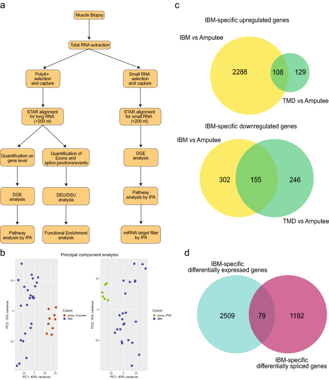
Methods: We performed RNA-seq on RNA extracted from skeletal muscle biopsies of clinically and histopathologically defined IBM (n = 24), tibial muscular dystrophy (n = 6), and histopathologically normal group (n = 9). In a comprehensive transcriptomics analysis, we analyzed the differential gene expression, differential splicing and exon usage, downstream pathway analysis, and the interplay between coding and non-coding RNAs (micro RNAs and long non-coding RNAs).
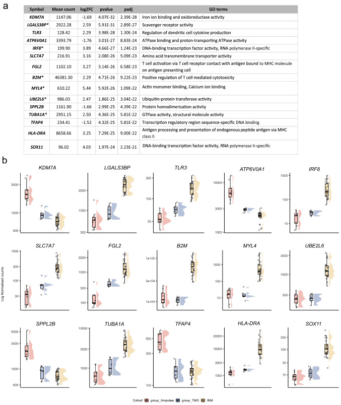
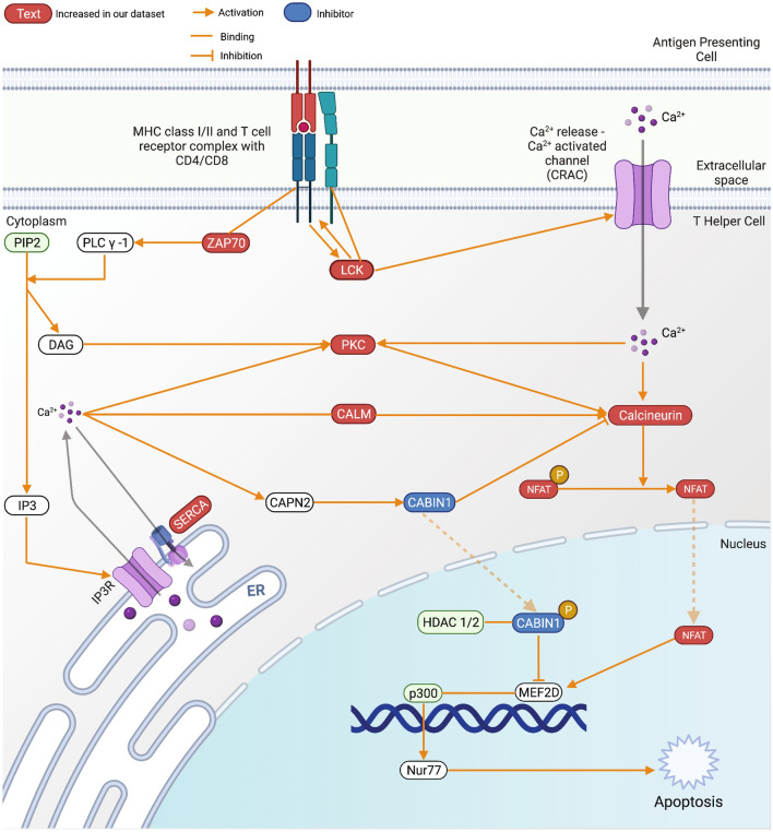
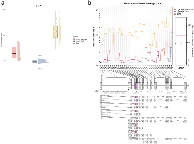
Results: We observe dysregulation of genes involved in calcium homeostasis, particularly affecting the T-cell activity and regulation, causing disturbed Ca2+-induced apoptotic pathways of T cells in IBM muscles. Additionally, LCK/p56, which is an essential gene in regulating the fate of T-cell apoptosis, shows increased expression and altered splicing usage in IBM muscles.
Interpretation: Our analysis provides a novel understanding of the molecular mechanisms in IBM by showing a detailed dysregulation of genes involved in calcium homeostasis and its effect on T-cell functioning in IBM muscles. Loss of T-cell regulation is hypothesized to be involved in the consistent observation of no response to immune therapies in IBM patients. Our results show that loss of apoptotic control of cytotoxic T cells could indeed be one component of their abnormal cytolytic activity in IBM muscles.
Keywords: Calcium; Differential expression; Differential splicing; Inclusion body myositis; T cells.
© 2022. The Author(s).
Conflict of interest statement
The authors report no conflicts of interest.
Figures
Similar articles
-
Calcium dysregulation, functional calpainopathy, and endoplasmic reticulum stress in sporadic inclusion body myositis.Acta Neuropathol Commun. 2017 Mar 22;5(1):24. doi: 10.1186/s40478-017-0427-7. Acta Neuropathol Commun. 2017. PMID: 28330496 Free PMC article.
-
Muscle Transcriptomics Shows Overexpression of Cadherin 1 in Inclusion Body Myositis.Ann Neurol. 2022 Mar;91(3):317-328. doi: 10.1002/ana.26304. Epub 2022 Feb 11. Ann Neurol. 2022. PMID: 35064929 Free PMC article.
-
Loss of TDP-43 function and rimmed vacuoles persist after T cell depletion in a xenograft model of sporadic inclusion body myositis.Sci Transl Med. 2022 Jan 19;14(628):eabi9196. doi: 10.1126/scitranslmed.abi9196. Epub 2022 Jan 19. Sci Transl Med. 2022. PMID: 35044790 Free PMC article.
-
Inclusion body myositis and associated diseases: an argument for shared immune pathologies.Acta Neuropathol Commun. 2022 Jun 3;10(1):84. doi: 10.1186/s40478-022-01389-6. Acta Neuropathol Commun. 2022. PMID: 35659120 Free PMC article. Review.
-
Familial inflammatory inclusion body myositis.Ann Rheum Dis. 2005 Apr;64(4):634-7. doi: 10.1136/ard.2004.025494. Ann Rheum Dis. 2005. PMID: 15769920 Free PMC article. Review.
Cited by
-
Inclusion Body Myositis and Neoplasia: A Narrative Review.Int J Mol Sci. 2022 Jul 1;23(13):7358. doi: 10.3390/ijms23137358. Int J Mol Sci. 2022. PMID: 35806366 Free PMC article. Review.
-
Revealing myopathy spectrum: integrating transcriptional and clinical features of human skeletal muscles with varying health conditions.Commun Biol. 2024 Apr 10;7(1):438. doi: 10.1038/s42003-024-06143-3. Commun Biol. 2024. PMID: 38600180 Free PMC article.
-
NLRP3 inflammasome activation and altered mitophagy are key pathways in inclusion body myositis.medRxiv [Preprint]. 2024 Jun 16:2024.06.15.24308845. doi: 10.1101/2024.06.15.24308845. medRxiv. 2024. Update in: J Cachexia Sarcopenia Muscle. 2025 Feb;16(1):e13672. doi: 10.1002/jcsm.13672. PMID: 38947067 Free PMC article. Updated. Preprint.
-
The Evolution of Complex Muscle Cell In Vitro Models to Study Pathomechanisms and Drug Development of Neuromuscular Disease.Cells. 2022 Apr 5;11(7):1233. doi: 10.3390/cells11071233. Cells. 2022. PMID: 35406795 Free PMC article. Review.
-
Idiopathic inflammatory myopathy and non-coding RNA.Front Immunol. 2023 Sep 6;14:1227945. doi: 10.3389/fimmu.2023.1227945. eCollection 2023. Front Immunol. 2023. PMID: 37744337 Free PMC article. Review.
References
-
- Askanas V, Engel WK. Sporadic inclusion-body myositis: a proposed key pathogenetic role of the abnormalities of the ubiquitin-proteasome system, and protein misfolding and aggregation. Acta Myol. 2005;24:17–24. - PubMed
MeSH terms
Substances
Grants and funding
LinkOut - more resources
Full Text Sources
Medical
Molecular Biology Databases
Research Materials
Miscellaneous
