Soluble CD137 as a dynamic biomarker to monitor agonist CD137 immunotherapies
- PMID: 35236742
- PMCID: PMC8896037
- DOI: 10.1136/jitc-2021-003532
Soluble CD137 as a dynamic biomarker to monitor agonist CD137 immunotherapies
Abstract
Background: On the basis of efficacy in mouse tumor models, multiple CD137 (4-1BB) agonist agents are being preclinically and clinically developed. The costimulatory molecule CD137 is inducibly expressed as a transmembrane or as a soluble protein (sCD137). Moreover, the CD137 cytoplasmic signaling domain is a key part in approved chimeric antigen receptors (CARs). Reliable pharmacodynamic biomarkers for CD137 ligation and costimulation of T cells will facilitate clinical development of CD137 agonists in the clinic.
Methods: We used human and mouse CD8 T cells undergoing activation to measure CD137 transcription and protein expression levels determining both the membrane-bound and soluble forms. In tumor-bearing mice plasma sCD137 concentrations were monitored on treatment with agonist anti-CD137 monoclonal antibodies (mAbs). Human CD137 knock-in mice were treated with clinical-grade agonist anti-human CD137 mAb (Urelumab). Sequential plasma samples were collected from the first patients intratumorally treated with Urelumab in the INTRUST clinical trial. Anti-mesothelin CD137-encompassing CAR-transduced T cells were stimulated with mesothelin coated microbeads. sCD137 was measured by sandwich ELISA and Luminex. Flow cytometry was used to monitor CD137 surface expression.
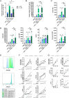
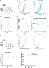
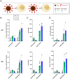
Results: CD137 costimulation upregulates transcription and protein expression of CD137 itself including sCD137 in human and mouse CD8 T cells. Immunotherapy with anti-CD137 agonist mAb resulted in increased plasma sCD137 in mice bearing syngeneic tumors. sCD137 induction is also observed in human CD137 knock-in mice treated with Urelumab and in mice transiently humanized with T cells undergoing CD137 costimulation inside subcutaneously implanted Matrigel plugs. The CD137 signaling domain-containing CAR T cells readily released sCD137 and acquired CD137 surface expression on antigen recognition. Patients treated intratumorally with low dose Urelumab showed increased plasma concentrations of sCD137.
Conclusion: sCD137 in plasma and CD137 surface expression can be used as quantitative parameters dynamically reflecting therapeutic costimulatory activity elicited by agonist CD137-targeted agents.
Keywords: biomarkers; costimulatory and inhibitory T-cell receptors; immunologic; receptors; tumor.
© Author(s) (or their employer(s)) 2022. Re-use permitted under CC BY-NC. No commercial re-use. See rights and permissions. Published by BMJ.
Conflict of interest statement
Competing interests: IM acknowledges grants from Roche, Alligator, Genmab, BMS, AstraZeneca, Pharmamar and Bioncotech, as well as consultancy fees from BMS, Roche, Genmab, Numab, Gossamer, Alligator, AstraZeneca and Pharmamar. MFS receives a grant from Roche.
Figures


Similar articles
-
New emerging targets in cancer immunotherapy: CD137/4-1BB costimulatory axis.ESMO Open. 2020 Jul;4(Suppl 3):e000733. doi: 10.1136/esmoopen-2020-000733. ESMO Open. 2020. PMID: 32611557 Free PMC article. Review.
-
Hypoxia-induced soluble CD137 in malignant cells blocks CD137L-costimulation as an immune escape mechanism.Oncoimmunology. 2015 Jun 24;5(1):e1062967. doi: 10.1080/2162402X.2015.1062967. eCollection 2016. Oncoimmunology. 2015. PMID: 26942078 Free PMC article.
-
CD137 Agonists Targeting CD137-Mediated Negative Regulation Show Enhanced Antitumor Efficacy in Lung Cancer.Front Immunol. 2022 Feb 7;13:771809. doi: 10.3389/fimmu.2022.771809. eCollection 2022. Front Immunol. 2022. PMID: 35197968 Free PMC article.
-
CD137 (4-1BB)-Based Cancer Immunotherapy on Its 25th Anniversary.Cancer Discov. 2023 Mar 1;13(3):552-569. doi: 10.1158/2159-8290.CD-22-1029. Cancer Discov. 2023. PMID: 36576322
-
Deciphering CD137 (4-1BB) signaling in T-cell costimulation for translation into successful cancer immunotherapy.Eur J Immunol. 2016 Mar;46(3):513-22. doi: 10.1002/eji.201445388. Epub 2016 Feb 9. Eur J Immunol. 2016. PMID: 26773716 Review.
Cited by
-
Soluble CD137: A Potential Prognostic Biomarker in Critically Ill Patients.Int J Mol Sci. 2023 Dec 15;24(24):17518. doi: 10.3390/ijms242417518. Int J Mol Sci. 2023. PMID: 38139346 Free PMC article.
-
CD137 (4-1BB) requires physically associated cIAPs for signal transduction and antitumor effects.Sci Adv. 2023 Aug 18;9(33):eadf6692. doi: 10.1126/sciadv.adf6692. Epub 2023 Aug 18. Sci Adv. 2023. PMID: 37595047 Free PMC article.
-
The emerging landscape of novel 4-1BB (CD137) agonistic drugs for cancer immunotherapy.MAbs. 2023 Jan-Dec;15(1):2167189. doi: 10.1080/19420862.2023.2167189. MAbs. 2023. PMID: 36727218 Free PMC article. Review.
-
Neoadjuvant enoblituzumab in localized prostate cancer: a single-arm, phase 2 trial.Nat Med. 2023 Apr;29(4):888-897. doi: 10.1038/s41591-023-02284-w. Epub 2023 Apr 3. Nat Med. 2023. PMID: 37012549 Free PMC article. Clinical Trial.
-
Short-term cultured tumor fragments to study immunotherapy combinations based on CD137 (4-1BB) agonism.Oncoimmunology. 2024 Jul 9;13(1):2373519. doi: 10.1080/2162402X.2024.2373519. eCollection 2024. Oncoimmunology. 2024. PMID: 38988823 Free PMC article.
References
-
- Pollok KE, Kim YJ, Zhou Z. Inducible T cell antigen 4-1BB. Analysis of expression and function. J Immunol 1993;150:771–81. - PubMed
Publication types
MeSH terms
Substances
LinkOut - more resources
Full Text Sources
Other Literature Sources
Research Materials
