

Restoring Ravaged Heart: Molecular Mechanisms and Clinical Application of miRNA in Heart Regeneration
- PMID: 35224063
- PMCID: PMC8866653
- DOI: 10.3389/fcvm.2022.835138
Restoring Ravaged Heart: Molecular Mechanisms and Clinical Application of miRNA in Heart Regeneration
Abstract
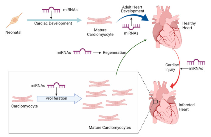
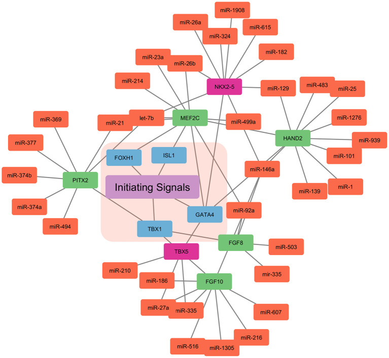
Human heart development is a complex and tightly regulated process, conserving proliferation, and multipotency of embryonic cardiovascular progenitors. At terminal stage, progenitor cell type gets suppressed for terminal differentiation and maturation. In the human heart, most cardiomyocytes are terminally differentiated and so have limited proliferation capacity. MicroRNAs (miRNAs) are non-coding single-stranded RNA that regulate gene expression and mRNA silencing at the post-transcriptional level. These miRNAs play a crucial role in numerous biological events, including cardiac development, and cardiomyocyte proliferation. Several cardiac cells specific miRNAs have been discovered. Inhibition or overexpression of these miRNAs could induce cardiac regeneration, cardiac stem cell proliferation and cardiomyocyte proliferation. Clinical application of miRNAs extends to heart failure, wherein the cell cycle arrest of terminally differentiated cardiac cells inhibits the heart regeneration. The regenerative capacity of the myocardium can be enhanced by cardiomyocyte specific miRNAs controlling the cell cycle. In this review, we focus on cardiac-specific miRNAs involved in cardiac regeneration and cardiomyocyte proliferation, and their potential as a new clinical therapy for heart regeneration.
Keywords: cardiac development; cardiomyocyte; cardiovascular diseases; heart regeneration; miRNA.
Copyright © 2022 Shah and Shah.
Conflict of interest statement
The authors declare that the research was conducted in the absence of any commercial or financial relationships that could be construed as a potential conflict of interest.
Figures
Similar articles
-
miRNA in cardiac development and regeneration.Cell Regen. 2021 Jun 1;10(1):14. doi: 10.1186/s13619-021-00077-5. Cell Regen. 2021. PMID: 34060005 Free PMC article. Review.
-
Applications of miRNAs in cardiac development, disease progression and regeneration.Stem Cell Res Ther. 2019 Nov 21;10(1):336. doi: 10.1186/s13287-019-1451-2. Stem Cell Res Ther. 2019. PMID: 31752983 Free PMC article. Review.
-
Extracellular vesicles and microRNAs in the regulation of cardiomyocyte differentiation and proliferation.Arch Biochem Biophys. 2023 Nov;749:109791. doi: 10.1016/j.abb.2023.109791. Epub 2023 Oct 18. Arch Biochem Biophys. 2023. PMID: 37858665 Review.
-
Cardiomyocyte maturation and its reversal during cardiac regeneration.Dev Dyn. 2024 Jan;253(1):8-27. doi: 10.1002/dvdy.557. Epub 2022 Dec 21. Dev Dyn. 2024. PMID: 36502296 Review.
-
Non-coding RNAs: emerging players in cardiomyocyte proliferation and cardiac regeneration.Basic Res Cardiol. 2020 Aug 3;115(5):52. doi: 10.1007/s00395-020-0816-0. Basic Res Cardiol. 2020. PMID: 32748089 Free PMC article. Review.
Cited by
-
A Thorough Navigation of miRNA's Blueprint in Crafting Cardiovascular Fate.Health Sci Rep. 2024 Nov 5;7(11):e70136. doi: 10.1002/hsr2.70136. eCollection 2024 Nov. Health Sci Rep. 2024. PMID: 39502130 Free PMC article.
-
Platelet-Rich Plasma for Heart Cell Regeneration Post-myocardial Infarction: A Propitious Therapeutic Approach.Cureus. 2024 Jan 9;16(1):e51951. doi: 10.7759/cureus.51951. eCollection 2024 Jan. Cureus. 2024. PMID: 38333505 Free PMC article. Review.
-
MicroRNA29B induces fetal hemoglobin via inhibition of the HBG repressor protein MYB in vitro and in humanized sickle cell mice.Front Med (Lausanne). 2022 Nov 25;9:1043686. doi: 10.3389/fmed.2022.1043686. eCollection 2022. Front Med (Lausanne). 2022. PMID: 36507536 Free PMC article.
-
miRNA Dysregulation in Cardiovascular Diseases: Current Opinion and Future Perspectives.Int J Mol Sci. 2023 Mar 8;24(6):5192. doi: 10.3390/ijms24065192. Int J Mol Sci. 2023. PMID: 36982265 Free PMC article.
References
-
- Roth GA, Abate D, Abate KH, Abay SM, Abbafati C, Abbasi N, et al. . Global, regional, and national age-sex-specific mortality for 282 causes of death in 195 countries and territories, 1980−2017: a systematic analysis for the Global Burden of Disease Study 2017. Lancet. (2018) 392:1736–88. 10.1016/S0140-6736(18)32203-7 - DOI - PMC - PubMed
Publication types
LinkOut - more resources
Full Text Sources