. 2022 Feb 5;14(2):327.
doi: 10.3390/v14020327.

Structural Insights into Alphavirus Assembly Revealed by the Cryo-EM Structure of Getah Virus

Affiliations

Structural Insights into Alphavirus Assembly Revealed by the Cryo-EM Structure of Getah Virus

Ming Wang et al. Viruses. .

Abstract

Getah virus (GETV) is a member of the alphavirus genus, and it infects a variety of animal species, including horses, pigs, cattle, and foxes. Human infection with this virus has also been reported. The structure of GETV has not yet been determined. In this study, we report the cryo-EM structure of GETV at a resolution of 3.5 Å. This structure reveals conformational polymorphism of the envelope glycoproteins E1 and E2 at icosahedral 3-fold and quasi-3-fold axes, which is believed to be a necessary organization in forming a curvature surface of virions. In our density map, three extra densities are identified, one of which is believed a "pocket factor"; the other two are located by domain D of E2, and they may maintain the stability of E1/E2 heterodimers. We also identify three N-glycosylations at E1 N141, E2 N200, and E2 N262, which might be associated with receptor binding and membrane fusion. The resolving of the structure of GETV provides new insights into the structure and assembly of alphaviruses and lays a basis for studying the differences of biology and pathogenicity between arthritogenic and encephalitic alphaviruses.

Keywords: Getah virus; alphavirus; block-based reconstruction; cryo-EM; viral assembly.


Conflict of interest statement

The authors declare no conflict of interest.

Figures

Figure 1
Figure 1
Cryo-EM reconstruction of GETV particles. (A) Radially colored 3D reconstruction of GETV particles. The 5-fold (i5) axis and 2-fold (i2) axis are indicated with a pentagon and a circle, respectively. The icosahedral 3-fold (i3) axis and quasi-3-fold (q3) axis are indicated with black triangles. (B) The central slice of the GETV virion structure. The lipid bilayer is marked with black dashed circles. (C) The 3D reconstruction of inner capsid shell. (D) Each asymmetric unit (ASU) is composed of four E1/E2 heterodimers. Left panel, one of the ASU is indicated with a black dashed quadrilateral. Right panel, a top view (left) and a side view (right) of one ASU. Each E1/E2/CP unit within an ASU is distinguished by different colors (pink, wheat, green, and blue).
Figure 2
Figure 2
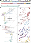
Structure of E1/E2 heterodimer. (A) Linear diagrams show the subdomain distributions in E1, E2, and CP with different colors (DI, light green; DII, light blue; DIII, purple; FL, peach puff; TM of E1, olive; domain A, Indian red; domain B, orchid; domain C, medium purple; domain D, orange; TM of E2, slate blue. CP, turquoise). (B,C) The atomic models of GETV heterodimer displayed with edged ribbon. Subdomains of E1 and E2 are color coded in the same way as (A). (DG) The primary hydrogen bonds formed between E1 and E2 are labeled with dashed lines.
Figure 3
Figure 3
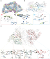
Assembly of envelope glycoproteins. (A,B) Five types of contacts between two adjacent E1. (A) Locations of the five contacts are labeled on the top view of three heterotrimers, indicated by i, ii, iii, iv, and v, respectively. The red dashed lines represent the interacting edges between two adjacent E1. Subdomains of E1 are color coded in the same way as Figure 2A. (B) Conformational changes between E1 from i3 and q3. We overlapped the two E1 from q3, and structural differences between the E1 from i3 (colored) and q3 (grey) are observed. Three contacts (R1, R2, and R3) are enlarged, showing the movement (indicated with dashed arrows) of the major residues. (C) Contacts between E2 from i3 and q3. Top view of E2 in the heterotrimers. E2 from q3 are labeled α (light blue), β (light green), and γ (wheat), and from the corresponding position of i3 are labeled α’ (pink), β’ (pink), and γ’ (pink), respectively. Interactions of α/β, β/γ, α/γ, and α’/β’ (β’/γ’ and α’/γ’ are identical with α’/β’) are enlarged. Dashed lines indicated hydrogen bonds.
Figure 4
Figure 4
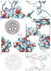
Structure and assembly of CP. (A) Interaction between CP and E2 is mediated by the cytoplasmic tail of E2 inserted into a hydrophobic pocket on the surface of CP. (B) The main residues involved in the interaction between CP and the cytoplasmic tail of glycoproteins (E1 and E2). The dashed lines represent hydrogen bonds. (C) Surface potential of CP. The enlarged images show that the electrostatic interaction between a pentamer and a hexamer is mediated by the complementary charged amino acids. The electrostatic potential ranges from negative (red) to positive (blue). (D) The overall atomic model of capsid. The four copies of CPs in each of the ASUs is colored light blue, light green, wheat, and pink, respectively. A pentamer and a hexamer are labeled with a pentagon and a hexagon, respectively. The enlarged image shows the interactions between CPs. The distances between D127 from pentamer/ hexamer and K158 from hexamer are labeled.
Figure 5
Figure 5
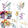
Hydrophobic pocket and three extra densities. (A) Three extra densities located by the E1/E2 transmembrane helix. The density responses to the previously identified “pocket factor” is showed as light green surface with an 18C model fitted in. The two newly identified extra densities are presented as wheat and olive surfaces with two 14C models fitted in, respectively. (B) The hydrophobic pocket with an 18C molecule modelled in. (C) A “lid-like” structure formed by P351 from E2 and W409 from E1 at the posterior of the pocket. (D) Two putative fatty acids are fitted in the positions of the two extra densities, showing they attach to two hydrophobic grooves on the side surface of domain D of E2 and transmembrane helix (TM), respectively. The two putative fatty acid with 14C are displayed as stick. (E,F) Major amino acids from E2 participated in the interaction with the two densities.
Figure 6
Figure 6
The N-linked glycans in E1 and E2. (A) Top view and side view of an asymmetric unit (ASU) showing the locations of the N-linked glycans. The E1/E2 heterodimers are colored blue, green, wheat, and pink, respectively. (BD) Local density and structure of the three N-linked glycans. The glycan linked on N141 from E1 is shown in (B); The glycan linked on N200 from E2 has a close contact with E99 by forming a hydrogen bond (C); The glycans linked on N262 from two adjacent E2 form a “hand shake”-like structure (D).

Similar articles

Cited by

References

    1. Esposito D.L.A., Fonseca B. Will Mayaro virus be responsible for the next outbreak of an arthropod-borne virus in Brazil? Braz. J. Infect. Dis. 2017;21:540–544. doi: 10.1016/j.bjid.2017.06.002. - DOI - PMC - PubMed
    1. Tjaden N.B., Cheng Y., Beierkuhnlein C., Thomas S.M. Chikungunya Beyond the Tropics: Where and When Do We Expect Disease Transmission in Europe? Viruses. 2021;13:1024. doi: 10.3390/v13061024. - DOI - PMC - PubMed
    1. Calisher C.H. Medically important arboviruses of the United States and Canada. Clin. Microbiol. Rev. 1994;7:89–116. doi: 10.1128/CMR.7.1.89. - DOI - PMC - PubMed
    1. Kril V., Aiqui-Reboul-Paviet O., Briant L., Amara A. New Insights into Chikungunya Virus Infection and Pathogenesis. Annu. Rev. Virol. 2021;8:327–347. doi: 10.1146/annurev-virology-091919-102021. - DOI - PubMed
    1. Lefkowitz E.J., Dempsey D.M., Hendrickson R.C., Orton R.J., Siddell S.G., Smith D.B. Virus taxonomy: The database of the International Committee on Taxonomy of Viruses (ICTV) Nucleic Acids Res. 2018;46:D708–D717. doi: 10.1093/nar/gkx932. - DOI - PMC - PubMed

Publication types

MeSH terms

Substances

LinkOut - more resources