A Brief History of Giant Viruses' Studies in Brazilian Biomes
- PMID: 35215784
- PMCID: PMC8875882
- DOI: 10.3390/v14020191
A Brief History of Giant Viruses' Studies in Brazilian Biomes
Abstract
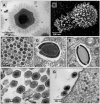
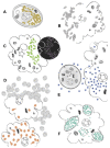
Almost two decades after the isolation of the first amoebal giant viruses, indubitably the discovery of these entities has deeply affected the current scientific knowledge on the virosphere. Much has been uncovered since then: viruses can now acknowledge complex genomes and huge particle sizes, integrating remarkable evolutionary relationships that date as early as the emergence of life on the planet. This year, a decade has passed since the first studies on giant viruses in the Brazilian territory, and since then biomes of rare beauty and biodiversity (Amazon, Atlantic forest, Pantanal wetlands, Cerrado savannas) have been explored in the search for giant viruses. From those unique biomes, novel viral entities were found, revealing never before seen genomes and virion structures. To celebrate this, here we bring together the context, inspirations, and the major contributions of independent Brazilian research groups to summarize the accumulated knowledge about the diversity and the exceptionality of some of the giant viruses found in Brazil.
Keywords: Brazilian isolates; NCLDV; amoebae viruses; giant virus; virosphere; virus diversity.
Conflict of interest statement
The authors declare no conflict of interest.
Figures


Similar articles
-
A long-term prospecting study on giant viruses in terrestrial and marine Brazilian biomes.Virol J. 2024 Jun 10;21(1):135. doi: 10.1186/s12985-024-02404-z. Virol J. 2024. PMID: 38858684 Free PMC article.
-
Discovery and Further Studies on Giant Viruses at the IHU Mediterranee Infection That Modified the Perception of the Virosphere.Viruses. 2019 Mar 30;11(4):312. doi: 10.3390/v11040312. Viruses. 2019. PMID: 30935049 Free PMC article. Review.
-
Giant Viruses of Amoebae: A Journey Through Innovative Research and Paradigm Changes.Annu Rev Virol. 2017 Sep 29;4(1):61-85. doi: 10.1146/annurev-virology-101416-041816. Epub 2017 Jul 31. Annu Rev Virol. 2017. PMID: 28759330 Review.
-
Amoebae: Hiding in Plain Sight: Unappreciated Hosts for the Very Large Viruses.Annu Rev Virol. 2022 Sep 29;9(1):79-98. doi: 10.1146/annurev-virology-100520-125832. Epub 2022 Jun 2. Annu Rev Virol. 2022. PMID: 35655338 Review.
-
Morphologic and Genomic Analyses of New Isolates Reveal a Second Lineage of Cedratviruses.J Virol. 2018 Jun 13;92(13):e00372-18. doi: 10.1128/JVI.00372-18. Print 2018 Jul 1. J Virol. 2018. PMID: 29695424 Free PMC article.
Cited by
-
Diversity of Surface Fibril Patterns in Mimivirus Isolates.J Virol. 2023 Feb 28;97(2):e0182422. doi: 10.1128/jvi.01824-22. Epub 2023 Feb 2. J Virol. 2023. PMID: 36728417 Free PMC article.
-
A long-term prospecting study on giant viruses in terrestrial and marine Brazilian biomes.Virol J. 2024 Jun 10;21(1):135. doi: 10.1186/s12985-024-02404-z. Virol J. 2024. PMID: 38858684 Free PMC article.
-
Eukaryogenesis: The Rise of an Emergent Superorganism.Front Microbiol. 2022 May 11;13:858064. doi: 10.3389/fmicb.2022.858064. eCollection 2022. Front Microbiol. 2022. PMID: 35633668 Free PMC article.
-
Surface fibrils on the particles of nucleocytoviruses: A review.Exp Biol Med (Maywood). 2023 Nov;248(22):2045-2052. doi: 10.1177/15353702231208410. Epub 2023 Nov 13. Exp Biol Med (Maywood). 2023. PMID: 37955170 Free PMC article. Review.
References
Publication types
MeSH terms
LinkOut - more resources
Full Text Sources

