Gelatin Methacrylate Hydrogel for Tissue Engineering Applications-A Review on Material Modifications
- PMID: 35215284
- PMCID: PMC8878046
- DOI: 10.3390/ph15020171
Gelatin Methacrylate Hydrogel for Tissue Engineering Applications-A Review on Material Modifications
Abstract
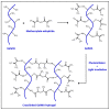
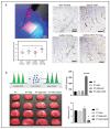
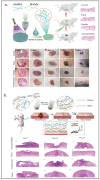
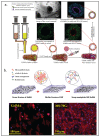
To recreate or substitute tissue in vivo is a complicated endeavor that requires biomaterials that can mimic the natural tissue environment. Gelatin methacrylate (GelMA) is created through covalent bonding of naturally derived polymer gelatin and methacrylic groups. Due to its biocompatibility, GelMA receives a lot of attention in the tissue engineering research field. Additionally, GelMA has versatile physical properties that allow a broad range of modifications to enhance the interaction between the material and the cells. In this review, we look at recent modifications of GelMA with naturally derived polymers, nanomaterials, and growth factors, focusing on recent developments for vascular tissue engineering and wound healing applications. Compared to polymers and nanoparticles, the modifications that embed growth factors show better mechanical properties and better cell migration, stimulating vascular development and a structure comparable to the natural-extracellular matrix.
Keywords: GelMA; biomaterials; hydrogel; material modifications; tissue engineering; vascularization.
Conflict of interest statement
The authors declare no conflict of interest.
Figures


Similar articles
-
Recent Advances on Bioprinted Gelatin Methacrylate-Based Hydrogels for Tissue Repair.Tissue Eng Part A. 2021 Jun;27(11-12):679-702. doi: 10.1089/ten.TEA.2020.0350. Epub 2021 Mar 9. Tissue Eng Part A. 2021. PMID: 33499750 Review.
-
Photo-crosslinkable amniotic membrane hydrogel for skin defect healing.Acta Biomater. 2021 Apr 15;125:197-207. doi: 10.1016/j.actbio.2021.02.043. Epub 2021 Mar 4. Acta Biomater. 2021. PMID: 33676048
-
Biohybrid methacrylated gelatin/polyacrylamide hydrogels for cartilage repair.J Mater Chem B. 2017 Jan 28;5(4):731-741. doi: 10.1039/c6tb02348g. Epub 2017 Jan 3. J Mater Chem B. 2017. PMID: 32263841
-
Calcium Silicate-Activated Gelatin Methacrylate Hydrogel for Accelerating Human Dermal Fibroblast Proliferation and Differentiation.Polymers (Basel). 2020 Dec 27;13(1):70. doi: 10.3390/polym13010070. Polymers (Basel). 2020. PMID: 33375390 Free PMC article.
-
Gelatin methacryloyl (GelMA)-based biomaterials for bone regeneration.RSC Adv. 2019 Jun 5;9(31):17737-17744. doi: 10.1039/c9ra02695a. eCollection 2019 Jun 4. RSC Adv. 2019. PMID: 35520570 Free PMC article. Review.
Cited by
-
Fabrication of Fish Scale-Based Gelatin Methacryloyl for 3D Bioprinting Application.Polymers (Basel). 2024 Feb 1;16(3):418. doi: 10.3390/polym16030418. Polymers (Basel). 2024. PMID: 38337307 Free PMC article.
-
Self-Organization of Long-Lasting Human Endothelial Capillary-Like Networks Guided by DLP Bioprinting.Adv Healthc Mater. 2024 Jun;13(14):e2302830. doi: 10.1002/adhm.202302830. Epub 2024 Feb 20. Adv Healthc Mater. 2024. PMID: 38366136 Free PMC article.
-
Enhancing the mechanical strength of 3D printed GelMA for soft tissue engineering applications.Mater Today Bio. 2023 Dec 30;24:100939. doi: 10.1016/j.mtbio.2023.100939. eCollection 2024 Feb. Mater Today Bio. 2023. PMID: 38249436 Free PMC article. Review.
-
Active Media Perfusion in Bioprinted Highly Concentrated Collagen Bioink Enhances the Viability of Cell Culture and Substrate Remodeling.Gels. 2024 May 5;10(5):316. doi: 10.3390/gels10050316. Gels. 2024. PMID: 38786233 Free PMC article.
-
MOF@platelet-rich plasma antimicrobial GelMA dressing: structural characterization, bio-compatibility, and effect on wound healing efficacy.RSC Adv. 2024 Sep 20;14(41):30055-30069. doi: 10.1039/d4ra04546g. eCollection 2024 Sep 18. RSC Adv. 2024. PMID: 39309655 Free PMC article.
References
-
- Camci-Unal G., Annabi N., Dokmeci M.R., Liao R., Khademhosseini A. Hydrogels for cardiac tissue engineering. NPG Asia Mater. 2014;6:e99. doi: 10.1038/am.2014.19. - DOI
Publication types
Grants and funding
LinkOut - more resources
Full Text Sources

