Oncological transformation in vitro of hepatic progenitor cell lines isolated from adult mice
- PMID: 35210455
- PMCID: PMC8873244
- DOI: 10.1038/s41598-022-06427-w
Oncological transformation in vitro of hepatic progenitor cell lines isolated from adult mice
Abstract
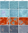
Colorectal cancer cells can transfer the oncogene KRAS to distant cells, predisposing them to malignant transformation (Genometastasis Theory). This process could contribute to liver metastasis; besides, hepatic progenitor cells (HPCs) have been found to be involved in liver malignant neoplasms. The objective of this study is to determine if mouse HPCs-Oval cells (OCs)-are susceptible to incorporate Kras GAT (G12D) mutation from mouse colorectal cancer cell line CT26.WT and if OCs with the incorporated mutation behave like malignant cells. To achieve this, three lines of OCs in different conditions were exposed to CT26.WT cells through transwell co-culture for a week. The presence of KrasG12D and capacity to form tumors were analyzed in treated samples by droplet digital PCR and colony-forming assays, respectively. The results showed that the KrasG12D mutation was detected in hepatic culture conditions of undifferentiated OCs and these cells were capable of forming tumors in vitro. Therefore, OCs are susceptible to malignant transformation by horizontal transfer of DNA with KrasG12D mutation in an undifferentiated condition associated with the liver microenvironment. This study contributes to a new step in the understanding of the colorectal metastatic process.
© 2022. The Author(s).
Conflict of interest statement
The authors declare no competing interests.
Figures


Similar articles
-
Nicotine promotes initiation and progression of KRAS-induced pancreatic cancer via Gata6-dependent dedifferentiation of acinar cells in mice.Gastroenterology. 2014 Nov;147(5):1119-33.e4. doi: 10.1053/j.gastro.2014.08.002. Epub 2014 Aug 12. Gastroenterology. 2014. PMID: 25127677
-
Mutant Hras(G12V) and Kras(G12D) have overlapping, but non-identical effects on hepatocyte growth and transformation frequency in transgenic mice.Liver Int. 2012 Apr;32(4):582-91. doi: 10.1111/j.1478-3231.2011.02732.x. Epub 2012 Jan 3. Liver Int. 2012. PMID: 22221894 Free PMC article.
-
[Mouse models for human colorectal cancer with liver metastasis].Zhonghua Yi Xue Za Zhi. 2019 Sep 10;99(34):2701-2705. doi: 10.3760/cma.j.issn.0376-2491.2019.34.013. Zhonghua Yi Xue Za Zhi. 2019. PMID: 31505723 Chinese.
-
ETS1 regulates Twist1 transcription in a KrasG12D/Lkb1-/- metastatic lung tumor model of non-small cell lung cancer.Clin Exp Metastasis. 2018 Mar;35(3):149-165. doi: 10.1007/s10585-018-9912-z. Epub 2018 Jun 16. Clin Exp Metastasis. 2018. PMID: 29909489
-
Review: KRAS mutations are influential in driving hepatic metastases and predicting outcome in colorectal cancer.Chin Clin Oncol. 2019 Oct;8(5):53. doi: 10.21037/cco.2019.08.16. Chin Clin Oncol. 2019. PMID: 31597434 Review.
Cited by
-
Tumor-derived cell-free DNA and circulating tumor cells: partners or rivals in metastasis formation?Clin Exp Med. 2024 Jan 17;24(1):2. doi: 10.1007/s10238-023-01278-9. Clin Exp Med. 2024. PMID: 38231464 Free PMC article. Review.
-
Efficacy and safety of different options for liver regeneration of future liver remnant in patients with liver malignancies: a systematic review and network meta-analysis.World J Surg Oncol. 2022 Dec 16;20(1):399. doi: 10.1186/s12957-022-02867-w. World J Surg Oncol. 2022. PMID: 36527081 Free PMC article. Review.
-
A Protocol for the Isolation of Oval Cells without Preconditioning.Int J Mol Sci. 2024 Sep 29;25(19):10497. doi: 10.3390/ijms251910497. Int J Mol Sci. 2024. PMID: 39408831 Free PMC article.
-
Differential presence of exons (DPE): sequencing liquid biopsy by NGS. A new method for clustering colorectal Cancer patients.BMC Cancer. 2023 Jan 3;23(1):2. doi: 10.1186/s12885-022-10459-w. BMC Cancer. 2023. PMID: 36593457 Free PMC article.
-
New and Old Key Players in Liver Cancer.Int J Mol Sci. 2023 Dec 5;24(24):17152. doi: 10.3390/ijms242417152. Int J Mol Sci. 2023. PMID: 38138981 Free PMC article. Review.
References
Publication types
MeSH terms
Substances
LinkOut - more resources
Full Text Sources
Medical
Miscellaneous

