. 2022 Feb 21;14(4):1091.
doi: 10.3390/cancers14041091.

Organotypic Epithelial Raft Cultures as a Three-Dimensional In Vitro Model of Merkel Cell Carcinoma

Affiliations

Organotypic Epithelial Raft Cultures as a Three-Dimensional In Vitro Model of Merkel Cell Carcinoma

Arturo Temblador et al. Cancers (Basel). .

Abstract

Merkel cell carcinoma (MCC) is a rare type of skin cancer for which an in vitro model is still lacking. MCC tumorigenesis is associated either with the integration of Merkel cell polyomavirus into the host genome, or with the accumulation of somatic mutations upon chronic exposure to UV light. Transgenic animals expressing the viral oncoproteins, which are constitutively expressed in virus-related MCC, do not fully recapitulate MCC. Although cell-line-derived xenografts have been established for the two subtypes of MCC, they still present certain limitations. Here, we generated organotypic epithelial raft cultures (OERCs) of MCC by using primary human keratinocytes and both virus-positive and virus-negative MCC cell lines. The primary human keratinocytes and the tumor cells were grown on top of a dermal equivalent. Histological and immunohistochemical examination of the rafts confirmed the growth of MCC cells. Furthermore, gene expression analysis revealed differences in the expression profiles of the distinct tumor cells and the keratinocytes at the transcriptional level. In summary, considering the limited availability of patient samples, OERCs of MCC may constitute a suitable model for evaluating the efficacy and selectivity of new drug candidates against MCC; moreover, they are a potential tool to study the oncogenic mechanisms of this malignancy.

Keywords: 3D cell culture model; Merkel cell carcinoma; Merkel cell polyomavirus; gene expression profile; non-melanoma skin cancer; raft culture.


Conflict of interest statement

The authors declare no conflict of interest.

Figures

Figure 1
Figure 1
Development of organotypic epithelial raft cultures of Merkel cell carcinoma cell lines. Different strategies were assayed for the development of organotypic epithelial raft cultures (OERCs). (A) In the first option, a matrix or dermal equivalent was prepared by mixing type 1 rat-tail collagen with murine fibroblasts, which functioned as feeder cells, in a well of a 24-well plate. Dermal equivalents were covered with medium and left to equilibrate for 6–24 h in the incubator. Then, the cell lines of choice—a determined MCC cell line or primary human keratinocytes (PHKs)—were seeded on top of the dermal equivalent and incubated at 37 °C with 5% CO2. The next day, dermal equivalents were lifted on stainless metal grids so that cell cultures could grow at the air–liquid interface for 10–12 days. Eventually, tumor cells were expected to show a dysplastic phenotype, while PHKs should differentiate in a stratified epithelium resembling human skin. (B) Dermal equivalents were prepared as described above. Then, PHKs and MCC cells were mixed at different ratios and seeded on top of the dermal equivalents. In this case, tumor cells were expected to proliferate in the stratified epithelium formed by the differentiation of PHKs. (C) MCC cells were also included in the matrix mixture for the preparation of the dermal equivalents. Once equilibrated, PHKs were added, and cultures were allowed to differentiate. Tumor cells were expected to proliferate in the dermal equivalent, whereas PHKs were expected to differentiate.
Figure 2
Figure 2
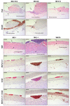
Organotypic epithelial raft cultures (OERCs) of primary human keratinocytes and tumor cells. (A) Hematoxylin and eosin (H&E)-stained section of an OERC of primary human keratinocytes (PHKs). The dermal equivalent is composed of collagen, and contains murine fibroblasts used as feeder cells. On top of the dermal equivalent, the PHKs form a differentiated epithelium. (B) The MCPyV tumor cell line MCC14/2 proliferates on top of the dermal equivalent, showing a dysplastic phenotype, without the presence of PHKs, as observed in the H&E-stained section. These cells express the MCC marker NCAM. (C) MCC14/2 cells also proliferate in OERCs when embedded into the dermal equivalents, as indicated by the arrow, while the PHKs form a differentiated epithelium on top. MCC14/2 cells growing into the dermal equivalent also express NCAM. All images were taken at an overall 200× magnification, and the bars equal 100 μm.
Figure 3
Figure 3
Histological analysis of organotypic epithelial raft cultures (OERCs) of MCC cell lines co-cultured with primary human keratinocytes (PHKs). MCPyV MCC cell lines (MCC14/2, MCC26 and MCC13) grow in OERCs (indicated by arrows in the H&E sections) when co-cultured with PHKs at a ratio of 1 to 5. Except MCC13, these cell lines express NCAM, a typical MCC marker. MCPyV+ MCC cell lines (MS-1, MKL-1 and WAGA) also proliferate in our 3-D culture model, as indicated by full arrows in the H&E sections, when co-cultured with PHKs. These cells express the LT of MCPyV (although the murine fibroblasts stain positive as well, likely due to cross-reactivity to the murine antibody) and the MCC marker CK20, as confirmed by IHC. Double IHC staining for MCPyV LT (brown) and CK20 (red) show co-localization of these signals. PHKs differentiate in a normal epithelium (pointed out by dashed arrows). All images were taken at 20× magnification and the bars equal 100 μm.
Figure 4
Figure 4
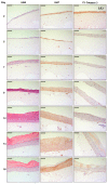
Morphological analysis of organotypic epithelial raft cultures (OERCs) of (A) PHKs and (B) MCC14/2 over time. Hematoxylin and eosin (H&E) staining and immunohistochemical (Ki67 and cleaved caspase 3 staining) analysis were performed at different time points after lifting the rafts. All images were taken at an overall 200× magnification, and the bars equal 100 μm.
Figure 4
Figure 4
Morphological analysis of organotypic epithelial raft cultures (OERCs) of (A) PHKs and (B) MCC14/2 over time. Hematoxylin and eosin (H&E) staining and immunohistochemical (Ki67 and cleaved caspase 3 staining) analysis were performed at different time points after lifting the rafts. All images were taken at an overall 200× magnification, and the bars equal 100 μm.
Figure 5
Figure 5
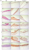
Morphological analysis of organotypic epithelial raft cultures (OERCs) of tumor cells co-cultured with primary human keratinocytes (PHKs) over time. (A) Hematoxylin and eosin (H&E) staining and immunohistochemical (Ki67 and cleaved caspase 3 staining) analysis of MCC14/2 cells (indicated by arrows in the H&E sections) co-cultured with PHKs at different time points after lifting the rafts. (B) H&E staining and immunohistochemical (Ki67 and cleaved caspase 3 staining) analysis of MKL-1 cells (indicated by arrows in the H&E sections) co-cultured with PHKs at different time points after lifting the rafts. All images were taken at an overall 200× magnification, and the bars equal 100 μm.
Figure 5
Figure 5
Morphological analysis of organotypic epithelial raft cultures (OERCs) of tumor cells co-cultured with primary human keratinocytes (PHKs) over time. (A) Hematoxylin and eosin (H&E) staining and immunohistochemical (Ki67 and cleaved caspase 3 staining) analysis of MCC14/2 cells (indicated by arrows in the H&E sections) co-cultured with PHKs at different time points after lifting the rafts. (B) H&E staining and immunohistochemical (Ki67 and cleaved caspase 3 staining) analysis of MKL-1 cells (indicated by arrows in the H&E sections) co-cultured with PHKs at different time points after lifting the rafts. All images were taken at an overall 200× magnification, and the bars equal 100 μm.
Figure 6
Figure 6
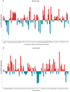
Gene expression profiles of co-cultures of MCC14/2 cells with primary human keratinocytes (PHKs). The plots depict the fold-change expression of (A) extracellular matrix and cell adhesion molecules and (B) growth factors relative to their expression in OERCs of PHKs. Data represent mean values ± SD of three independent experiments.
Figure 7
Figure 7
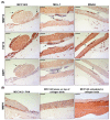
Immunohistochemical analysis of organotypic epithelial raft cultures (OERCs). (A) MCC14/2, MS-1, MKL-1, or WAGA cells co-cultured with primary human keratinocytes (PHKs) expressing matrix metalloproteinase 13 (MMP13), fibroblast growth factor 13 (FGF13), and bone morphogenic protein 7 (BMP7), as indicated by arrows. (B) Matrix metalloproteinase 9 (MMP9) is highly expressed by proliferating basal keratinocytes, and occasionally by MCC14/2 cells. All images were taken at an overall 200× magnification, and the bars equal 100 μm.

Similar articles

Cited by

References

    1. Harms P.W., Harms K.L., Moore P.S., DeCaprio J.A., Nghiem P., Wong M.K.K., Brownell I. International Workshop on Merkel Cell Carcinoma Research Working, G. The biology and treatment of Merkel cell carcinoma: Current understanding and research priorities. Nat. Rev. Clin. Oncol. 2018;15:763–776. doi: 10.1038/s41571-018-0103-2. - DOI - PMC - PubMed
    1. Paulson K.G., Park S.Y., Vandeven N.A., Lachance K., Thomas H., Chapuis A.G., Harms K.L., Thompson J.A., Bhatia S., Stang A., et al. Merkel cell carcinoma: Current US incidence and projected increases based on changing demographics. J. Am. Acad. Dermatol. 2018;78:457–463.e452. doi: 10.1016/j.jaad.2017.10.028. - DOI - PMC - PubMed
    1. Feng H., Shuda M., Chang Y., Moore P.S. Clonal integration of a polyomavirus in human Merkel cell carcinoma. Science. 2008;319:1096–1100. doi: 10.1126/science.1152586. - DOI - PMC - PubMed
    1. White M.K., Khalili K. Interaction of retinoblastoma protein family members with large T-antigen of primate polyomaviruses. Oncogene. 2006;25:5286–5293. doi: 10.1038/sj.onc.1209618. - DOI - PubMed
    1. Hafner C., Houben R., Baeurle A., Ritter C., Schrama D., Landthaler M., Becker J.C. Activation of the PI3K/AKT pathway in Merkel cell carcinoma. PLoS ONE. 2012;7:e31255. doi: 10.1371/journal.pone.0031255. - DOI - PMC - PubMed

LinkOut - more resources