

Detection of Hypoxia in Cancer Models: Significance, Challenges, and Advances
- PMID: 35203334
- PMCID: PMC8869817
- DOI: 10.3390/cells11040686
Detection of Hypoxia in Cancer Models: Significance, Challenges, and Advances
Abstract
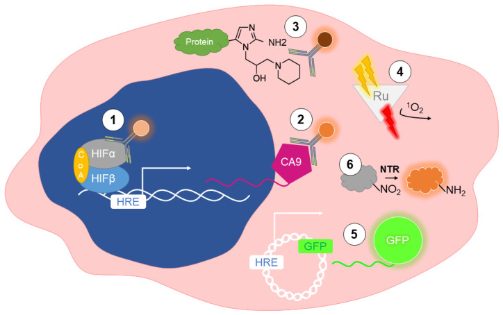
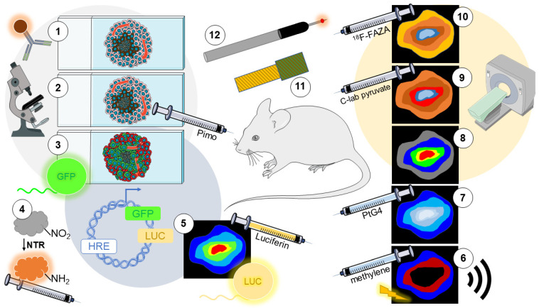
The rapid proliferation of cancer cells combined with deficient vessels cause regions of nutrient and O2 deprivation in solid tumors. Some cancer cells can adapt to these extreme hypoxic conditions and persist to promote cancer progression. Intratumoral hypoxia has been consistently associated with a worse patient prognosis. In vitro, 3D models of spheroids or organoids can recapitulate spontaneous O2 gradients in solid tumors. Likewise, in vivo murine models of cancer reproduce the physiological levels of hypoxia that have been measured in human tumors. Given the potential clinical importance of hypoxia in cancer progression, there is an increasing need to design methods to measure O2 concentrations. O2 levels can be directly measured with needle-type probes, both optical and electrochemical. Alternatively, indirect, noninvasive approaches have been optimized, and include immunolabeling endogenous or exogenous markers. Fluorescent, phosphorescent, and luminescent reporters have also been employed experimentally to provide dynamic measurements of O2 in live cells or tumors. In medical imaging, modalities such as MRI and PET are often the method of choice. This review provides a comparative overview of the main methods utilized to detect hypoxia in cell culture and preclinical models of cancer.
Keywords: HIF; cancer; detection; hypoxia.
Conflict of interest statement
The authors declare no conflict of interest.
Figures
Similar articles
-
Detection and impact of hypoxic regions in multicellular tumor spheroid cultures formed by head and neck squamous cell carcinoma cells lines.SLAS Discov. 2022 Jan;27(1):39-54. doi: 10.1016/j.slasd.2021.10.008. Epub 2021 Oct 13. SLAS Discov. 2022. PMID: 35058175
-
Multi-Parametric Imaging of Hypoxia and Cell Cycle in Intestinal Organoid Culture.Adv Exp Med Biol. 2017;1035:85-103. doi: 10.1007/978-3-319-67358-5_6. Adv Exp Med Biol. 2017. PMID: 29080132
-
Preclinical development and current status of the fluorinated 2-nitroimidazole hypoxia probe N-(2-hydroxy-3,3,3-trifluoropropyl)-2-(2-nitro-1-imidazolyl) acetamide (SR 4554, CRC 94/17): a non-invasive diagnostic probe for the measurement of tumor hypoxia by magnetic resonance spectroscopy and imaging, and by positron emission tomography.Anticancer Drug Des. 1998 Sep;13(6):703-30. Anticancer Drug Des. 1998. PMID: 9755726 Review.
-
Imaging of neurosphere oxygenation with phosphorescent probes.Biomaterials. 2013 Dec;34(37):9307-17. doi: 10.1016/j.biomaterials.2013.08.065. Epub 2013 Sep 6. Biomaterials. 2013. PMID: 24016849
-
Regulation of Cancer-Associated miRNAs Expression under Hypoxic Conditions.Anal Cell Pathol (Amst). 2024 May 10;2024:5523283. doi: 10.1155/2024/5523283. eCollection 2024. Anal Cell Pathol (Amst). 2024. PMID: 38766303 Free PMC article. Review.
Cited by
-
Beyond 2D cell cultures: how 3D models are changing the in vitro study of ovarian cancer and how to make the most of them.PeerJ. 2024 Aug 29;12:e17603. doi: 10.7717/peerj.17603. eCollection 2024. PeerJ. 2024. PMID: 39221267 Free PMC article. Review.
-
EPR and Related Magnetic Resonance Imaging Techniques in Cancer Research.Metabolites. 2023 Jan 1;13(1):69. doi: 10.3390/metabo13010069. Metabolites. 2023. PMID: 36676994 Free PMC article. Review.
-
Tumor hypoxia unveiled: insights into microenvironment, detection tools and emerging therapies.Clin Exp Med. 2024 Oct 3;24(1):235. doi: 10.1007/s10238-024-01501-1. Clin Exp Med. 2024. PMID: 39361163 Free PMC article. Review.
-
Antitumour effect of the mitochondrial complex III inhibitor Atovaquone in combination with anti-PD-L1 therapy in mouse cancer models.Cell Death Dis. 2024 Jan 11;15(1):32. doi: 10.1038/s41419-023-06405-8. Cell Death Dis. 2024. PMID: 38212297 Free PMC article.
-
Oxygen Sensor-Guided Fine Needle Biopsy Studies of Human Cancer Xenografts in Mice.bioRxiv [Preprint]. 2024 Jun 1:2024.05.27.596060. doi: 10.1101/2024.05.27.596060. bioRxiv. 2024. PMID: 38854036 Free PMC article. Preprint.
References
-
- Vaupel P., Mayer A., Briest S., Höckel M. Oxygenation gain factor: A novel parameter characterizing the association between hemoglobin level and the oxygenation status of breast cancers. Cancer Res. 2003;63:7634–7637. - PubMed
Publication types
MeSH terms
Grants and funding
LinkOut - more resources
Full Text Sources
