Review
. 2022 Jun 1:205:114101.
doi: 10.1016/j.bios.2022.114101. Epub 2022 Feb 17.

Isothermal amplification-assisted diagnostics for COVID-19

Affiliations
Review

Isothermal amplification-assisted diagnostics for COVID-19

Mariarita De Felice et al. Biosens Bioelectron. .

Abstract

The scenery of molecular diagnostics for infectious diseases is rapidly evolving to respond to the COVID-19 epidemic. The sensitivity and specificity of diagnostics, along with speed and accuracy, are crucial requirements for effective analytical tools to address the disease spreading around the world. Emerging diagnostic devices combine the latest trends in isothermal amplification methods for nucleic acids with state-of-the-art biosensing systems, intending to bypass roadblocks encountered in the last 2 years of the pandemic. Isothermal nucleic acid amplification is a simple procedure that quickly and efficiently accumulates nucleic acid sequences at a constant temperature, without the need for sophisticated equipment. The integration of isothermal amplification into portable biosensing devices confers high sensitivity and improves screening at the point of need in low-resource settings. This review reports the latest trends reached in this field with the latest examples of isothermal amplification-powered biosensors for detecting SARS-CoV-2, with different configurations, as well as their intrinsic advantages and disadvantages.

Keywords: Biosensors; High throughput; Isothermal amplification; SARS-CoV-2 diagnostics; Sensitive and specific screening.


Conflict of interest statement

The authors declare that they have no known competing financial interests or personal relationships that could have appeared to influence the work reported in this paper.

Figures

Fig. 1
Fig. 1
Schematic of recommended SARS-CoV-2 PAND workflow. MB: molecular beacon; gn: newly generated guide; Q: quencher; F: fluorophore. Reprinted with permission fromWang et al. (2021)for Microalgal-bacterial consortia: PfAgo-based detection of SARS-CoV-2. Biosensors and Bioelectronics. Copyright (2021) Elsevier.
Fig. 2
Fig. 2
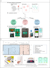
a) SARS-CoV-2 diagnostic workflow. A) Sample collection and preparation illustrating nasopharyngeal swab and RNA extraction. B) Nucleic acid amplification methods for SARS-CoV-2 RNA detection used in this study (RT-qPCR, RT-qLAMP, and RT-eLAMP). Thermal profiles are illustrated for comparison of the assays. C) Point-of-care diagnostics by RT-eLAMP showing the proposed handheld LOC platform including the microfluidic cartridge with control and sample inlets, and the smartphone-enabled application for geolocation and real-time visualization of results. Reprinted with permission ofRodriguez-Manzano et al. (2021) for Handheld Point-of-Care System for Rapid Detection of SARS-CoV-2 Extracted RNA in under 20 min. ACS central science. Copyright (2021) American chemical society. b) Illustration of a small van-sized mobile COVID-19-LAMP diagnostic unit. A drawn-to-scale layout of a van-sized mobile COVID-19-LAMP diagnostic unit, with sample processing and LAMP reactions compartments have been illustrated. A cargo van/lorry can be modified quickly to become a mobile diagnostic unit for rapid deployment in any region. Reprinted with permission fromChow et al. (2020)for A Rapid, Simple, Inexpensive, and Mobile Colorimetric Assay COVID-19-LAMP for Mass On-Site Screening of COVID-19. Int. J. Mol. Sci. Copyright (2020) MDPI. c) Sensitivities of the loop-mediated isothermal amplification (LAMP) assay. A) Seven different dilutions of in vitro transcribed RNA were run for quantitative measurement using qRT-PCR. Relative fluorescence units show a gradient decrease in signals. B) The corresponding PCR products on the electrophoresis gel. C) qRT-PCR standard curve based on the Ct value and dilution factor. D) The serially diluted synthetic RNAs were run in the LAMP assay and colour change represents positive (yellow) or negative (pink). The lower panel shows the LAMP gradient products. Reprinted with permission from Rohaim et al. (2020) for Artificial Intelligence-Assisted Loop Mediated Isothermal Amplification (AI-LAMP) for Rapid Detection of SARS-CoV-2. Viruses. Copyright (2020) MDPI. d) The schematic of specific detection of LAMP products using the Proofman probe. A) LAMP reaction process including the initial amplification phase and exponential amplification phase. Primer design was based on the target sequence: FIP and BIP were the inner primers; F3 and B3 were the outer primers; LF was the loop primer. B) The principle of sequence-specific detection using the Proofman probe. The Proofman probe was designed based on the target sequence and a deliberate mismatch at the 3′ end of the probe was needed to trigger the cleavage activity of the proofreading enzyme (Pfu). Once the Proofman probe binds to the target loop of LAMP products, the Pfu can cleave the probe at the mismatching nucleotide, releasing the fluorophore. Then, the cleaved probe serves as a loop primer to enhance the amplification efficiency. Reprinted with permission from Ding et al. (2021) for Sequence-specific and multiplex detection of COVID-19 virus (SARS-CoV-2) using proofreading enzyme-mediated probe cleavage coupled with isothermal amplification. Biosensors and Bioelectronics. Copyright (2021) Elsevier. e) Outline of COVID-19 RT-LAMP-LFB design. A) Outline of LAMP assay with LF* and LB*. B) Mechanistic description of the COVID-19 RT-LAMP-LFB assay. C) The principle of LFB for visualization of COVID-19 RT-LAMP products. D) Primer design of COVID-19 mRT-MCDA-LFB assay. Up row, SARS-CoV-2 genome organization (GenBank: MN908947, Wuhan-Hu-1). Reprinted with permission from Zhu et al. (2020) for Multiplex reverse transcription loop-mediated isothermal amplification combined with nanoparticle-based lateral flow biosensor for the diagnosis of COVID-19. Biosensors and Bioelectronics. Copyright (2020) Elsevier.
Fig. 2
Fig. 2
a) SARS-CoV-2 diagnostic workflow. A) Sample collection and preparation illustrating nasopharyngeal swab and RNA extraction. B) Nucleic acid amplification methods for SARS-CoV-2 RNA detection used in this study (RT-qPCR, RT-qLAMP, and RT-eLAMP). Thermal profiles are illustrated for comparison of the assays. C) Point-of-care diagnostics by RT-eLAMP showing the proposed handheld LOC platform including the microfluidic cartridge with control and sample inlets, and the smartphone-enabled application for geolocation and real-time visualization of results. Reprinted with permission ofRodriguez-Manzano et al. (2021) for Handheld Point-of-Care System for Rapid Detection of SARS-CoV-2 Extracted RNA in under 20 min. ACS central science. Copyright (2021) American chemical society. b) Illustration of a small van-sized mobile COVID-19-LAMP diagnostic unit. A drawn-to-scale layout of a van-sized mobile COVID-19-LAMP diagnostic unit, with sample processing and LAMP reactions compartments have been illustrated. A cargo van/lorry can be modified quickly to become a mobile diagnostic unit for rapid deployment in any region. Reprinted with permission fromChow et al. (2020)for A Rapid, Simple, Inexpensive, and Mobile Colorimetric Assay COVID-19-LAMP for Mass On-Site Screening of COVID-19. Int. J. Mol. Sci. Copyright (2020) MDPI. c) Sensitivities of the loop-mediated isothermal amplification (LAMP) assay. A) Seven different dilutions of in vitro transcribed RNA were run for quantitative measurement using qRT-PCR. Relative fluorescence units show a gradient decrease in signals. B) The corresponding PCR products on the electrophoresis gel. C) qRT-PCR standard curve based on the Ct value and dilution factor. D) The serially diluted synthetic RNAs were run in the LAMP assay and colour change represents positive (yellow) or negative (pink). The lower panel shows the LAMP gradient products. Reprinted with permission from Rohaim et al. (2020) for Artificial Intelligence-Assisted Loop Mediated Isothermal Amplification (AI-LAMP) for Rapid Detection of SARS-CoV-2. Viruses. Copyright (2020) MDPI. d) The schematic of specific detection of LAMP products using the Proofman probe. A) LAMP reaction process including the initial amplification phase and exponential amplification phase. Primer design was based on the target sequence: FIP and BIP were the inner primers; F3 and B3 were the outer primers; LF was the loop primer. B) The principle of sequence-specific detection using the Proofman probe. The Proofman probe was designed based on the target sequence and a deliberate mismatch at the 3′ end of the probe was needed to trigger the cleavage activity of the proofreading enzyme (Pfu). Once the Proofman probe binds to the target loop of LAMP products, the Pfu can cleave the probe at the mismatching nucleotide, releasing the fluorophore. Then, the cleaved probe serves as a loop primer to enhance the amplification efficiency. Reprinted with permission from Ding et al. (2021) for Sequence-specific and multiplex detection of COVID-19 virus (SARS-CoV-2) using proofreading enzyme-mediated probe cleavage coupled with isothermal amplification. Biosensors and Bioelectronics. Copyright (2021) Elsevier. e) Outline of COVID-19 RT-LAMP-LFB design. A) Outline of LAMP assay with LF* and LB*. B) Mechanistic description of the COVID-19 RT-LAMP-LFB assay. C) The principle of LFB for visualization of COVID-19 RT-LAMP products. D) Primer design of COVID-19 mRT-MCDA-LFB assay. Up row, SARS-CoV-2 genome organization (GenBank: MN908947, Wuhan-Hu-1). Reprinted with permission from Zhu et al. (2020) for Multiplex reverse transcription loop-mediated isothermal amplification combined with nanoparticle-based lateral flow biosensor for the diagnosis of COVID-19. Biosensors and Bioelectronics. Copyright (2020) Elsevier.
Fig. 3
Fig. 3
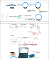
b) Overview of the detection platform. a Detection workflow of SARS-CoV-2 from clinical samples using the electrochemical biosensor with RCA of the N and S genes. b The detection setup for electrochemical analysis by using a portable potentiostat device connected to a laptop. Reprinted with permission fromChaibun et al. (2021)for Rapid electrochemical detection of coronavirus SARS-CoV-2. Nature Communications. Copyright (2021) Nature.c) Schematic illustration of homogeneous circle-to-circle amplification. In the first round of RCA, polymerases act tandemly to generate intermediate amplicons. Intermediate amplicons anneal to CT2 for the second round of RCA, generating amplicon coils that lead to the assembly of MNPs. After a ligation step, all processes of amplification, hybridization, and detection take place simultaneously on-chip at 37 °C.
Fig. 3
Fig. 3
b) Overview of the detection platform. a Detection workflow of SARS-CoV-2 from clinical samples using the electrochemical biosensor with RCA of the N and S genes. b The detection setup for electrochemical analysis by using a portable potentiostat device connected to a laptop. Reprinted with permission fromChaibun et al. (2021)for Rapid electrochemical detection of coronavirus SARS-CoV-2. Nature Communications. Copyright (2021) Nature.c) Schematic illustration of homogeneous circle-to-circle amplification. In the first round of RCA, polymerases act tandemly to generate intermediate amplicons. Intermediate amplicons anneal to CT2 for the second round of RCA, generating amplicon coils that lead to the assembly of MNPs. After a ligation step, all processes of amplification, hybridization, and detection take place simultaneously on-chip at 37 °C.
Fig. 4
Fig. 4
A) Schematic of Toehold switches. Toehold RNA switches consist of a central stem loop structure that harbours a ribosome binding site (RBS, blue) and a translation start site (AUG, pink) with a downstream reporter gene (such as lacZ, grey). A variable region with the toehold (green) are designed to specifically base-pair with a trigger RNA (dark green). In the absence of trigger RNA (left), the RBS and AUG are sequestered within the sensor structure and inaccessible to the ribosome. Presence of the trigger RNA (right) induces intermolecular interactions between the toehold and the trigger RNA, resulting in an alternate conformation wherein the RBS and AUG are accessible to the ribosome, enabling translation of the downstream LacZ enzyme. Production of LacZ is easily monitored with colour, using a chromogenic substrate. The concept is modular and allows the use of alternate reporter genes and modes of detection. B) Schematic showing our assay development pipeline. RNA extracted from viral particles is amplified isothermally using NASBA (Nucleic Acid Sequence-Based Amplification) and detected with specifically designed toehold-based biosensors in an in vitro transcription-translation (IVTT) assay. The NASBA coupled IVTT assay leads to production of colour that can be easily visualized by eye or with cell phone cameras or luminescence that can be quantified by luminometry. Our assay development pipeline focused on identifying targetable regions of the SARS-CoV-2 genome, design of specific biosensors, optimized primers for efficient NASBA and overall sensitivity and response of the assay. Reprinted with permission fromChakravarthy et al. (2021)for Engineered RNA biosensors enable ultrasensitive SARS-CoV-2 detection in a simple colour and luminescence assay. Copyright (2021) medRxiv.
Fig. 5
Fig. 5
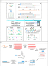
a) Schematic representation of the rapid and multiplex RT-RPA assay with real-time fluorescence and dipstick detection. A) One-pot RT-RPA assay including reverse transcription of the viral RNA and amplification by RPA at constant temperature (37–39 °C). B) Sequences of the primers/probe sets used for SARS-CoV-2 E gene and RdRP gene in the multiplex RT-RPA assay with real-time detection (blue) and sequences of the modified primers used for the multiplex dipstick detection (orange). C) Real-time fluorescence detection by exonuclease cleavage of the probes for E gene and RdRP gene at their THF residue. D) Design of the dipstick for multiplexed detection of the E gene and the RdRP gene. Reprinted with permission fromCherkaoui et al. (2021)for Harnessing recombinase polymerase amplification for rapid multi-gene detection of SARS-CoV-2 in resource-limited settings. Biosensors and Bioelectronics. Copyright (2021) Elsevier. b) Combining RPA with a rkDNA-GO system for the detection of COVID-19. Reprinted with permission fromChoi et al. (2021)for Combined recombinase polymerase amplification/rkDNA–graphene oxide probing system for detection of SARS-CoV-2. Analytica Chimica Acta. Copyright (2021) Elsevier. c) RT-RPA-Coupled Cas12a for Colorimetric Detection of SARS-CoV-2; (A) Schematic Illustration of the Strategy Design. The Whole Process Consists of Three Steps: RT-RPA of the Selected SARS-CoV-2 Genome Region, Cas12a Activation and Colorimetric Detection; (B) SARS-CoV-2 Genome Alignment of the Selected Target Region in the ORF1ab Gene and the N Protein gene; The Accession Numbers of SARS-CoV-2, SARS-CoV, and MERS-CoV Genomes Were NC_045512.2, AY278741.1, and NC_019843.3, Respectively. Reprinted with permission fromZhang et al. (2021). Reverse Transcription Recombinase Polymerase Amplification Coupled with CRISPR-Cas12a for Facile and Highly Sensitive Colorimetric SARS-CoV-2 Detection. Copyright (2021) American Chemical Society. d) All-inclusive portable suitcase lab for deployment at the point of need. Reprinted with permission fromEl Wahed et al. (2021). Reverse Transcription Recombinase Polymerase Amplification Coupled with CRISPR-Cas12a for Suitcase Lab for Rapid Detection of SARS-CoV-2 Based on Recombinase Polymerase Amplification Assay. Copyright (2021) American Chemical Society.
Fig. 5
Fig. 5
a) Schematic representation of the rapid and multiplex RT-RPA assay with real-time fluorescence and dipstick detection. A) One-pot RT-RPA assay including reverse transcription of the viral RNA and amplification by RPA at constant temperature (37–39 °C). B) Sequences of the primers/probe sets used for SARS-CoV-2 E gene and RdRP gene in the multiplex RT-RPA assay with real-time detection (blue) and sequences of the modified primers used for the multiplex dipstick detection (orange). C) Real-time fluorescence detection by exonuclease cleavage of the probes for E gene and RdRP gene at their THF residue. D) Design of the dipstick for multiplexed detection of the E gene and the RdRP gene. Reprinted with permission fromCherkaoui et al. (2021)for Harnessing recombinase polymerase amplification for rapid multi-gene detection of SARS-CoV-2 in resource-limited settings. Biosensors and Bioelectronics. Copyright (2021) Elsevier. b) Combining RPA with a rkDNA-GO system for the detection of COVID-19. Reprinted with permission fromChoi et al. (2021)for Combined recombinase polymerase amplification/rkDNA–graphene oxide probing system for detection of SARS-CoV-2. Analytica Chimica Acta. Copyright (2021) Elsevier. c) RT-RPA-Coupled Cas12a for Colorimetric Detection of SARS-CoV-2; (A) Schematic Illustration of the Strategy Design. The Whole Process Consists of Three Steps: RT-RPA of the Selected SARS-CoV-2 Genome Region, Cas12a Activation and Colorimetric Detection; (B) SARS-CoV-2 Genome Alignment of the Selected Target Region in the ORF1ab Gene and the N Protein gene; The Accession Numbers of SARS-CoV-2, SARS-CoV, and MERS-CoV Genomes Were NC_045512.2, AY278741.1, and NC_019843.3, Respectively. Reprinted with permission fromZhang et al. (2021). Reverse Transcription Recombinase Polymerase Amplification Coupled with CRISPR-Cas12a for Facile and Highly Sensitive Colorimetric SARS-CoV-2 Detection. Copyright (2021) American Chemical Society. d) All-inclusive portable suitcase lab for deployment at the point of need. Reprinted with permission fromEl Wahed et al. (2021). Reverse Transcription Recombinase Polymerase Amplification Coupled with CRISPR-Cas12a for Suitcase Lab for Rapid Detection of SARS-CoV-2 Based on Recombinase Polymerase Amplification Assay. Copyright (2021) American Chemical Society.

Similar articles

Cited by

References

    1. Augustine R., Hasan A., Das S., Ahmed R., Mori Y., Notomi T., Kevadiya B.D., Thakor A.S. Loop-mediated isothermal amplification (LAMP): a rapid, sensitive, specific, and cost-effective point-of-care test for coronaviruses in the context of COVID-19 pandemic. Biology. 2020;9:182. doi: 10.3390/biology9080182. - DOI - PMC - PubMed
    1. Böger B., Fachi M.M., Vilhena R.O., Cobre A.F., Tonin F.S., Pontarolo R. Systematic review with meta-analysis of the accuracy of diagnostic tests for COVID-19. Am. J. Infect. Control. 2021;49:21–29. doi: 10.1016/j.ajic.2020.07.011. - DOI - PMC - PubMed
    1. Bru D., Martin-Laurent F., Philippot L. Quantification of the detrimental effect of a single primer-template mismatch by real-time PCR using the 16S rRNA gene as an example. Appl. Environ. Microbiol. 2008 doi: 10.1128/AEM.02403-07. - DOI - PMC - PubMed
    1. Chaibun T., Puenpa J., Ngamdee T., Boonapatcharoen N., Athamanolap P., O'Mullane A.P., Vongpunsawad S., Poovorawan Y., Lee S.Y., Lertanantawong B. Rapid electrochemical detection of coronavirus SARS-CoV-2. Nat. Commun. 2021;12:802. doi: 10.1038/s41467-021-21121-7. - DOI - PMC - PubMed
    1. Chakravarthy A., Nandakumar A., George G., Ranganathan S., Umashankar S., Shettigar N., Palakodeti D., Gulyani A., Ramesh A. Engineered RNA biosensors enable ultrasensitive SARS-CoV-2 detection in a simple color and luminescence assay. Life Sci. Alliance. 2021;4 doi: 10.26508/lsa.202101213. - DOI - PMC - PubMed